Introduction
The ability to receive, integrate, and respond to light stimuli is essential for insect survival. Photoreception and its downstream physiological effects can directly or indirectly influence insect life cycles and behaviour, with outcomes dependent on the developmental and physiological state of the insect during exposure (Gilbert Reference Gilbert1994; Bradshaw and Holzapfel Reference Bradshaw and Holzapfel2007). For example, insects use photoperiodic time measurement for circadian entrainment and developmental synchronisation (Bradshaw and Holzapfel Reference Bradshaw and Holzapfel2007), and phototaxis behaviour to facilitate dispersal, resource location, and threat avoidance (Gilbert Reference Gilbert1994; Jackowska et al. Reference Jackowska, Bao, Liu, McDonald, Cook and Friedrich2007; Xiang et al. Reference Xiang, Yuan, Vogt, Looger, Jan and Jan2010; Chen et al. Reference Chen, Luo, Kuang, Li, Chen and Liu2013). The mechanisms by which cryptic insects that lack external photoreceptor organs (i.e., are “eyeless”) regulate these and other light-dependent physiological processes are not well understood. The term “cryptic”, which has multiple meanings in the biological sciences, is used in this context to connote the niche or habit of a physically concealed organism.
A range of behavioural, genetic, and developmental evidence indicates that cryptic insects, including beetles, with reduced or absent photoreceptor organs are able to perceive light (Shintani et al. Reference Shintani, Ishikawa and Tatsuki1996; Fox et al. Reference Fox, Coyle-Thompson, Bellinger and Cohen2007; Xiang et al. Reference Xiang, Yuan, Vogt, Looger, Jan and Jan2010; Friedrich et al. Reference Friedrich, Chen, Daines, Bao, Caravas and Rai2011; Shintani Reference Shintani2011; Hustert and Mashaly Reference Hustert and Mashaly2013; Tierney et al. Reference Tierney, Cooper, Saint, Bertozzi, Hyde, Humphreys and Austin2015). Opsins, the G protein-coupled receptors that form the basis of all animal vision, have been detected in many extraocular animal tissues (summarised in Porter et al. Reference Porter, Blasic, Bok, Cameron, Pringle, Cronin and Robinson2012; Porter Reference Porter2016) including the insect brain, where the proteins function independently from image formation (Shimizu et al. Reference Shimizu, Yamakawa, Shimazaki and Iwasa2001; Lampel et al. Reference Lampel, Briscoe and Wasserthal2005; Spaethe and Briscoe Reference Spaethe and Briscoe2005; Velarde et al. Reference Velarde, Sauer, Walden, Fahrbach and Robertson2005). Long-wavelength (LW) visible light (480–600 nm) is particularly important to the functional ecology of insects living in low-light environments, and LW sensitive opsin expression is prevalent among cryptic insects (Jackowska et al. Reference Jackowska, Bao, Liu, McDonald, Cook and Friedrich2007; Friedrich et al. Reference Friedrich, Chen, Daines, Bao, Caravas and Rai2011; Tierney et al. Reference Tierney, Cooper, Saint, Bertozzi, Hyde, Humphreys and Austin2015).
In this study, we used the mountain pine beetle, Dendroctonus ponderosae Hopkins (Coleoptera: Curculionidae: Scolytinae), an eruptive bark beetle species with extensive ecological and economic impacts in North America (Safranyik et al. Reference Safranyik, Carroll, Régnière, Langor, Riel and Shore2010) to investigate photosensitivity in a cryptic insect species. Bark beetles, which are eyeless (lacking stemmata (Jordal Reference Jordal2014)) as larvae, develop primarily within subcortical tree tissues where they are shielded from direct illumination in the immature stages. Dispersing D. ponderosae adults attack host pine trees, boring into the phloem layer beneath the bark where they construct galleries and lay eggs (Safranyik and Carroll Reference Safranyik and Carroll2006). The eggs hatch and the larvae construct galleries in the phloem, passing through four instars before pupating and eclosing into young teneral adults. Once mature, the adults emerge from the natal host and disperse in search of new host trees. Thus, the entire life cycle of D. ponderosae, except for adult dispersal, is completed beneath the bark of pine trees. Based upon existing observations of light sensitivity in cryptic beetles (Friedrich et al. Reference Friedrich, Chen, Daines, Bao, Caravas and Rai2011; Tierney et al. Reference Tierney, Cooper, Saint, Bertozzi, Hyde, Humphreys and Austin2015), including the immature stages of some subcortical tree-tissue developing species (Shintani et al. Reference Shintani, Ishikawa and Tatsuki1996; Doležal and Sehnal Reference Doležal and Sehnal2007; Shintani Reference Shintani2011; Hustert and Mashaly Reference Hustert and Mashaly2013), we hypothesised that D. ponderosae larvae could receive and respond behaviourally to light.
Phototaxis assays were used to assess the ability of D. ponderosae larvae to react to light. After evaluating phototactic behaviour in larvae, we investigated the potential for photoreception in D. ponderosae larvae at the molecular level by examining immature insects for opsin gene expression. We exposed trends in opsin gene transcription across developmental stages and between larval body parts. Our results expand upon current knowledge of extraocular photoreception in insects to include behavioural and genetic evidence of light sensitivity in larvae of a bark beetle.
Methods
Phototaxis experiment
We used phloem sandwich phototaxis assays to test whether D. ponderosae larvae are able to receive and respond behaviourally to light. Larvae were sourced from four lodgepole pine (Pinus contorta Douglas ex Loudon var. latifolia Engelmann (Pinaceae)) trees, felled on 24 September 2015 near Grande Prairie, Alberta, Canada (54.55396°N, 118.71654°W) and on 21 September 2016 outside Whitecourt, Alberta (54.22197°N, 116.59433°W and 54.28255°N, 116.59732°W). Infested logs were held in a walk-in cold room at 0 °C or below until, as required for experimentation, larvae were extracted from beneath the bark. Extracted larvae were held at 22 °C for up to three days so that the insects had resumed activity before use in experiments. To obtain fresh phloem for the experiment, we collected logs from a shore pine (Pinus contorta Douglas ex Loudon var. contorta (Pinaceae)) from Jordan River, Vancouver Island, British Columbia, Canada (48.40119°N, 123.88644°W) in late July 2016. The logs were held at 5.0–6.0 °C until needed for phloem sandwich construction.
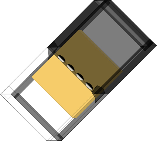
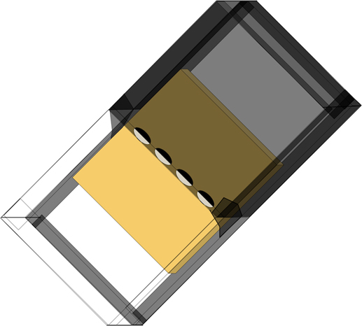
To construct a sandwich (Fig. 1), a 14×14 cm phloem square was peeled away from the sapwood and placed phloem side up in the centre of a large pane of glass (15×30 cm) edged with black duct tape. Four 1.2×0.5 cm oval-shaped punches were made horizontally along the midline of the phloem square, and a third or fourth-instar D. ponderosae was placed into each punch. Two 15×15 cm glass panes, one unaltered and the other coated with black duct tape, were placed on top of the phloem so that the edges butted against one another directly across the punches, and the completed sandwich was tightly secured. Larval movement within the small punches was unconstrained in assembled sandwiches, and thus larvae were distributed and oriented haphazardly in the punches at the time of experiment commencement. The 0.6 cm thick tempered glass panes (Oldcastle Glass, Langley, British Columbia, Canada) used to construct the sandwiches did not filter ultraviolet (UV) light.
Diagram illustrating the layering of the main components of a 15×30 cm phloem sandwich that was designed as a phototaxis assay for third-instar and fourth-instar Dendroctonus ponderosae. Each sandwich was constructed using 0.6 cm thick tempered glass panes, a square of shore pine (Pinus contorta Douglas ex Loudon var. contorta (Pinaceae)) phloem (yellow), clear packing tape, black duct tape, and masking tape (translucent black). The completed sandwiches exposed one half of each larva-containing phloem punch to light (white), and the other to dark (black).
Eight sandwiches were run simultaneously beneath a 35×57 cm bank of two broad-spectrum fluorescent lights (SunBlaster 2' T5 HO 24 W 6400 K with NanoTech T5 Reflector Combo (item number 0900302); SunBlaster Lighting, Langley, British Columbia, Canada). The bulbs mimic sunlight, and emit small amounts of both UVB (280–320 nm) and UVA (320–400 nm) (Sang et al. Reference Sang, He, Wang, Zhu-Salzman and Lei2015) radiation. The light bank was suspended 33.5 cm above the sandwiches and produced a maximum light intensity, or illuminance, of 4740 lux. The phloem sandwiches were arranged in four different grid configurations to account for any geotaxis or directional preference of the larvae beneath the light bank, and each configuration was run twice for a total of eight experimental runs. After 96 hours, sandwiches were disassembled and larvae were individually scored for dark or light microhabitat selection, which we determined from the position of each larva after mining or wandering beneath the treatment panels. Larval galleries were measured to the nearest 0.25 cm. An experimental control of four runs was conducted in the absence of light to assess the expected “light” (unmodified glass) to “dark” (duct-tape covered glass) panel selection ratio of 1:1.
Phloem temperatures beneath the dark and light panels were measured in each sandwich from half of the experimental and control runs. A temperature logger (HOBO Pro v2 2× External Temperature Data Logger (part no. U23-003); Onset Computer Corporation, Bourne, Massachusetts, United States of America), recording the temperature every five minutes, was inserted into each treatment panel. The sensor end of each temperature probe was fit tightly against the phloem, and a small piece of white duct tape on the glass prevented light from transferring to the metal probe directly. The accuracy and resolution of each temperature logger are reported to be ±0.21 °C and ±0.02 °C, respectively (Onset Computer Corporation 2017); using standard deviations of phototaxis control temperature data, we determined the precision of the loggers to be ±0.05 °C.
Phototaxis data analysis
A χ 2 analysis was used to test whether the observed experimental larva microhabitat selection frequencies differed significantly from the expected light to dark selection ratio under the null hypothesis (no effect of light treatment on larval behaviour). Expected frequencies were calculated using data from control larvae assayed in the absence of light. A total of 256 test larvae (four larvae per sandwich) were used in the experiment, of which 65 individuals failed to make a selection (either remaining in the punch and not mining (54 larvae), or, in a few (11 larvae) cases, mining along or returning to the midline), and another 33 either died (14 larvae) or went missing (19 larvae) during the experiment. These 98 larvae were excluded from the analysis, while the 158 insects that did display discrete microhabitat selections were evaluated statistically. Following the same criteria, 57 out of the 128 control larvae (28 dead or missing and 39 that displayed no microhabitat selection) were excluded from the analysis, yielding 71 larval observations suitable for analysis.
A Wilcoxon rank-sum test was used to assess the effect of light treatment on larval gallery lengths, and a Wilcoxon signed-rank test was used to determine whether the mean temperatures (calculated using HOBOware Pro Version 3.7.4; Onset Computer Corporation) of the dark and light treatment panels differed. All statistical analyses were performed in RStudio (RStudio Team 2012; R Core Team 2015).
Opsin gene expression
We first detected the presence of two insect visual opsin orthologs in the D. ponderosae genome (Keeling et al. Reference Keeling, Yuen, Liao, Roderick Docking, Chan and Taylor2013): a LW sensitive and an UV sensitive opsin. This is consistent with National Center for Biotechnology Information amino acid sequence annotations (rhodopsin-like, XP_019757680.1; UV-sensitive-like opsin, XP_019755582.1 (National Center for Biotechnology Information Resource Coordinators 2017)) and published research on opsin diversity in beetles (Sharkey et al. Reference Sharkey, Fujimoto, Lord, Shin, McKenna and Suvorov2017). A bioinformatics server terminal at the University of Victoria (Victoria, British Columbia, Canada) was used to compare a complete opsin sequence dataset for Danio rerio (Hamilton) (Cypriniformes: Cyprinidae) (41 sequences) against the D. ponderosae genome database, using a BLAST (tBLASTx) (Altschul et al. Reference Altschul, Gish, Miller, Myers and Lipman1990) search, and hits were analysed for opsin homology.
We used quantitative polymerase chain reaction and nucleotide sequencing to evaluate the expression of the LW and UV opsins across D. ponderosae life stages (fourth instars, pupae, and teneral and emerged adults), followed by analysis of LW opsin expression in the younger stages (eggs, and first, second, and third instars) (n=4 individuals per life stage, except for eggs where n=2). To assess the location(s) of LW opsin transcription in the larval body, we quantified relative expression in the heads versus bodies of fourth instars (n=7). Insect samples originated from a source population near Grande Prairie, Alberta, and were reared from either field-collected (older life stages) or laboratory-infested logs (younger life stages) under constant temperature (18–19 °C) and photoperiod (17:7 or 10:14 light:dark hours).
RNA extraction and cDNA production
Insect specimens were processed individually using phenol-chloroform RNA extractions (TRIzol; Invitrogen, Thermo Fisher Scientific, Waltham, Massachusetts, United States of America), with the addition of glycogen as needed to increase pellet visibility. RNA was assessed for quality (NanoDrop 2000 Spectrophotometer; Thermo Fisher Scientific), and concentration standardised RNA solutions were subject to genomic DNA deactivation using DNAse 1 (Thermo Fisher Scientific), following manufacturer protocol. Reverse transcription was performed using 5× All-In-One RT MasterMix (Applied Biological Materials, Richmond, British Columbia, Canada), according to the instructions of the manufacturer. The cDNA products were diluted to either 1:2, 1:3, or 1:5 for the quantitative polymerase chain reaction analysis.
Quantitative polymerase chain reaction
Quantitative real-time polymerase chain reaction analyses were performed using 16 μL EvaGreen 2× qPCR MasterMix reactions (SYBR) in a CFX96 Touch Real-Time PCR Detection System (C1000 Touch Thermal Cycler; Bio-Rad, Mississauga, Ontario, Canada), according to manufacturer instructions (Applied Biological Materials). Each cDNA sample was analysed in duplicate or triplicate, alongside no-template negative controls. We analysed LW and UV opsin expression across life stages using presence/absence of transcription only, as the samples were not representative of an independent cohort.
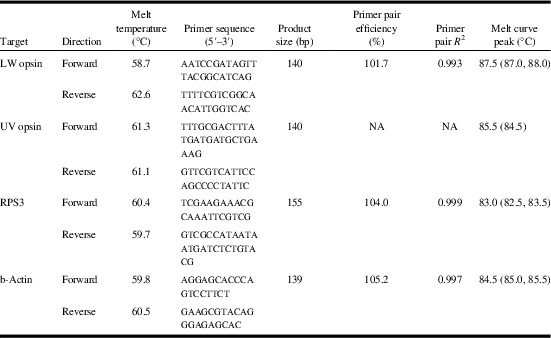
Long-wavelength opsin expression data were normalised using two reference genes, ribosomal protein subunit 3 (RPS3) and beta-actin (bAct) (Sang et al. Reference Sang, He, Wang, Zhu-Salzman and Lei2015). Primers (LW, UV, RPS3, bAct; summarised in Table 1) (Integrated DNA Technologies, Coralville, Iowa, United States of America) were designed using Geneious version 9.1.5 (Kearse et al. Reference Kearse, Moir, Wilson, Stones-Havas, Cheung and Sturrock2012, www.geneious.com), Integrated DNA Technologies OligoAnalyzer 3.1 (www.idtdna.com/calc/analyzer), and Primer-BLAST (Ye et al. Reference Ye, Coulouris, Zaretskaya, Cutcutache, Rozen and Madden2012). Amplified sequence identity was confirmed for each target using Sanger nucleotide sequencing (Macrogen, Maryland or Sequetech, California, United States of America). Standard curves (LW, RPS3, bAct) spanning six orders of magnitude were generated from four 10-fold dilutions of adult beetle cDNA.
Summary of primer melt temperatures and nucleotide sequences, primer pair product sizes (bp, base pairs), efficiencies (%), and R 2 values, and amplified product melt curve peak temperatures (°C), for each of the genes targeted in this study.

LW, long-wavelength; UV, ultraviolet; RPS3, ribosomal protein subunit 3; b-Actin, beta-actin.
Gene expression data analysis
A maximum quantitation-cycle value variation threshold of 0.5 cycles for technical replicates was enforced for each sample. For samples run in triplicate, the two replicates with the closest quantitation-cycle values were selected. Relative expression of the LW opsin was quantified using methods outlined by Brookman-Amissah et al. (Reference Brookman-Amissah, Packer, Prediger, Sabel, Gunstream and Hellemans2015: 57–58, equation (4)). Calculations and subsequent analyses were performed separately for the LW opsin relative to either reference gene (RPS3 or bAct). Relative-expression values were log transformed before analysis, and normality of the logR distributions confirmed. A paired t-test was implemented to evaluate expression levels in the heads versus bodies of fourth instars. The presence/absence of LW and UV opsin transcripts was determined by comparing technical replicate quantitation-cycle values (at least two replicates P to EA<0.5 cycles apart), and melt curve peaks with the known values for the previously sequenced products. Analyses were executed in RStudio (RStudio Team 2012; R Core Team 2015), and data were plotted using ggplot2 (Wickham Reference Wickham2009).
Results
Phototaxis experiment
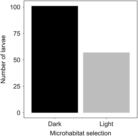
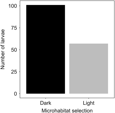
Dendroctonus ponderosae larvae were negatively phototactic, with 101 larvae recovered from the dark microhabitats and 57 from the light microhabitats (χ 2 1=8.83; P<0.01) (Fig. 2) at the time of experiment completion. Control larvae did not deviate from the 1:1 “light” (unmodified glass) to “dark” (duct-tape covered glass) panel selection ratio within phloem sandwich units assayed in the absence of light (dark n=37, light n=34). The dark to light microhabitat selection ratio of the test larvae (101/57) was compared with the panel selection frequency calculated from the control larvae (37/34). There was no difference in the mean temperature of the light and dark microhabitats (mean±standard deviation: dark panel 23.8±1.06 °C, light panel 23.8±1.09 °C) (Wilcoxon signed-rank test: V=219.5; P=0.41). Light treatment had no effect on larval gallery lengths (dark 2.51±1.41 cm, light 2.91±1.65 cm) (Wilcoxon rank-sum test: W=1532.5; P=0.16).
Counts of 158 mature Dendroctonus ponderosae larvae by light or dark microhabitat selection across eight phototaxis experiment runs (dark n=101, light n=57). The χ 2 analysis reveals a significant effect of light treatment on larval behaviour (χ 2 1=8.83; P<0.01). An expected microhabitat selection ratio was obtained from 71 control larvae assayed in the absence of light (37/34 dark/light).
Opsin gene expression analysis
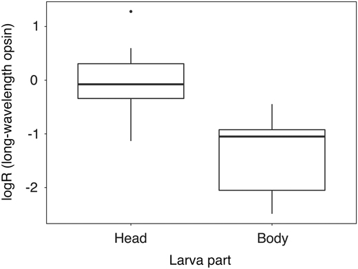
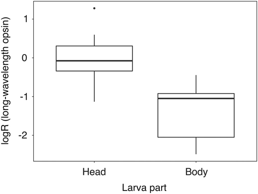
Long-wavelength opsin transcripts were recovered from each sample and every D. ponderosae life stage surveyed, including eggs. All technical replicates amplified before cycle 32, and the majority of products exhibited distinct melt curve peaks (87.5 °C). Ultraviolet opsin transcripts were recovered from pupae, teneral, and emerged adults, but not from larvae. At least two technical replicates for each post-larval sample were <0.5 cycles apart with consistent melt peaks (85.5 °C), in contrast to the lack of specific product amplification in fourth instars. The LW opsin was expressed throughout the bodies of mature larvae, and transcript levels were significantly elevated in the head compared with the body (RPS3 paired t-test: t 6=2.71; P<0.05 (Fig. 3); bAct paired t-test: t 6=2.91; P<0.05). Efficiencies for each of three primer sets (LW, RPS3, bAct), and melt curve peaks for each amplified product (LW, UV, RPS3, bAct), are summarised in Table 1. There was no genomic DNA amplification in any of our RNA control (DNAse-treated) samples, indicating that all quantitative polymerase chain reaction analysis reflected levels of gene expression only (data not shown). Additionally, Sanger sequencing confirmed that the RPS3 product was consistent with cDNA and not genomic amplification, as it lacked the intronic region spanned by the RPS3 primers.
Boxplot of logR values for long-wavelength opsin expression in the heads versus bodies of fourth-instar Dendroctonus ponderosae (n=7), showing significantly higher transcript levels in the head than the body (paired t-test: t 6=2.71; P<0.05 (RPS3 reference gene)).
Discussion
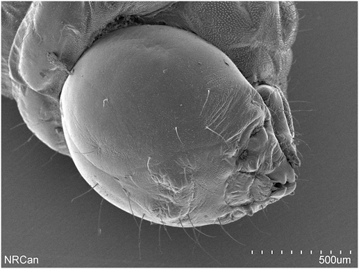
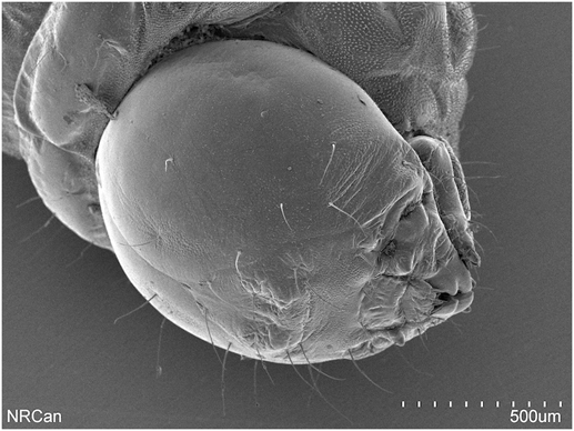
This study revealed that mature D. ponderosae larvae are photosensitive and respond behaviourally to light. We demonstrated negative phototaxis in mature larvae using a phloem sandwich assay. While positive phototaxis, and reversal to negative phototaxis after a period of flight, is known in adult D. ponderosae and other bark beetles (Reid Reference Reid1962; Shepherd Reference Shepherd1966; Safranyik Reference Safranyik1978; Groberman and Borden Reference Groberman and Borden1982; Safranyik et al. Reference Safranyik, Silversides, McMullen and Linton1989), our results represent what we believe to be the first description of negative phototaxis among larval Scolytinae. Long-wavelength opsin transcripts were recovered from all life stages, including eggs (potentially late embryos), whereas UV opsin transcripts were recovered from pupae and adults but not from larvae. The heads of mature larvae had higher levels of LW opsin transcription than the bodies. All immature Scolytinae, including D. ponderosae (Fig. 4), lack stemmata (Jordal Reference Jordal2014), and therefore our data provide support for an opsin-mediated mechanism of extraocular photorception in D. ponderosae larvae.
Scanning electron micrograph of a fourth-instar Dendroctonus ponderosae showing the lack of exterior photoreceptor organs (stemmata). The larval specimen was processed by critical point drying and white gold sputter coating, and was imaged using a Hitachi S-3400N Scanning Electron Microscope (Pacific Forestry Centre, Canadian Forest Service, Natural Resources Canada; Victoria, British Columbia, Canada).
Our finding of negative phototactic behaviour in bark beetle larvae is consistent with other reports of light avoidance by eyeless invertebrates including Caenorhabditis elegans Maupas (Rhabditida: Rhabditidae) (Edwards et al. Reference Edwards, Charlie, Milfort, Brown, Gravlin, Knecht and Miller2008; Gong et al. Reference Gong, Yuan, Ward, Kang, Zhang and Wu2016), the springtail Folsomia candida Willem (Collembola: Isotomidae) (Fox et al. Reference Fox, Coyle-Thompson, Bellinger and Cohen2007), and larval Drosophila melanogaster Meigen (Diptera: Drosophilidae) (Xiang et al. Reference Xiang, Yuan, Vogt, Looger, Jan and Jan2010; Kane et al. Reference Kane, Gershow, Afonso, Larderet, Klein and Carter2013), and by cryptic beetles with reduced ocular organs including the troglomorphic beetle Ptomaphagus hirtus (Tellkampf) (Coleoptera: Leiodidae: Cholevinae) (Friedrich et al. Reference Friedrich, Chen, Daines, Bao, Caravas and Rai2011) and larvae of Rhynchophorus ferrugineus (Olivier) (Coleoptera: Curculionidae: Dryophthorinae) (Hustert and Mashaly Reference Hustert and Mashaly2013). Negative phototaxis in Drosophila Fallén larvae is essential to survival, as, analogous to the habits of bark beetle larvae, the insects must remain submerged in their food to avoid exposure to threats (Xiang et al. Reference Xiang, Yuan, Vogt, Looger, Jan and Jan2010). The discovery by Hustert and Mashaly (Reference Hustert and Mashaly2013) of negative phototaxis and LW light sensitivity in the ventral nerve chord of R. ferrugineus larvae lends support to our results for larval D. ponderosae. Immature R. ferrugineus do possess stemmata (Anderson and Marvaldi Reference Anderson and Marvaldi2014), however, both R. ferrugineus and D. ponderosae inhabit subcortical tree tissues as larvae and likely encounter comparable light microhabitats.
We suggest that negative phototaxis in D. ponderosae larvae functions to maintain the placement of the insect within the phloem layer, the subcortical habitat that provides access to essential resources and physical protection from threats. Woodpeckers (Piciformes: Picidae: Picinae) are highly efficient predators of developing bark beetles, and are responsible for extensive removal of pine bark in natural systems (Amman and Cole Reference Amman and Cole1983; reviewed in Safranyik and Carroll Reference Safranyik and Carroll2006). Woodpeckers may therefore exert direct (i.e., predation) and indirect (i.e., exposure to other biotic and abiotic threats) selection pressures favouring light avoidance behaviour in D. ponderosae larvae developing beneath the bark. Negative phototaxis may also contribute to larval distribution within intact bark tissues, as LW light (green, 550 nm) is known to penetrate farther into leaf tissues than blue and red light (Vogelmann Reference Vogelmann1993) and could theoretically produce a low-light microhabitat gradient within the phloem layer.
Dendroctonus ponderosae larvae are sensitive to light despite the lack of stemmata, which likely indicates extraocular photoreceptive abilities. The most obvious candidate extraocular photoreceptor was the LW sensitive opsin that we found expressed throughout the D. ponderosae larval body. Our interpretation of the detectable expression levels in both the head and body is that the LW opsin is likely transcribed throughout the central nervous system, in both the brain (probably in the optic lobes (Lampel et al. Reference Lampel, Briscoe and Wasserthal2005; Spaethe and Briscoe Reference Spaethe and Briscoe2005; Buschbeck and Friedrich Reference Buschbeck and Friedrich2008)) and the ventral nerve chord (Hustert and Mashaly Reference Hustert and Mashaly2013). The identification of LW opsin expression in D. ponderosae larvae is consistent with other studies of cryptic beetle opsins, including P. hirtus (Friedrich et al. Reference Friedrich, Chen, Daines, Bao, Caravas and Rai2011) and Limbodessus palmulaoides Watts (Coleoptera: Dytiscidae) (Tierney et al. Reference Tierney, Cooper, Saint, Bertozzi, Hyde, Humphreys and Austin2015).
The recovery of LW opsin transcripts from all D. ponderosae life stages likely reflects the importance of the low-level light detection throughout the life cycle of the beetle (Jackowska et al. Reference Jackowska, Bao, Liu, McDonald, Cook and Friedrich2007). In contrast, UV opsin transcripts were recovered from D. ponderosae pupae and adults and not from fourth instars, which is coincident with the development of compound eyes. While both LW and UV opsins expressed in the compound eyes are likely to function in adult D. ponderosae dispersal, UV wavelengths are more directly linked to navigation in weevils and other insects (Chen et al. Reference Chen, Luo, Kuang, Li, Chen and Liu2013; Wang et al. Reference Wang, Xiao, Bian, Niu, Murphy and Huang2013; Stone et al. Reference Stone, Mangan, Ardin and Webb2014; Ilić et al. Reference Ilić, Pirih and Belušič2016). Ultraviolet opsins orthologs are absent in a number of cryptic beetles (Friedrich et al. Reference Friedrich, Chen, Daines, Bao, Caravas and Rai2011; Tierney et al. Reference Tierney, Cooper, Saint, Bertozzi, Hyde, Humphreys and Austin2015; Sharkey et al. Reference Sharkey, Fujimoto, Lord, Shin, McKenna and Suvorov2017) that presumably do not require UV sensitivity for adult navigation.
In addition to the reception of phototaxis-inducing light stimuli, the LW sensitive opsin could also mediate circadian regulation and photoperiodism in D. ponderosae larvae. Both cerebral and visual opsins have been speculated to function in circadian entrainment and the reception of photoperiodic information in the brains of insects (Shimizu et al. Reference Shimizu, Yamakawa, Shimazaki and Iwasa2001; Lampel et al. Reference Lampel, Briscoe and Wasserthal2005; Spaethe and Briscoe Reference Spaethe and Briscoe2005; Velarde et al. Reference Velarde, Sauer, Walden, Fahrbach and Robertson2005). Furthermore, the internalised adult stemmata of endopterygote insects, exemplified by the Hofbauer–Buchner eyelets in Drosophila that originate from larval Bolwig organs, function as deep-brain photoreceptor organs that are implicated in circadian regulation (reviewed in Buschbeck and Friedrich Reference Buschbeck and Friedrich2008). It is therefore possible that photoperiod and light-mediated entrainment of the larval circadian clock influences D. ponderosae phenology (Shintani et al. Reference Shintani, Ishikawa and Tatsuki1996; Doležal and Sehnal Reference Doležal and Sehnal2007; Shintani Reference Shintani2011), metabolism, and/or activity patterns (Friedrich Reference Friedrich2013; Tierney et al. Reference Tierney, Friedrich, Humphreys, Jones, Warrant and Wcislo2017). Future studies may identify if and how D. ponderosae uses light information during early development to regulate daily and seasonal rhythms.
Acknowledgements
The authors thank J. Taylor, M. Ballinger, G. Smith, B. Van Hezewijk, W. Strong, C. Dunbar, T. Holmes, S. Pokorny, and J. Wertman for valuable insight and technical assistance. Support for this research was provided by Natural Resources Canada (K.P.B.) and the Natural Sciences and Engineering Research Council of Canada (S.J.P.).