Introduction
Infertility is a common and worrying issue for couples. Therefore, it is necessary to find methods to prevent infertility. Statistical studies have shown that 50% of infertility cases in couples are due to female factors (Turner, Reference Turner2003; Fathi et al., Reference Fathi, Valojerdi, Ebrahimi, Eivazkhani, Akbarpour and Tahaei2017). Some women develop premature ovarian failure (POF) so that, before the age of 40, the production of estrogen in the ovary stops and no ovulation takes place. The etiology of POF is unknown yet; however, it may be related to genetic disorders, autoimmune diseases, infections, enzyme deficiencies, metabolic syndromes and smoking. In some cases, it is due to medical interventions such as ovarian surgery or long-term use of GnRH or chemotherapy drugs (Van Der Voort et al., Reference Van Der Voort, van Der Weijer and Barentsen2003; Okeke et al., Reference Okeke, Anyaehie and Ezenyeaku2013). Cyclophosphamide (Cy) is described as an alkylating compound utilized for the therapy of various malignant and non-malignant disorders; however, it can have detrimental impacts, such as pulmonary fibrosis, gastrointestinal disorder, kidney infection, mutagenesis, and impaired fertility and could stimulated POF (Meirow et al., Reference Meirow, Assad, Dor and Rabinovici2004). The Cy also causes atrophy of the ovaries, shrinkage of the ovarian follicles, and reduction of the number of follicles in the stages of primary, secondary, and antral. This paves the way for ovarian infertility and infertility (Jiang et al., Reference Jiang, Zhao, Qi, Li, Zhang, Song, Yu and Gao2013). Utilizing platelet-rich plasma (PRP) in clinics has gained particular interest since the 1970s. The PRP is derived from autologous blood whose platelet concentration is significantly high and is rich in growth factors, for example, connective tissue growth factor, platelet-derived growth factor (PDGF), keratinocyte growth factor, interleukin 8 (IL-8), transforming growth factor beta (TGF-β), fibroblast growth factor (FGF), epidermal growth factor (EGF), and vascular endothelial growth factor (VEGF) (Sugiura et al., Reference Sugiura, Su, Li, Wigglesworth, Matzuk and Eppig2009; Lin et al., Reference Lin, Jia and Zhang2011; Nagashima et al., Reference Nagashima, Kim, Li, Lydon, DeMayo, Lyons and Matzuk2011; Dhillon et al., Reference Dhillon, Schwarz and Maloney2012). As many people with autoimmune diseases and cancer are at risk of losing their ovarian reserve or for whatever reason oocytes are unable to reach puberty in vivo, obtaining a protocol to improve the developmental process and ovarian reserve recovery is needed.
In this study, an attempt was made to provide an environment that improves the development process and restore the ability of damaged ovary. According to recent studies, conditioned medium (CM) or the optimal culture medium derived from cells includes a variety of enzymes, cytokines, growth factors, and hormones involved in regulating cell growth, differentiation, repair, and angiogenesis (Chen et al., Reference Chen, Zhuang, Chen and Huang2011; Srivastava et al., Reference Srivastava, Sengupta, Kriplani, Roy and Ghosh2013; Hosseini et al., Reference Hosseini, Shirazi, Naderi, Shams-Esfandabadi, Borjian Boroujeni, Sarvari, Sadeghnia, Behzadi and Akhondi2017). Given that PRP contains many of the factors required for folliculogenesis, it seems that PRP and CM can be effective in improving the developmental process and restoring the ability of the damaged ovary (Malekshah et al., Reference Malekshah, Moghaddam and Daraka2006; Hosseini et al., Reference Hosseini, Shirazi, Naderi, Shams-Esfandabadi, Borjian Boroujeni, Sarvari, Sadeghnia, Behzadi and Akhondi2017; Ahmadian et al., Reference Ahmadian, Sheshpari, Pazhang, Bedate, Beheshti, Abbasi, Nouri, Rahbarghazi and Mahdipour2020). Growth differentiation factor 9 (GDF9) and bone morphogenetic protein 15 (BMP15) genes, released by the oocytes, are capable of modulating primordial follicle recruitment, granulosa cell differentiation and proliferation, and steroids synthesis, and increasing preantral follicles growth (Fenwick et al., Reference Fenwick, Mora, Mansour, Baithun, Franks and Hardy2013). Bone morphogenetic proteins (BMPs), as one of the members of superfamily of the transforming growth factor-β (TGF-β) in the ovary, regulate cell proliferation, migration, and stem cell differentiation. The GDF-9 and BMP15 are expressed in each phase of follicular growth and have a role in monitoring and steroidogenesis and proliferation of granulosa cells. The Smad family is a subgroup of proteins belonging to signal transduction molecules responsible for the transmission of TGF-β signals from the cell surface into the nucleus. Smad1, Smad2, and Smad3 are receptor-activated Smads related to both activin and TGF-β signalling (Xu et al., Reference Xu, Oakley and McGee2002). This is the first report to introduce the development of mice ovarian follicles using CM derived from endometrial cells and PRP.
Materials and methods
Animals
In total, 65 healthy BALB/c female mice (∼28–30 g and 6-8 weeks old) in five groups were studied. There were 10 mice in each group and adult female mice were used for blood sampling, preparation of PRP, and conditioned medium (CM). Animals were kept in normal conditions (temperature: 21 ± 2°C and light and dark cycle: 12 h) with free accessibility to water and food in standard cages. All procedures were done according to the instruction of the Ethics Committee of Shahid Beheshti University of Medical Sciences (Reference number: IR.SBMU.RETECH.REC.1399.247).
Experimental groups
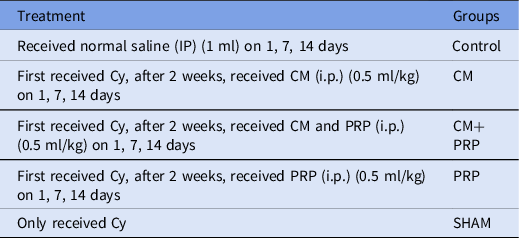
Control: received normal saline intraperitoneally, Sham (CYC): They only received cyclophosphamide (CYC), (i.p.). CM: First received cyclophosphamide and after a week of infertility, received conditioned medium i.p. (0.5 ml) (Pouya et al., Reference Pouya, Heidari, Baghaei, Asadzadeh Aghdaei, Moradi, Namaki, Zali and Hashemi2018) on 1st, 7th, and 14th days. PRP: First received cyclophosphamide and after a week of infertility, received PRP (0.5 ml) i.p. on 1st, 7th, and 14th days.CM+PRP: First, received cyclophosphamide and after a week of infertility, received (0.5 ml) i.p. on 1st, 7th, and 14th days. The list of experimental groups is given in Table 1.
Experimental groups

Preparation of conditioned media
The required CM was obtained from endometrial cells (in the prime phase of the menstrual cycle [menstrual phase]). The culture of newly extracted stromal cells and epithelial cells (30,000 cells) was performed in the growth medium in six-well plates for 1 day, and then they were washed with phosphate-buffered saline (PBS) and subsequently substituted with growth medium (3 ml). In the next step, collecting, filtering, and sterilizing of the CM were carried out (After 2 days of culture), and finally, it was stored at −80 °C until the experiment. For concentrating the factors secreted from the cells of endometrial niche, the stromal and epithelial cells were cultured in serum-free DMEM/F-12 medium (1 ml). After that, the secreted agents in the CM (after 48 h) were concentrated (CCM) by centrifuging (4000 g for 20 min at 4 °C) through the use of Amicon ultra-15 centrifugal filter devices (EMD Millipore) with a molecular weight cutoff of 10 kDa. The amount of the concentrated protein originated from one culture well was assumed as one unit and 1 ml of concentrated (CCM) was used (Srivastava, et al; Reference Srivastava, Sengupta, Kriplani, Roy and Ghosh2013).
Preparation of platelet-rich plasma
After collecting venous blood (10 ml) from models of induced disc-degeneration by syringes (10 ml), including 3000 U of heparin, the obtained blood sample was transferred to a tube (10 ml) and subsequently centrifuged at 2400 rpm for 10 min. Next, the whole buffy coat and supernatant in the tube were collected and transferred into another tube (10 ml). After centrifuging the tube at 3600 rpm for 15 min, upper three-quarters of the supernatant in the tube were removed, and the remaining section was considered as the PRP. The PRP solution was activated with calcium gluconate to a final concentration of 1 × 10⁸ cells/ml. (Dehghani, et al; Reference Dehghani, Aboutalebi, Esmaeilpour, Panjehshahin and Bordbar2018, Aflatoonian, etal; Reference Aflatoonian, Lotfi, Saeed and Tabibnejad2021)
Preparation of cyclophosphamide
75 mg/kg of Cy powder (Sigma, USA) was dissolved in 0.9% normal saline. Then, the injection was performed based on the animal’s weight (Dehghani, et al; Reference Dehghani, Aboutalebi, Esmaeilpour, Panjehshahin and Bordbar2018).
Induction of infertility in mice using cyclophosphamide
Mice weight was measured and Cyclophosphamide was prescribed at 75 mg/kg by i.p. injection irrespective of the reproductive cycle (single dose). (Dehghani, et al; Reference Dehghani, Aboutalebi, Esmaeilpour, Panjehshahin and Bordbar2018.)
Tissue processing
The fixation of ovarian tissue samples was conducted (overnight) in 4% (w/v) paraformaldehyde/0.02 M PBS (pH 7.2) at 4°C. After that, their washing in running water, dehydrating by gradient alcohol (70, 80, 90, 95, and 100% alcohol I, II), and transparentizing in xylene were performed. Following embedding in paraffin, each paraffinized ovary sample was sectioned (serially) at a 4–5 μM thickness. Totally, 20 slides were evaluated, these were selected from every 30 slides to prevent the evaluation of the same follicle more than once.
Immunohistochemistry
The sections were fixed in PBS comprising 4% paraformaldehyde for 20 min. In the next step, the non-specific binding sites were blocked with PBS comprising 10% horse serum, 0.3% Triton X-100, and 1% BSA for 45 min. The primary antibodies, i.e., GDF-9 (sc-12244 Santa Cruz) and GDF-9B (sc-28911 Santa Cruz), were diluted at 1:200 and 1:100 ratios, respectively. Then, the sections were incubated (overnight) at 4° C in the suitable dilutions of the mentioned antibodies. Subsequently, incubation with secondary antibody rabbit anti-goat SABC Kit was performed. After washing with PBS comprising BSA, the cells were counter stained with 40,6-diamidino-2-phenylindole (DAPI) (Sigma) for 5 min and observed under a fluorescence microscope (Zeiss, Germany) with the appropriate excitation wavelength filters.
Quantitative real-time polymerase chain reaction
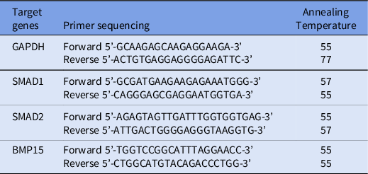
Gene expression (SMAD1, SMAD2, and BMP15) was evaluated using the real-time PCR technique. GAPDH was assumed as the housekeeping gene. RNA extraction was carried out by a RNeasy Mini kit (Qiagen, Germany) and obtained RNAs were converted to cDNA using a cDNA synthesis kit (Qiagen, Germany). For each gene, the reaction mixture possessed 10 μl Master Mix SYBR Green (Biofact, Korea), 7 μl nuclease free water, 1 μl each of forward and reverse primers and 1 μl cDNA. Table 2 listed the sequences for each primer. Over 45 cycles of PCR were conducted by the Applied Biosystems™ 7500 Real-Time PCR System (95°C for 10 min, 95°C for 25s, 5°C for 50s and 60°C for 45s). In the end, the 2−ΔΔCT method was used for data analysis.
Primer sequences

Assessment of fertility rate
The monogamous system was used for evaluation the live-birth. In this system, a pair of male and female mice from each group were kept together in a cage. A separate cage was used for each pair of mice. Mating could be established within 24 h by the formation of a waxy vaginal plug and pregnancy lasted for 19–21 days. Then, the live-birth rate was evaluated.
Statistical analysis
Statistical analysis was accomplished using Statistical and GraphPad Prism software. The differences between the groups were measured by the Welch two-sample unpaired t-test. Moreover, the statistical significance between more than two groups was investigated using the Kruskal–Wallis test. All obtained data were reported as mean ± SEM (P-value < 0.05).
Results
Expression of GDF-9 in ovaries
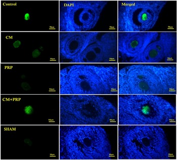
The GDF9 expression significantly decreased in the CYC group compared with the control group. Also, the expression level in the PRP-treated group was very low. Albeit in the CM+PRP group, GDF9 expression was lower than in the control group, it was considerably higher than in the CYC group and the PRP group (Figure 1).
Immunohistochemical analysis of mouse ovary tissue labelling of GDF 9, Positive staining on follicles of mouse ovary is observed (green florescent). The nuclei were counterstained and observed using 40,6-diamidino-2-phenylindole (DAPI) staining.

Gene expression assessment
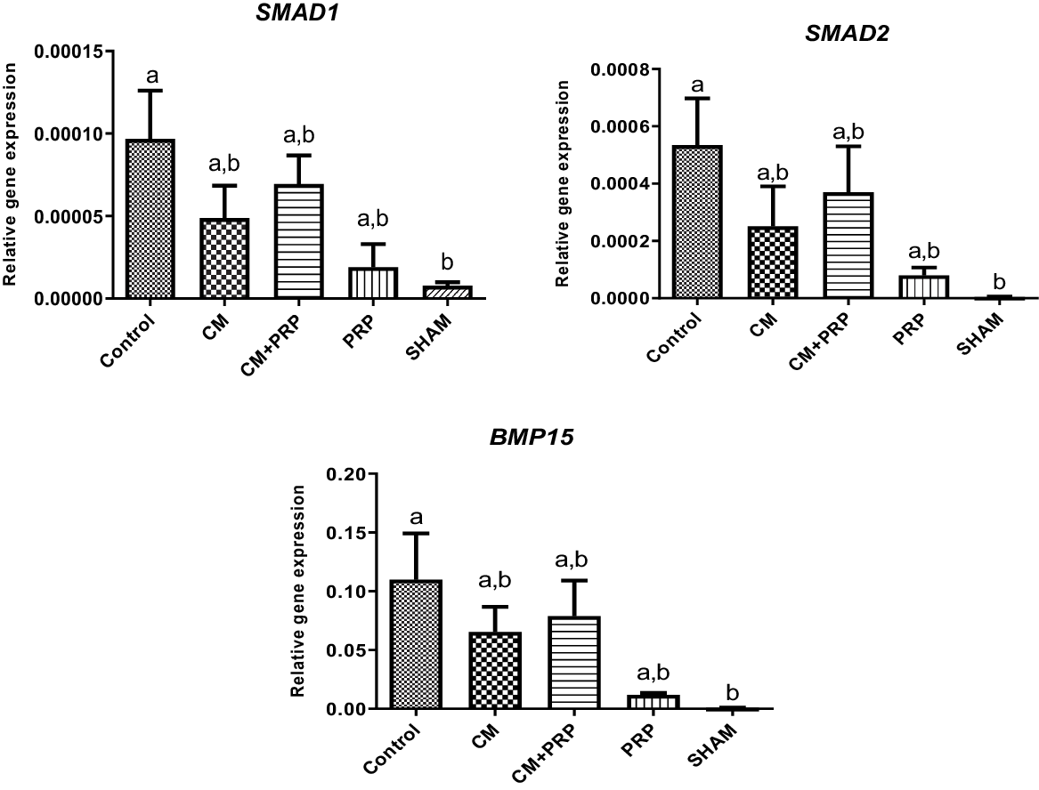
The expression levels of SMAD1, SMAD2, and BMP15 in the CYC group were remarkably lower compared with the control group. In the CM+PRP group, the expression levels of the mentioned genes were higher than in the CM group and the PRP group; however, the differences between them were not significant. The expression levels of SMAD1, SMAD2, and BMP15 in the CM group were more than in the PRP and CYC groups (Figure 2).
mRNA expression levels of SMAD1, SMAD2 and BMP15 in experimental groups.

Live birth rate evaluation
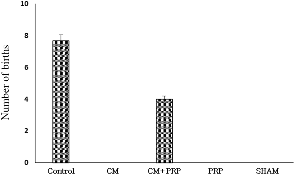
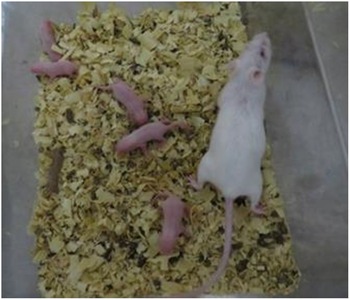
In CYC, PRP, and CM groups, fertility and live birth were not observed. But, in the CM+PRP and control groups, live birth was observed (Figures 3 and 4).
Number of live births in the experimental groups.

Treatment with conditioned medium and PRP in infertile mice resulted in successful fertility and live birth

Discussion
The mixture of PRP and CM was more effective than other treatment groups in mice treated with cyclophosphamide. Also, live birth in the mixed group of PRP and CM was observed. The abundance of PRP-rich therapeutic molecules is beneficial for cell proliferation and injury repair (Atashi et al., Reference Atashi, Jaconi, Pittet-Cuénod and Modarressi2015). Plus, the creation of PRP-containing scaffolds for stem cell transplantation can dramatically promote the therapeutic efficacy of mesenchymal stem cell (MSC) (Chang et al., Reference Chang, Li, Chen, Wei, Yang, Shi and Liang2015; Wu et al., Reference Wu, Zhou, Liu, Zhang, Xiong, Lv, Liu, Wang, Du, Zhang and Liu2017). Recent documents have recommended that the exploitation of PRP to improve the expression of adhesion molecules could be useful for the treatment of the endometrium (Colombo et al., Reference Colombo, Fanton, Sosa, Criado Scholz, Lotti, Aragona and Lotti2017; Liu et al., Reference Liu, Yuan, Fernandes, Dziak, Ionita, Li and Yang2017). Several published articles have shown that PRP provides an inhibitory effect on mating endometritis (Reghini et al., Reference Reghini, Ramires Neto, Segabinazzi, Castro Chaves, Dell’Aqua, Bussiere, Dell’Aqua, Papa and Alvarenga2016; Segabinazzi et al., Reference Segabinazzi, Friso, Correal, Crespilho, Dell’Aqua, Miró, Papa and Alvarenga2017).
Conversely, PRP decreased IL-6 expression in menstrual blood-derived stromal cells (MenSCs) in vitro over a short time (Zhang et al., Reference Zhang, Li, Yuan and Tan2018). WNT/ β-catenin signalling has a role in adjusting the function of endometrial stem cells during menopause. Specific cytokines during menopause can elevate endometrial MSC (eMSC) proliferation. Understanding the mechanism of eMSC regulation may help in the treatment of endometrial proliferation disorders, such as Asherman’s syndrome (Xu et al., Reference Xu, Chan, Li, Ng and Yeung2020).
The results of the current work implicated that the process of inhibiting inflammation was very weak in the PRP group. PRP alone may not be effective for treating severe intrauterine adhesions; although it can dramatically regenerate the endometrium. Kim et al. (Reference Kim, Park, Paek, Lee, Song and Lyu2020) studied the human PRP injection efficacy for endometrial regeneration in a mouse model of AS damage and showed that human PRP can ameliorate endometrial morphology and diminish fibrosis degree in an AS mouse model. In addition, human PRP treatment was associated with more implantation sites (IS) and live births (Kim et al., Reference Kim, Park, Paek, Lee, Song and Lyu2020).
The results of the study by Kim and colleagues (2020) were consistent with the present study. Human PRP-based therapy may be a useful approach for improving damaged endometrial regeneration and for fertility and pregnancy rates.
In a study, Pantos et al. (Reference Pantos, Simopoulou, Pantou, Rapani, Tsioulou, Nitsos, Syrkos, Pappas, Koutsilieris and Sfakianoudis2019) evaluated postmenopausal women and premature menopause after treatment with PRP. They showed that menstrual repair after harnessing autologous ovarian PRP remedy, as well as enhancement of hormonal characteristics, reduction in follicle-stimulating hormone (FSH) levels and there was a simultaneous elevation in anti-mullerian hormone (AMH) levels. In addition, patients were reported to have normal pregnancies within 2–6 months after PRP treatment (Pantos et al., Reference Pantos, Simopoulou, Pantou, Rapani, Tsioulou, Nitsos, Syrkos, Pappas, Koutsilieris and Sfakianoudis2019).
It has been offered that randomized controlled trials (RCTs) can also be helpful in monitoring the effectiveness of PRP. This may be a good clue to the successful therapy for a particular group of patients who have considered reproductive treatment methods after menopause.
Zhang et al. (Reference Zhang, Li, Yuan and Tan2019) studied the curative effect of transplantation of MenSCs in combination with PRP in a rat model of intrauterine adhesion (IUA) and MenSCs mechanisms in endometrial regeneration. They showed a significantly improved endometrial proliferation on days 9 and 18 after MenSC transplantation treatment. They also reported an angiogenesis and morphological improvement and reduction of inflammation and collagen fibrosis in the uterus. Also, MenSCs had lesion chemotaxis and were observed around the endometrial glands. Human secretory protein gene expression of TSP-1, SDF-1, and IGF-1 were perceived in uterus. The three treatments promoted fertility rate in rats with IUA. Furthermore, the expression of genes of cell proliferation and growth, and other biological occurrences were induced in the MenSC transplant group. The signal pathway was altered and Gdf5, Wnt5a, and CTGF were significantly modulated in the treatment groups. PRP potentiated these parameters by exerting synergistic influences. MenSCs could effectively ameliorate the uterus and significantly accelerate the healing rate of endometrial damage and enhance fertility regeneration in IUA rats (Zhang et al., Reference Zhang, Li, Yuan and Tan2019).
Genes for BMP15 and GDF9, secreted by the oocytes, can regulate granulosa cell differentiation and proliferation, primordial follicle recruitment, and steroid synthesis, and increase preantral follicle growth (Fenwick et al., Reference Fenwick, Mora, Mansour, Baithun, Franks and Hardy2013).
Previous studies have reported that BMP15 in the follicular fluid of mature follicles might be synthesized by oocytes from mature follicles and regulate the expansion of cumulus cell (Yoshino et al., Reference Yoshino, McMahon, Sharma and Shimasaki2006; Sun et al., Reference Sun, Lei, Cheng, Jin, Zu, Shan, Wang, Zhang and Liu2010). Moreover, evidence suggested that GDF9 was found in the primary follicle onwards (Aaltonen et al., Reference Aaltonen, Laitinen, Vuojolainen, Jaatinen, Horelli-Kuitunen, Seppa, Louhio, Tuuri, Sjoberg, Butzow and Hovatta1999). Our study demonstrated that the levels of BMP15 and GDF9 gene expression in the PRP+CM group was increased in comparison with the CYC, PRP, and CM groups.
In summary, the results of this study showed that fertility decreased in people undergoing chemotherapy with cyclophosphamide. Treatment with CM+PRP can stimulate the growth of ovarian follicles.
Acknowledgements
This work was supported by the Department of anatomy, Shiraz University of medical sciences (grant no. 16689). This article was extracted from PhD thesis written by Neda Taghizabet. This study was supported by the Shahid Beheshti University of Medical Sciences and Shiraz University of medical sciences.
Conflict of interest
The authors have no conflict of interest to declare.