Implications
The main implication of this study is to investigate whether environmental enrichment and herbal compound provision reduce the effect of stressful factors, especially those of chronic exposure. Non-invasive techniques and different physiological indicators were contrasted. This study supports environmental enrichment and herbal compounds as strategies to reduce stress in commercial pig husbandries.
Introduction
Growing pigs in modern pig husbandries are exposed to many stress factors which have a negative effect on their health and welfare. Basically, the environmental conditions suitable for intensive husbandry systems, although promoting several management tasks and caring for some welfare aspects of the pigs, do not always meet the behavioural requirements of the individuals. More precisely, the lack of materials to forage has been widely signalled as a stressful factor for pigs, as they would spend most of their active time rooting if given the possibility (Stolba and Wood-Gush, Reference Stolba and Wood-Gush1989). Thus, the usual barren environment found in commercial piggeries have been said to prevent pigs from expressing their foraging species-specific behaviour, and leading to frustration (for a review see van de Weerd and Day, Reference Van de Weerd and Day2009). Furthermore, enriched environments enhance the well-being of the animals by increasing ‘desirable’ behaviours such as exploration and by reducing the frequency of abnormal or ‘undesirable’ behaviours (Chamove, Reference Ciepielewski, Stojek, Borman, Myślińska, Glac and Kamyczek1989; Averós et al., Reference Averós, Brossard, Dourmad, de Greef, Edge, Edwards and Meunier-Salaün2010). As a consequence, the European Legislation requires farmers to provide enrichment materials to pigs, in an attempt to create a more proper production environment and prevent tail biting outbreaks.
Another described strategy to reduce stress is by means of plants with sedative and tranquilizer properties such as Valeriana officinalis and Passiflora incarnata (in mice: Soulimani et al., Reference Soulimani, Younos, Jarmouni and Bousta1997; Nam et al., Reference Nam, Choi, Yoo, Kim, Jung, Kim, Kang, Park, Kim, Kim, Yoon and Hwang2013; in pigs: Peeters et al., Reference Peeters, Driessen, Steegmans, Henot and Geers2004; in rats: Murphy et al., Reference Murphy, Kubin, Shepherd and Ettinger2010). The mechanism involved in the sedative and anxiolytic properties of V. officinalis seems to be mediated by the interaction of valerian acid with the γ-amino butyric acid receptors type A (GABAA). The stimulation of GABAA, opens the permeability of chloride channels, producing neural inhibition (Khom et al., Reference Khom, Baburin, Timin, Hohaus, Trauner, Kopp and Hering2007; Murphy et al., Reference Murphy, Kubin, Shepherd and Ettinger2010). Moreover, different bioactive compounds have been detected in P. incarnata such as flavonoids, maltol, cynogenic glycosides and indole alkaloids, without a consensus regarding the most important one for the sedative properties. Flavonoids, which are beyond the most studied components, have a similar effect than valerian acid, increasing the membrane permeability by means of the modulation of GABAA (for a review see: Miroddi et al., Reference Miroddi, Calapai, Navarra, Minciullo and Gangemi2013).
Stress response produces biochemical changes by the activation of the sympathetic adrenomedullary (SAM) system in a first phase and the hypothalamic–pituitary–adrenal (HPA) axis after a short delay (Manteuffel, Reference Manteuffel2002). As a consequence of the activation of the SAM system, chromogranin A (CgA) is co-released with catecholamines (adrenaline and noradrenaline) (O’Connor and Frigon, Reference O’Connor and Frigon1984), whereas the main output of the HPA axis in pigs is cortisol (Mormède et al., Reference Mormède, Andanson, Aupérin, Beerda, Guémené, Malmkvist, Manteca, Manteuffel, Prunet, van Reenen, Richard and Veissier2007). Thus, CgA and cortisol have been used in pigs as physiological indicators of the SAM system and HPA axis activity, respectively (e.g. Escribano et al., Reference Escribano, Fuentes and Cerón2012 and Reference Escribano, Soler, Gutiérrez and Martínez-Subiela Sand Cerón2013). On the other hand, stress-induced immunosuppression has also been widely reported, affecting the release of pro-inflammatory cytokines such as tumour necrosis factor-α (TNF-α) and suggesting neuroendocrine mechanisms somehow meditated by glucocorticoids and catecholamines (Connor et al., Reference Connor, Brewer, Kelly and Harkin2005). Chronic stress has been associated with dysregulated immunity and subsequent low-grade inflammation (Kemeni and Schedlowski, Reference Kemeni and Schedlowski2007). Studies carried out with some stress protocols (physical, psychological or mixed) showed a pro-inflammatory response, mainly characterized by release of inflammatory mediators, including interleukin (IL)-1, IL-6 and TNF-α (Tuchscherer et al., Reference Tuchscherer, Kanitz, Otten and Tuchscherer2002).
Some studies have demonstrated that barren conditions, compared with enriched environments, are associated with signs of chronic stress (Munsterhjelm et al., Reference Munsterhjelm, Valros, Heinonen, Hälli, Siljander-Rasi and Peltoniemi2010), having effects on physiological, behavioural and performance parameters. The assessment of chronic stress, especially by means of non-invasive techniques is an important field of investigation. The increase of cortisol levels in biological samples such as plasma, saliva, urine, faeces and milk between a few minutes and several hours post stress (Mormède et al., Reference Mormède, Andanson, Aupérin, Beerda, Guémené, Malmkvist, Manteca, Manteuffel, Prunet, van Reenen, Richard and Veissier2007) has been associated with acute or sub-acute stress. On the contrary, hair cortisol has been suggested as a long-term indicator of stress exposure (Davenport et al., Reference Davenport, Tiefenbacher, Lutz, Novak and Meyer2006; Gow et al., Reference Gow, Thomson, Rieder and Van Uum Sand Koren2010). Although there are still some gaps to completely understand the mechanism for cortisol incorporation into hair, the most accepted theory is the model suggested by Henderson (Reference Henderson1993). On the other hand, CgA is an acidic soluble protein which is the major member of the granin family (Hendry et al., Reference Hendry, Bevan, Mattei and Mouland1995). Chromogranin A is stored and co-released with catecholamines to the blood from the vesicles of the adrenal medulla, the sympathetic nerve endings and neuroendocrine tissues (O’Connor and Frigon, Reference O’Connor and Frigon1984; Obayashi, Reference Obayashi2013). Furthermore, Saruta et al. (Reference Saruta, Tsukinoki, Sasaguri, Ishii, Yasuda, Osamura, Watanabe and Sato2005) described the production of CgA from the submandibular glands of humans, and different studies have found a relation between physical or psychological stress and CgA levels in saliva (e.g. Kanamaru et al., Reference Kanamaru, Kikukawa and Shimamura2006; Ott et al., Reference Ott, Soler, Moons, Kashiha, Bahr, Vandermeulen, Janssens, Gutiérrez, Escribano, Cerón, Berckmans, Tuyttens and Niewold2014; Escribano et al., Reference Escribano, Gutiérrez and Tecles Fand Cerón2015). Salivary CgA seems to peak after a few minutes, but it lasts for up to 1 h in humans (Obayashi, Reference Obayashi2013) and does not present a circadian rhythm in pigs (Escribano et al., Reference Escribano, Gutiérrez, Fuentes-Rubio and Cerón2014). However, CgA has not been, up to now, investigated as a possible chronic stress indicator.
The aim of this study was to evaluate the effect of providing environmental enrichment and/or an herbal compound with sedative effects on physiological indicators of stress. For this purpose, hair and saliva cortisol, salivary CgA and TNF-α in blood were chosen as biomarkers of either acute or chronic stress and evaluated over time after the implementation of the expected stress reducing strategies. Other indicators of pig welfare such as behaviour and performance were also recorded and reported in another paper.
Material and methods
Animals and housing conditions
In total, 56 crossbred ((Landrace×Large white)×Pietrain) entire males from the same genetic company were used in this experiment. No more than two piglets per litter were selected in order to ensure maximum genetic diversity. Weaners arrived at the experimental farm in December at the age of 10 weeks and an average weight of 25 kg. The facilities where the experiment was carried out had an automatic control system for temperature and ventilation (to ensure a range of temperatures in between 22±5°C), total slatted floor, and bowl-type drinkers. At the age of 16 weeks, and a mean live weight of 49.8 kg, pigs were randomly distributed into eight groups of seven animals, located in two modules. Each module had four pens, two on the left and two on the right separated by a corridor. Pens were divided using metallic-bar fences to allow direct visual contact. Each pen was 13.67 m2, allowing a floor space availability of 1.95 m2/pig. All pens were provided with one drinker and two hoppers, and water and food were supplied ad libitum. Feed diet consisted of a commercial concentrate provided with a phase feeding regime (i.e. adjusting protein and energy throughout time).
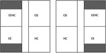
Pigs were reared in either an enriched environment consisting of sawdust, natural hemp ropes and rubber balls (all materials were provided at the same time) or in a barren environment. Half of the groups were also supplemented with an herbal compound containing Valerian (V. officinalis) and Maypop (P. incarnata), both having sedative effects (Sedafit; Phytosynthèse, Saint-Bonnet de Rochefort, France), whereas the others were not supplemented. Thus, four different treatments were assessed in both modules in a 2×2 factorial design: (1) pigs supplemented with both environmental enrichment and herbal compound (named as EEHC pigs from this point), (2) pigs supplemented with environmental enrichment (EE), (3) pigs supplemented with the herbal compound (HC) and (4) control group (CG), consisting in pigs kept in a barren environment and without herbal compound supplementation. (Figure 1). The slats of the pens supplied with EE were partially covered (1/3) with polypropylene sticks (Click-in®; Rotenca, Agramunt, Spain) to support the sawdust. New sawdust was added every 2 days. The hemp ropes were attached on the walls of EE pens, and were periodically substituted when the length of the rope after its use by the pigs was <30 cm. A single rubber ball with a diameter of 15 cm was provided in pens supplied with environmental enrichment. Sedafit was manually added to the HC and EEHC groups in a concentration of 2000 mg/kg to the food concentrate, whereas EE and CG pigs were supplied with normal food concentrate without supplementation. Blood, saliva and hair samples were taken before starting the experiment (T0) (16 weeks of age), at the middle (T1) (20 weeks of age) and at the end (T2) (24 weeks of age). Furthermore, at the ages of 15, 18, 20, 22 and 24 weeks, all animals were weighed and body lesions were assessed as part of a broader investigation, which also included weekly behavioural observations. All procedures carried out during this experiment were approved by the Institut de Recerca i Tecnologia Agroalimentàries (IRTA) ethical committee.
Schematic representation of the experimental pig farm and distribution of the two rooms used: EEHC= provided with enrichment material and herbal product (Sedafit); HC=provided with herbal product (Sedafit); EE=provided with enrichment material; CG= control group. The grey area represents the slats covered to support sawdust.

Hair collection and analysis
Hair was sampled in the morning taking advantage of the restraint provided by a scale with a cage and a two-door system for access and exit used for the regular weighing carried out every two weeks. Clean hair was carefully shaved using a hair trimmer, trying not to include the hair follicles, which have been described as producing cortisol themselves (Ito et al., Reference Ito, Ito, Kromminga, Bettermann, Takigawa, Kees, Straub and Paus2005). The samples were obtained from the lumbar region, which was suggested in a previous study as the most appropriate region (Casal et al., Reference Casal, Manteca, Bassols, Peña and Fàbrega2014). The lumbar region plus a security margin was shaved when the animals arrived at the experimental farm and after every sample procedure in order to ensure that the period of time analyzed was comprised between the shaving and the sampled day.
Pigs were gently accompanied to the scales, and the hair trimmer was cleaned after each animal. Once sampled, hair was stored at room temperature inside hermetically sealed bags until analysis. Although 56 pigs were sampled, five samples in T0, and three in T1 and T2 were discarded because of the small amount of sample.
Cortisol extraction was performed following a method developed by Tallo-Parra et al. (Reference Tallo-Parra, Manteca, Sabes-Alsina, Carbajal and Lopez-Bejar2015), based on the method of Davenport et al. (Reference Davenport, Tiefenbacher, Lutz, Novak and Meyer2006). Approximately 250 mg of hair from each sample were washed in 2.5 ml of 2-propanol, vortexed at room temperature for 2.5 min, and the supernatant was discarded by decantation. The same washing process was repeated twice more. Hair samples were then allowed to dry completely at room temperature for ~36 h; then, samples were powdered using a ball mill (Mixer mill MM200; Retsch, Haan, Germany) for 5 min at 25 Hz. In total, 50 mg of powdered hair per sample were placed in 2-ml Eppendorf tubes with 1.5 ml of methanol and moderately shaken at 30°C for 18 h (G24 Environmental Incubator Shaker; New Brunswick Scientific Co. Inc., Edison, NJ, USA) to extract the steroids. Then, the sample was spun in a micro centrifuge for 2 min at 7000 g, and 0.750 ml of each sample were aliquoted into a new 2-ml Eppendorf and dried at 38°C in an oven. The dried extracts were reconstituted with 0.2 ml of the solution provided with the cortisol assay kit, and stored at −20°C until the analysis.
Hair cortisol concentration was analyzed using an ELISA kit (Cortisol ELISA KIT; Neogen ® Corporation Europe, Ayr, UK) following the instructions provided by the manufacturer. All samples were analyzed in duplicate. Validation was necessary as this kit is not specific for pigs and it had not been tested before. The sensitivity of the kit was of 0.32 pg cortisol/mg of hair. Validation tests were performed using pools of 10 different samples. According to the manufacturer, cross-reactivity of the ELISA antibody with other steroids was as follows: prednisolone 47.4%, cortisone 15.7%, 11-deoxycortisol 15.0%, prednisone 7.83%, corticosterone 4.81%, and <2% for the rest of the steroids not presented. Specificity was also evaluated comparing slopes from the straight lines resulting from the standard curve (m standard) and a new pool curve (m pool) with the same serial dilutions (1 : 1, 1 : 5, 1 : 10, 1 : 25, 1 : 50 and 1 : 100). Linearity was evaluated with pooled samples serially diluted (1 : 1, 1 : 2, 1 : 5 and 1 : 10) in the buffer kit, showing a R 2=0.972 and a mean percentage error of −18.9% (as in Tallo-Parra et al., Reference Tallo-Parra, Manteca, Sabes-Alsina, Carbajal and Lopez-Bejar2015). Accuracy was assessed through the spike-and-recovery test: 50, 100 and 200 μl of pure standard cortisol solutions were added to 200, 100 and 50 μl of pool sample, respectively; this process was repeated at different standard cortisol concentrations (20, 2 and 0.2 ng/ml), and the percentage of recovery was 113.67±14.15% SEM. Assay precision was assessed by calculating the intra-assay and inter-assay CV. The intra-assay CV was calculated from all duplicated samples, with a result of 2.33%, whereas for the inter-assay it was calculated from a pool of 10 high-variability samples duplicated in each ELISA plate, with a result of 11.83%.
Saliva collection
All saliva samples were taken between 0800 and 1100 h allowing the animals to chew on one cotton bud (Salivette®; SARSTEDT AG & Co., Nürbrecht, Germany) for >30 s and randomising the animals from different treatments. Cotton buds were offered using a clamp. Both cortisol and CgA were assessed from the same samples. If the amount of saliva collected was not sufficient for both analyses, CgA was considered the priority. At the age of 16 weeks, saliva was collected from outside the pen as a first option. For those animals who refused to chew the cotton, samples were obtained from inside the pen offering the cotton bud, with no restraint. Sampling time was fixed up to 5 min. Although all animals were correctly sampled, in 10 pigs (four EEHC, one EE, three HC and two CG) the amount of saliva was not sufficient for both cortisol and CgA, and hence, cortisol was not assessed. For the second and third samples, saliva was collected at the same moment as was blood by two different operators, taking advantage of the pigs being restrained. All animals had enough sample for CgA analysis, but at the age of 20 weeks, two pigs did not have enough sample for cortisol determination (one HC and one CG). Once sampled, and before the analysis, salivettes® were centrifuged at 3500 r.p.m. for 10 min, cotton buds were removed, and tubes were frozen at −18°C.
Salivary cortisol analysis
Salivary cortisol concentration was measured using a solid-phase competitive chemoluminescent enzyme immunoassay kit (Immulite 1000 cortisol; Siemens Medical Solutions Diagnostics, Los Angeles, CA, USA) validated for porcine saliva (Escribano et al., Reference Escribano, Fuentes and Cerón2012).
Salivary chromogranin A analysis
Salivary CgA was measured using a validated time-resolved immunofluorometric assay (Escribano et al., Reference Escribano, Soler, Gutiérrez and Martínez-Subiela Sand Cerón2013), with a lower limit of detection of 4.27 ng/ml and a high level of precision (inter-assay CV of 6.23% and intra-assay CV of 5.82%). The coefficient of correlation for accuracy was r=0.968. The calibration curve covered a range from 46.8 to 1500 ng of CgA/ml. Fluorescence was measured using a VICTOR2 1420 multi-label counter (Perkin Elmer Life and Analytical Science; Wallac Oy, Turku, Finland).
Blood collection and analysis
All blood samples were collected in ethylenediaminetetraacetic acid (1 mg/ml) coated test-tubes via jugular vein puncture between 0800 and 1100 h, randomising pigs from the different treatments. As previously said, blood and saliva from second and third samples were collected at the same moment by two different operators, whereas for the first sample, blood was collected 2 days before saliva. Once sampled, blood was centrifuged for 10 min at 3500 r.p.m., aliquoted and stored at −18°C until analyses were performed. tumour necrosis factor-α levels were measured using a validated porcine TNF-α ELISA kit (Quantikine®; R&D Systems, Minneapolis, MN, USA).
Statistical analysis
All statistical analyses were conducted using the Statistical Analysis System (SAS version 9.2; SAS Institute Inc., Cary, NC, USA). The level of significance was established at P<0.05 for all the analyses, and a tendency was considered between P>0.05 and <0.1. Results are presented as mean±SEM unless otherwise indicated. The Shapiro-Wilk’s test (with Proc univariate in SAS) was used to examine the normality of distributions. For data without a Gaussian distribution, a logarithmic transformation was performed, and normality was evaluated again. One outlier was eliminated from the second sampled day for CgA.
Data were analyzed using generalized linear mixed models for repeated measurements (Glimmix procedure). The experimental unit was the subject. Use or not of enrichment material, and herbal compound, sample day and their interactions were considered as fixed effects. Subject nested with pen (and, thus, EE and HC) was considered as a random effect, after testing that the pen was not significant as a fixed effect. Sample day provided the repeated measurement. Least square means of fixed effects (i.e. estimated marginal means or means adjusted by a covariate) with Tukey adjustment were used for comparisons when ANOVA indicated significant differences. Parametric Pearson’s rank correlations were calculated between different physiological indicators and days using the Proc Corr procedure of SAS. Correlations between the physiological indicators according to experimental groups were also carried out, and the same results as for the overall correlations were found.
Results
Pigs did not significantly differ in live weight at 16 weeks of age (mean of 49.62 kg and SEM 1.01), whereas at 24 weeks of age CG pigs presented a significantly lower live weight (105.11±1.17 kg) compared with the EE (111.79±2.49 kg), HC (110.04±3.04 kg) and EEHC (112.35±2.39 kg) (unpublished results).
Hair cortisol
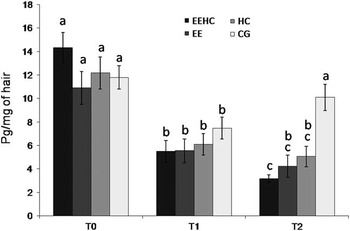
At T2, hair cortisol was significantly lower for pigs raised with EE compared with pigs raised in barren conditions (3.72±0.52 v. 7.58±0.84 pg/mg hair, respectively), and for pigs supplemented with the herbal compound compared with pigs not supplemented with Sedafit (4.19±0.51 v. 7.27±0.92 pg/mg hair, respectively). Hair cortisol concentrations and differences between treatments for each sampled day are presented in Figure 2. No significant differences between treatments were observed for T0 and T1, whereas the control group presented significantly higher levels in relation to the other treatments at T2 (P=0.004 for HC and P<0.001 for EE and EEHC). Comparing the initial and final levels of hair cortisol, a significant decrease was observed between T0 and T2 for all three treatments (P<0.001 for HC, EE and EEHC). These differences were significant between T0 and T1 for all groups, including the control group (P=0.01 for control group and P<0.001 for the other treatments), but between T1 and T2, the only group with a significant decrease was the one provided with both enrichment and herbal product (P=0.006).
Hair cortisol concentrations (mean and SE) of pigs supplemented with both environmental enrichment and herbal compound (EEHC), supplemented with environmental enrichment (EE), supplemented with the herbal compound (HC) and control group (CG) before starting the experiment (T0, 16 weeks of age), after 1 month of treatment (T1, 20 weeks of age) and after 2 months of treatment (T2, 24 weeks of age). a,b,cDifferent letters represent significant differences between groups (P<0.05).

Chromogranin A
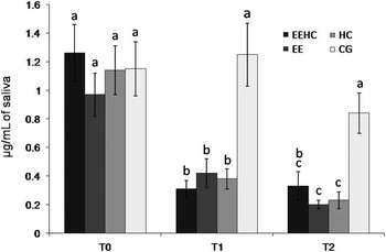
Salivary CgA was significantly lower for pigs with EE compared with BE at both T1 (0.37±0.06 v. 0.82±0.14 μg/ml, respectively) and T2 (0.26±0.05 v. 0.54±0.09 μg/ml, respectively), and for pigs with herbal compound compared with without herbal compound at both T1 (0.35±0.04 v. 0.84±0.14 μg/ml, respectively) and T2 (0.28±0.05 v. 0.52±0.09 μg/ml, respectively). Salivary CgA concentrations for each treatment and day are expressed in Figure 3. No significant differences between treatments were observed for T0, whereas CgA concentrations were different at T1 and T2 for the control group, when compared with the rest of the treatments (at T1: P<0.001 for EE and EEHC, and P=0.002 for HC; at T2: P<0.001 for EE and HC, and P=0.02 for EEHC). When comparing over time, significant differences were observed between T0 and T2 and between T0 and T1 for all treatments (P<0.001) except for the control group. The only differences observed between T1 and T2 were a slight decrease for HC (P=0.036) and a tendency to decrease for EE (P=0.10).
Salivary chromogranin A (CgA) concentrations (mean and SE) of pigs supplemented with both environmental enrichment and herbal compound (EEHC), supplemented with environmental enrichment (EE), supplemented with the herbal compound (HC) and control group (CG) before starting the experiment (T0, 15 weeks of age), after 1 month of treatment (T1, 20 weeks of age) and after 2 months of treatment (T2, 24 weeks of age). a,b,cDifferent letters represent significant differences between groups (P<0.05).

Salivary cortisol and serum tumour necrosis factor-α
Neither enrichment nor Sedafit resulted in significant differences for salivary cortisol and TNF-α concentration (Table 1). However, a significant decrease was observed between T0 and T2 in both salivary cortisol (P=0.02) and TNF-α (P<0.001).
Salivary cortisol and tumour necrosis factor-α (TNF-α) concentrations of the different sampled days for the growing pigs supplemented or not with environmental enrichment (EE) or herbal compound (HC)

Day: T0=before experiment, week 16; T1=after 1 month of treatment, week 20; T2=after 2 months of treatment, week 24.
a,b,cLSMEANS with different superscripts in a row differ significantly (day effect). EE×day interaction not significant for any parameter.
Correlations
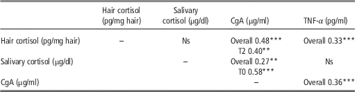
A summary of the significant correlations found is presented in Table 2. Hair cortisol was positively correlated with CgA overall (P<0.0001) and at T2 (P=0.0024) and with overall TNF-α (P<0.0001). Chromogranin A was also correlated with salivary cortisol overall (P=0.0007) and at T0 (P<0.0001) and with overall TNF-α (P<0.0001). Contrasting the different sampled days, hair cortisol correlations were found between T0 and T1 (r=0.41, P=0.0031) and T1 and T2 (r=0.57, P<0.0001), and CgA presented a significant correlation between T1 and T2 (r=0.42, P=0.0016).
Significant correlations between the different physiological indicators of stress at an overall level or at each sampled day (only significant correlations presented)

CgA=chromogranin A; TNF-α=tumour necrosis factor α.
Day: T0=before experiment, week 16; T1=after 1 month of treatment, week 20; T2=after 2 months of treatment, week 24.
***P<0.001, **P<0.01.
Discussion
Intensification of production systems has been linked to both certain advantages and drawbacks for pig welfare. Among the disadvantages, the impossibility to perform some behaviours, like foraging, and the lack of stimuli in the barren environments has been associated with stress. The present research focussed on the effect of two strategies aiming at stress reduction on physiological indicators of both chronic and acute stress, with a special emphasis on non-invasive techniques. With this purpose, pigs were reared in different conditions including enrichment material and supplementation of a herbal product (Sedafit), both suggested as potential reducers of stress. Along that line, higher salivary cortisol levels had been previously reported in pigs reared in barren environments (e.g. Merlot et al., Reference Merlot, Vincent, Thomas, Meunier-Salaün, Damon, Robert, Dourmad and Lebret Band Prunier2012). Concerning the herbal products, the sedative and tranquilizing properties of V. officinalis and P. incarnata (the main components of Sedafit) have previously been described (Soulimani et al., Reference Soulimani, Younos, Jarmouni and Bousta1997; Nam et al., Reference Nam, Choi, Yoo, Kim, Jung, Kim, Kang, Park, Kim, Kim, Yoon and Hwang2013). Furthermore, Peeters et al. (Reference Peeters, Driessen, Steegmans, Henot and Geers2004) reported a reduction of the stress response in pigs receiving this product. Therefore, in this experiment the hypothesis formulated was that pigs in barren environments and/or without the sedative effects of the herbal product would be subjected to a more chronically stressful environment, and, thus, an effect on physiological biomarkers of stress was expected. It has to be pointed out that stress response can be influenced by other environmental factors such temperature or activity itself and this has to be considered when interpreting the results. For that reason, in the present experiment the different treatment groups were kept under the same environmental conditions with regards to temperature and humidity, but the effect of environmental enrichment on the physiological indicators could vary under other environmental conditions. Each indicator is discussed separately as follows.
Hair cortisol
Hair has been used as an indicator of HPA axis activity in multitude of species including farm species (e.g. dairy cattle Tallo-Parra et al., Reference Tallo-Parra, Manteca, Sabes-Alsina, Carbajal and Lopez-Bejar2015). To the knowledge of the authors, only a limited number of studies have focussed on hair cortisol in pigs: Casal et al. (Reference Casal, Manteca, Bassols, Peña and Fàbrega2014) in growing pigs, Martelli et al. (Reference Martelli, Sardi, Stancampiano, Govoni, Zannoni, Nannoni, Forni and Bacci2014) in transgenic pigs and their relatives, and Bacci et al. (Reference Bacci, Nannoni, Govoni, Scorrano, Zannoni, Forni, Martelli and Sardi2014) in sows. Cortisol levels found in the present study (Figure 2) when the animals were aged 16 weeks were slightly lower to those reported at the age of 17 weeks in a previous experiment using growing pigs subjected to a weekly remixing procedure (Casal et al., Reference Casal, Manteca, Bassols, Peña and Fàbrega2014) and, similar concentrations were also reported in sows by Bacci et al. (Reference Bacci, Nannoni, Govoni, Scorrano, Zannoni, Forni, Martelli and Sardi2014). Furthermore, whilst hair cortisol concentration of the pigs belonging to this previous experiment on the effects of weekly remixing increased significantly throughout time (Casal et al., Reference Casal, Manteca, Bassols, Peña and Fàbrega2014), in the present experiment hair cortisol concentration of pigs supplied with the herbal compound, enrichment material and both decreased at the age of 24 weeks (T2), when compared with the initial concentration. In contrast, for the control group, a significant (but smaller, as compared with treatment groups) decrease was observed between T0 and T1, and levels increased again at T2.
Our hypothesis is that the lower levels of cortisol over time in the treatment groups may indicate an effect of enrichment and herbal compound on chronic stress levels. This decrease could be associated with an age effect as suggested by de Jong et al. (Reference De Jong, Prelle, van de Burgwal, Lambooij, Korte, Blokhuis and Koolhaas2000) in cortisol levels in saliva, as time was also found to have a significant effect, but in this case a decrease would have been expected for the control group also, and this was not the case for T2 levels. Furthermore, the fact that in the previous experiment levels were found to increase over time when subjecting pigs to a chronic stressor, like weekly remixing, add further support to the finding that both environmental enrichment and the herbal compound may act on the reduction of chronic stress. As mentioned previously, few studies have used hair cortisol in pigs as chronic stress biomarker, and those published had not implemented neither environmental enrichment nor herbal compounds. However, the present findings would be in line with those reported previously in other investigations based on other behavioural or physiological indicators of stress on the effects of both using enrichment materials (de Jong et al., Reference De Jong, Prelle, van de Burgwal, Lambooij, Korte, Blokhuis and Koolhaas2000) or herbal compounds (Peeters et al., Reference Peeters, Driessen, Steegmans, Henot and Geers2004). Furthermore, the results presented elsewhere (Casal et al., unpublished) on behavioural patterns and performance indicators of the same pigs used in this study indicated a higher percentage of negative social behaviours and a lower final weight of control pigs compared with those offered enrichment, herbal compound or both.
Other confounding factors should be also considered when using hair as stress biomarker. Although hair growth patterns can be studied, growth rates may be affected by a number of intrinsic and extrinsic factors (Koren et al., Reference Koren, Mokady, Karaskov, Klein, Koren and Geffen2002). As suggested by Mowafy and Cassens (Reference Mowafy and Cassens1976) the depth of the hair in growing pigs is about 3 to 4 mm. This could partly explain why no differences between treatments were found at 20 weeks of age (T1) on hair cortisol levels. Before each sample, hair was shaved to ensure a similar length (and age) of the hair comprised between two consecutive samples. However, part of the hair analyzed was not belonging to the in-between samples’ periods, as the hair under the skin was not considered. As a consequence, assuming that cortisol can be incorporated into growing hair cells via passive diffusion (Stalder and Kirschbaum, Reference Stalder and Kirschbaum2012), 3 to 4 mm of hair remained in the skin and were analyzed in the next period. According to Bacci et al. (Reference Bacci, Nannoni, Govoni, Scorrano, Zannoni, Forni, Martelli and Sardi2014), the proportion of hair remaining in the skin belongs to 15 days approximately. Thus, the sample taken at the age of 20 weeks, was composed by 3 to 4 mm of hair probably containing cortisol information from ~15 days before starting the experiment (<16 weeks of age), masking the effect of the treatments.
No correlations between salivary and hair cortisol were reported in this study. Sauvé et al. (Reference Sauvé, Koren, Walsh, Tokmakejian and Van Uum2007) did not report a significant correlation between hair cortisol and salivary cortisol in humans (r=0.31, P=0.12) with a single time-point sample either. On the contrary, Van Holland et al., (Reference Van Holland, Frings-Dresen and Sluiter2012) found a moderate correlation (r=0.41, P=0.03) in humans, but taking six samples per day for 3 days. D’Anna-Hernandez et al. (Reference D’Anna-Hernandez, Ross, Natvig and Laudenslager2011) reported correlations when samples taken over a long period of time were considered. Therefore, more saliva samples over a longer period may be required and are strongly recommended to study the correlation between saliva and hair in pigs.
Chromogranin A
Salivary CgA has been reported as being a good acute stress indicator in humans (Kanamaru et al., Reference Kanamaru, Kikukawa and Shimamura2006) and in pigs (Escribano et al., Reference Escribano, Soler, Gutiérrez and Martínez-Subiela Sand Cerón2013 and Reference Escribano, Gutiérrez and Tecles Fand Cerón2015; Ott et al., Reference Ott, Soler, Moons, Kashiha, Bahr, Vandermeulen, Janssens, Gutiérrez, Escribano, Cerón, Berckmans, Tuyttens and Niewold2014). Although no studies regarding the relation between CgA and chronic stress were found, the authors hypothesize that the storage described in the secretory granules of the neuroendocrine tissues may produce increased CgA levels in chronically stressed animals. Thus, in this experiment, the differences between treatments may be explained because both enrichment material and the herbal compound affected the production and storage of CgA, as compared with the control group. Furthermore, steady levels of CgA in the control group may indicate again, as for cortisol that the decrease over time may not be associated with age itself, but rather with the differential stress conditions of the two treatments.
No other studies evaluating the effect of implementing these two potentially stress reducing strategies on CgA have been found. However, the levels of CgA reported in this experiment are similar to those previously obtained in pigs by Escribano et al. (Reference Escribano, Soler, Gutiérrez and Martínez-Subiela Sand Cerón2013), when first presenting CgA as a stress indicator in this species. These authors reported higher CgA concentrations after acute stress (immobilization). Similar results were observed 30 min after isolation and 30 min after regrouping the isolated animals (Escribano et al., Reference Escribano, Gutiérrez and Tecles Fand Cerón2015). Increased CgA levels were also reported after feed deprivation, but surprisingly, not after mixing animals (Ott et al., Reference Ott, Soler, Moons, Kashiha, Bahr, Vandermeulen, Janssens, Gutiérrez, Escribano, Cerón, Berckmans, Tuyttens and Niewold2014). A hypothesis to explain why CgA levels can be affected by acute and chronic stress situations could be that while in acute stress there is a punctual, increased production and release of CgA, in chronically stressed animals, the basal levels of CgA may be increased due to a higher number of molecules stored in the vesicles of neuroendocrine tissues. The correlation observed between hair cortisol and CgA, although moderate, adds further support to CgA as a chronic stress indicator because of the more widely reported increase of herbal compounds under chronic stress situations.
Salivary cortisol and tumour necrosis factor-α
The authors expected an effect of environmental enrichment and herbal compound on both salivary cortisol and TNF-α, which was not found.
Salivary cortisol was chosen as biomarker of HPA response to be compared with hair levels, and inconsistent results were found. No differences were observed at the beginning or at the end of the experiment, but after 1 month of starting the treatment, lower, although non-significantly different, salivary cortisol levels were observed in the animals raised in an enriched environment, as compared with the animals raised in a barren environment. Previous studies have reported discrepancies on the effect of providing environmental enrichment on salivary cortisol levels. Although de Leeuw and Ekkel (Reference De Leeuw and Ekkel2004) and Merlot et al. (Reference Merlot, Vincent, Thomas, Meunier-Salaün, Damon, Robert, Dourmad and Lebret Band Prunier2012) reported lower levels of salivary cortisol on enriched pigs, de Jong et al. (Reference De Jong, Ekkel, Van De Burgwal, Lambooi, Korte, Ruis, Koolhaas and Blokhuis1998) and de Groot et al. (Reference De Groot, De Jong, Prelle and Koolhaas2000) found higher levels, and Morrison et al. (Reference Morrison, Johnston and Hilbrands2007) did not report differences between housing environments. The positive effect of enriched housing has been attributed to a higher behavioural stimulation of the animals, leading to more environmental investigation and physical activity and less manipulation of conspecifics (de Jong et al., Reference De Jong, Ekkel, Van De Burgwal, Lambooi, Korte, Ruis, Koolhaas and Blokhuis1998). Thus, several explanations can explain the discrepancies between studies. First, in the present study, as mentioned before, the results on behaviour indicated a higher activity and less social negative interactions in enriched pigs compared to those grown in barren environments (Casal et al., unpublished). Thus, the levels of activity and interactions around the sampling of saliva could have a momentary influence on salivary cortisol levels. Second, the age when enrichment is provided has been found to influence both HPA reactivity and future social skills. Along that line, piglets offered enrichment from birth have been reported to increase salivary cortisol secretion during daytime compared with those reared in barren environments (Munsterhjelm et al., Reference Munsterhjelm, Valros, Heinonen, Hälli, Siljander-Rasi and Peltoniemi2010). In the present study, enrichment was offered after weaning, at around 16 weeks of age, and maybe an earlier exposure could have produced different results. Finally, the type of enrichment material provided may stimulate more or less exploration and influence cortisol release as a result of activity. Overall, the results on salivary cortisol of the present study reinforce the need of integrating multiple indicators to evaluate animal welfare and interpreting with caution some biomarkers, especially when evaluating factors like environmental enrichment which may trigger the HPA axis sometimes with different valences (i.e. acute increases of activity associated with positive exploratory behaviours and salivary cortisol levels, but decreased basal cortisol levels reflected in hair cortisol and less social negative behaviours).
Immune function has also been said to be impaired after exposure to stress, especially those of chronic exposure. In the present study, it was not possible to analyze the cellular immune response, and only TNF-α levels between treatments could be compared, with no significant differences. Acute stress can induce the release in plasma of pro-inflammatory cytokines like TNF-α. Ciepielewski et al. (Reference Chamove2013) reported higher levels in pigs during prolonged restraint. On the contrary, Tuchscherer et al. (Reference Tuchscherer, Kanitz, Puppe and Tuchscherer2010) reported decreased TNF-α levels in isolated neonatal piglets. Similar results were observed by Pearce et al. (Reference Pearce, Mani, Boddicker, Johnson, Weber, Ross, Rhoads, Baumgard and Gabler2013), who found lower levels in pigs under heat stress. In the present study, the lack of influence of environmental enrichment and herbal compound supplementation may be indicating either that there was no effect, or that this biomarker was not valid to study changes on the immune response related to a more chronic exposure stress factor. Further studies using cellular immune response would be required to understand the effect of environmental enrichment and herbal compound administration on the immune system.
In conclusion, two of the physiological indicators obtained in the present study, hair cortisol and chromogranin A, suggested that the environmental enrichment and herbal compound treatments implemented reduced the stress levels in pigs in the described thermal environment. Results on behaviour and performance of the same pigs not published yet are in agreement with these physiological data.
Acknowledgements
This study was funded by the Instituto Nacional de Investigación y Tecnología Agraria y Alimentaria (INIA) (RTA2010-00034-CO2-01), FEDER funds and Ministerio de Economia y Competitividad (AGL2011-30598-C03-02). The study was approved by the Institutional Animal Care and Use Committee (IACUC) of IRTA. The authors like to thank the technical staff in CAP-Monells, Albert and Carlos for their help during the sample procedure and animal care during the experiment.