Introduction
Oncogenic osteomalacia, or tumour-induced osteomalacia, is an uncommon cause of osteomalacia and its related symptoms. It has been reported to occur in patients with hypophosphataemia due to excess renal phosphate excretion secondary to several different types of mesenchymal tumour (including giant cell tumour, reparative granuloma, haemangioma, fibroma and others). Serum 1,25 (OH)2 vitamin D levels are often low, although this can be corrected by supplementation. However, such treatment does not correct the hypophosphataemia. Recent research has pointed toward fibroblast growth factor 23 as the most likely mediator in this process.Reference Jan de Beur1
We present a recent patient with tumour-induced osteomalacia caused by a pterygopalatine haeman-giopericytoma.
Case report
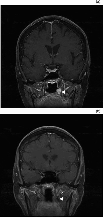
A 73-year-old woman was initially referred for evaluation of a thyroid nodule. Her history dated back to early 2000, when a screening bone density scan had been found to be normal (with the lowest T score (a measurement expressed in standard deviation units from a given mean used in assessment of osteoporosis, equal to a patient's bone mineral density measurement by DEXA minus the value in a young healthy person, divided by the standard deviation of the measurement in the population), of −0.8, being found in the right femoral neck). In 2002, she had tripped and fractured her left foot and toes. In 2003, she had been diagnosed with lower extremity osteoarthritis, causing pain and difficulty walking, and had been prescribed prednisone and Fosamax®. In August 2004, she had experienced sudden, left-sided chest pain without any antecedent trauma, and had been diagnosed with an eighth rib fracture. In July 2004, prior to undergoing a cholecystectomy, the patient had been noted to have a serum calcium concentration of 8.3 mg/dl (normal range 8.5–10.5 mg/dl) and a phosphorus concentration of 1.8 mg/dl (normal range 2.5–4.5 mg/dl). Later laboratory investigation had revealed persistent hypophosphataemia (with the phosphate concentration down to 1.3 mg/dl) and vitamin D deficiency (with a 25-OH vitamin D concentration of 20 ng/dl (normal range 25–50 ng/ml)). A technetium 99 scan undertaken in August 2005 had been of concern, owing to the presence of multiple lesions with a scintigraphic pattern highly suspicious for widespread metastatic disease. An indium-111 pentetreotide scan in August 2006 had shown increased uptake in the sella turcica, thyroid and left posterior maxillary sinus. Subsequent computed tomography (CT) (Figure 1) and magnetic resonance imaging (MRI) scans (Figure 2) had revealed a 5 mm pituitary microadenoma and a 0.8 × 1.0 × 1.3 cm, intraosseous lesion involving the posterior aspect of the left maxillary sinus at the junction with the hard palate.
Axial computed tomography image at the level of the hard palate, demonstrating a left pterygoid mass (arrow).

(a) Coronal magnetic resonance image at the level of the pterygoid plates, demonstrating a left pterygoid mass (arrow). (b) Coronal magnetic resonance image, with contrast, at the level of the pterygoid plates, one year after surgery, demonstrating no enhancing lesion (arrow).

The patient was initially referred back to our clinic for evaluation of the thyroid nodule, which was found to be thyroiditis on fine needle aspiration. During this time, the left pterygoid mass was identified and the patient was referred to our skull base surgeon. The patient's hypophosphataemia and vitamin D deficiency were felt to be related to her tumour, with presumed tumour-induced osteomalacia. She remained symptomatic with calcium, phosphorus and vitamin D supplementation.
In January 2007, the tumour was endoscopically resected via a transnasal, transpterygoid approach. Histopathological examination revealed neoplastic fragments comprising closely packed ovoid to spindle cells with scant to moderate amounts of eosinophilic cytoplasm interspersed with many vessels (Figure 3). Some of the vessels had a prominent, perivascular hyalinisation, whereas others were small and thin-walled. The neoplastic cells were vimentin-positive and pancytokeratin-negative, supporting a mesenchymal rather than epithelial phenotype. This was most consistent with a haemangiopericytoma.
Photomicrograph of the resected tumour, showing prominent vessels with perivascular hyalinisation (arrows) in a field of ovoid to spindle cells with scant to moderate amounts of eosinophilic cytoplasm (H&E; ×200).

The patient did well after surgery, and her hypophosphataemia and vitamin D deficiency resolved.
Discussion
Tumour-induced osteomalacia is a relatively uncommon entity. Recognition of the syndrome is traditionally attributed to McCrance in 1947.Reference McCrance2 In 1995, Crouzet et al. reviewed 100 cases reported in the medical literature.Reference Crouzet, Mimoune, Beraneck and Juan3 Since that time, there have been many other case reports, including a 2004 pathological review of 32 cases by Folpe et al. Reference Folpe, Fanburg-Smith, Billings, Bisceglia, Bertoni and Cho4 They discussed the phosphaturic mesenchymal tumour (mixed connective tissue variant) as a distinctive type of tumour, with the thought that previously diagnosed tumours may have been reported as other types of soft tissue tumours. In this study, two sinonasal tumours were identified and noted to have characteristics which would make them appear to be variants of haemangiopericytoma.
In 1998, Gonzalez-Compta et al. reviewed 21 cases of head and neck tumour induced osteomalacia.Reference Gonzalez-Compta, Manos-Pujol, Foglia-Fernandez, Peral, Condom and Claveguera5 They found that 57 per cent of tumours were located in the sinonasal area, and that patients had a mean age of diagnosis of 45 years. All of the patients had been previously diagnosed with osteomalacia a mean of 4.7 years prior to tumour diagnosis. These authors also reported that sinonasal tumours occurred in men and women in a ratio of 1:5, and that 58 per cent of these tumours resembled haemangiopericytomas.Reference Gonzalez-Compta, Manos-Pujol, Foglia-Fernandez, Peral, Condom and Claveguera5
There have been three recent reports of osteomalacia induced by paranasal sinus tumours, all of which presented with symptoms of bone demineralisation and a nasal tumour.Reference Inokuchi, Tanimoto, Ishida, Sugimoto, Yamauchi and Miyauchi6–Reference Fuentealba, Pinto, Ballesteros, Pacheco, Boettiger and Soto8 The same presentation was seen in our patient. Generally, patients have hypophosphataemia, normal calcium levels, low vitamin D levels and hyperphosphaturia. Biochemical analysis often triggers patient investigation, but imaging typically identifies the causative agent. Patients may undergo multiple types of imaging, including technetium-99 scintigraphy and indium-111 octreotide scintigraphy. The latter relies on binding of the somatostatin analogue octreotide to somatostatin receptors, which have been found on many tumours involved in tumour-induced osteomalacia.Reference Rhee, Lee, Shin, Lee, Huh and Lim9, Reference Jan de Beur, Streeten, Civelek, McCarthy, Uribe and Marx10 Symptoms typically resolve after surgical excision of the tumour.
There continues to be discussion on the pathogenesis of tumour-induced osteomalacia, which is though to be secondary to inhibition of the renal tubule ability to reabsorb phosphorus and to activate calcitriol synthesis. This leads to hypophosphataemia and hyperphosphaturia, with decreased calcitriol. Renal tubule inhibition has been attributed to a humoral product of the causative tumour, which has been termed phosphatonin.Reference Jan de Beur1 However, the most likely candidate for this inhibitor role is fibroblast growth factor 23. This growth factor has been shown to be over-expressed in tumour-induced osteomalacia,Reference Jonsson, Zahradnik, Larsson, White, Sugimoto and Imanishi11–Reference Bowe, Finnegan, Jan de Beur, Cho, Levine and Kumar13 and has been shown to inhibit phosphorus transport in cultures.Reference Bowe, Finnegan, Jan de Beur, Cho, Levine and Kumar13 In addition, Shimada et al. have demonstrated similar skeletal changes in transgenic mice with over-expressed fibroblast growth factor 23, further supporting its role in tumour-induced osteomalacia.Reference Shimada, Urakawa, Yamazaki, Hasegawa, Hino and Yoneya14
• Oncogenic osteomalacia, also known as tumour-induced osteomalacia, is an uncommon cause of osteomalacia and its related symptoms
• This paper describes tumour-induced osteomalacia secondary to a haemangiopericytoma arising in the pterygopalatine fossa
• The osteomalacia resolved following endoscopic excision of the neoplasm
Conclusion
Here, we present a case report of a skull base tumour causing tumour-induced osteomalacia. The tumour was successfully removed via an endoscopic, transnasal, transpterygoid approach. One year post-resection, the patient was free of the adverse effects of osteomalacia.