

Introduction
In Australia, about 28,000 individuals of working age sustain an acquired brain injury (ABI) every year. Of these injuries, 5–8% are classified as severe (Glasgow Coma Scale score 3–8) (Teasdale & Jennet, Reference Teasdale and Jennet1974) and are associated with long-lasting or permanent disability (Fortune & Wen, Reference Fortune and Wen1999). Patients with severe ABI are typically functionally dependent and a small amount of limb movement is often the best motor ability observed during the acute phase of care (Turner-Stokes, Nair, Sedki, Disler, & Wade, Reference Turner-Stokes, Nair, Sedki, Disler and Wade2005). During recovery such patients face a multitude of challenges, requiring treatment from many different disciplines. Physiotherapy is considered to be a key discipline for rehabilitation following ABI (Hellweg & Johannes, Reference Hellweg and Johannes2008; New Zealand Guidelines Group, 2007; Teasell et al., Reference Teasell, Bayona, Marshall, Cullen, Bayley, Chundamala and Tu2007; Tolfts & Stiller, Reference Tolfts and Stiller1997). Although there is limited robust research evaluating rehabilitation interventions in the ABI population (New Zealand Guidelines Group, 2007; Teasell et al., Reference Teasell, Bayona, Marshall, Cullen, Bayley, Chundamala and Tu2007; Zitnay et al., Reference Zitnay, Zitnay, Povlishock, Hall, Marion, Trudel and Barth2008), the delivery of allied health interventions, including physiotherapy, has been shown to reduce length of inpatient stay, optimise motor function at discharge and decrease overall disability (Chestnut, Reference Chestnut1990; Gray, Reference Gray2000; Hall & Cope, Reference Hall and Cope1995; Turner-Stokes et al., Reference Turner-Stokes, Nair, Sedki, Disler and Wade2003; Zhu, Poon, Chan, & Chan, Reference Zhu, Poon, Chan and Chan2007). In fact, increasing the intensity of rehabilitation has resulted in accelerated rates of recovery of personal independence and improved physical outcomes (Cifu, Kreutzer, Kolakowsky-Hayner, Marwitz, & Englander, Reference Cifu, Kreutzer, Kolakowsky-Hayner, Marwitz and Englander2003; Slade, Tennant, & Chamberlain, Reference Slade, Tennant and Chamberlain2002; Spivack, Spettell, Ellis, & Ross, Reference Spivack, Spettell, Ellis and Ross1992; Turner-Stokes et al., Reference Turner-Stokes, Nair, Sedki, Disler and Wade2003). Despite emerging evidence for the benefits of physiotherapy for ABI management, a specific scale to monitor early incremental changes in neuro-motor function during the early stages following ABI, when patients are functionally dependent, remains absent from the field.
A recent systematic review (Laxe, Tschiesner, Zasler, López-Blazquez, Tormos, & Bernabeu, Reference Laxe, Tschiesner, Zasler, López-Blazquez, Tormos and Bernabeu2012) identified the most frequent outcome measures in brain injury research as the Functional Independence Measures (FIM) (50%), Glasgow Outcome Scale (34%) and Disability Rating Scale (DRS) (32%). Of these, only the DRS incorporates motor function as variables or items within the scale. In the acute stages following severe ABI, few scales, including the DRS, are capable of assessing incremental changes in neuro-motor function that may occur at this time. The brain-injury-specific outcome measure database (Wright, Bushnik, & O’Hare, Reference Wright, Bushnik and O’Hare2000) highlights that scales typically in use during this stage evaluate consciousness, cognitive function, behaviour, social participation and functional limitations (Wright et al., Reference Wright, Bushnik and O’Hare2000). However, these scales fail to capture the incremental changes in neuro-motor function in the early stages of recovery important to physiotherapy management following severe ABI (Canedo, Grix, & Nicoletti, Reference Canedo, Grix and Nicoletti2002; O’Dell et al., Reference O’Dell, Jasin, Stivers, Lyons, Schmidt and Moore1996; Pape et al., Reference Pape, Lundgren, Heinemann, Guernon, Giobbie-Hurder, Wang and Williams2006; Teasdale & Jennet, Reference Teasdale and Jennet1974).
Some well-known assessment scales of motor function used specifically by physiotherapists include the Clinical Outcome Variable Scale (COVS) (Seaby & Torrance, Reference Seaby and Torrance1989), the, Motor Assessment Scale (MAS) (Carr, Shepherd, & Nordholm, Reference Carr, Shepherd and Nordholm1985) and the Functional Independence Measure–Motor component (FIM-motor) (Kidd, Stewart, & Baldry, Reference Kidd, Stewart and Baldry1996). These scales monitor important motor tasks such as walking, transfers, wheelchair mobility and fine motor upper limb skills, but most patients with severe ABI are not capable of attempting these tasks in the earliest stage of recovery (Pilon, Sullivan, & Coulombe, Reference Pilon, Sullivan and Coulombe1995). While valid and reliable for the assessment of patients’ motor function, these scales are more applicable when dealing with the patient who is able to actively participate in practising a range of motor tasks at more advanced stages of rehabilitation. Other well-known scales commonly used in the acute care setting include the Glasgow Coma Scale (GCS) (Chieregato et al., Reference Chieregato, Martino, Pransani, Nori, Russo, Nato and Simini2010), Glasgow Outcome Scale (GOS) (Weir et al., Reference Weir, Steyerberg, Butcher, Lu, Lingsma, McHugh and Murray2012) and Full Outline of UnResponsiveness (FOUR) scale (Fischer et al., Reference Fischer, Rüegg, Czaplinski, Strohmeier, Lehmann, Tschan and Marsch2010). These tools provide a measure of consciousness or responsiveness in the early stages following severe ABI but do not address the potential for incremental changes in neuro-motor recovery relevant to physiotherapy, such as muscle tone, head and trunk alignment, sitting balance, posture and movement.
A new scale that captures early changes in neuro-motor recovery following severe ABI is required. Four experienced physiotherapists from Princess Alexandra Hospital, Brain Injury Rehabilitation Unit (BIRU) sought to develop an outcome measure suitable for measuring incremental neuro-motor recovery during the early stage following severe ABI. The goal was to develop a quantitative assessment scale, informed by empirical evidence that would be responsive to change and incorporate the important items required to capture the incremental changes in neuro-motor recovery that underpin a physiotherapy assessment for the severely brain injured.
The study was undertaken in two parts. The first part involved the identification of relevant items to measure incremental changes in neuro-motor recovery that may be associated with the early physiotherapy management of people following severe ABI – that is, it identified the content of the Acquired Brain Injury Physiotherapy Assessment (ABIPA). The second part investigated the responsiveness of the ABIPA to measure change in neuro-motor recovery in the early stages of recovery following severe ABI, as a first step in determining concurrent validity of the tool for use in the clinical setting. Thus, our aims were: (1) to identify the items and develop scoring criteria for the ABIPA, a new measurement scale that could be used by physiotherapists to assess neuro-motor recovery of people in the early stages following severe ABI; (2) to compare the responsiveness to change of the ABIPA to measures of motor function (COVS and MAS) and a measure of consciousness (GCS); and (3) to determine the concurrent validity of the ABIPA with these tools at initial and discharge assessments in the acute hospital setting.
Methods
PART A: ABIPA Development – Item Selection
Literature Search
In order to inform item selection and establish practical assessment guidelines, a systematic approach to a literature review and an expert consensus panel of experienced clinical physiotherapists was employed. A literature search was undertaken of relevant databases including Cochrane, Pedro, PubMed, Medline, Cinahl, Embase, COMBI (Centre for Outcome Measurement in Brain Injury) and ABIEBR (Acquired Brain Injury Evidence Based Review). Search terms included ‘brain injury or head injury or CVA or stroke or cerebrovascular accident’ AND ‘physical therapy or physiotherapy’ AND ‘outcome assessment or outcome measure’ AND ‘motor recovery’. Search limits of human, English language and age-related 19 years+ were used. Studies were included if participants were in the acute phase of recovery following moderate or severe ABI (GCS < 12). All study types, including systematic reviews, meta-analysis studies and practical guidelines, were included. Studies were excluded if the focus was on spinal injury or other neurological diseases, such as Parkinson's disease or multiple sclerosis, if community based or if high-level function or mobility was being measured. Studies were also excluded if treatment focused, investigating the chronic phase of recovery, pharmacological studies, focused on cognitive or psychosocial interventions, if they were conference proceedings or were unavailable in full text.
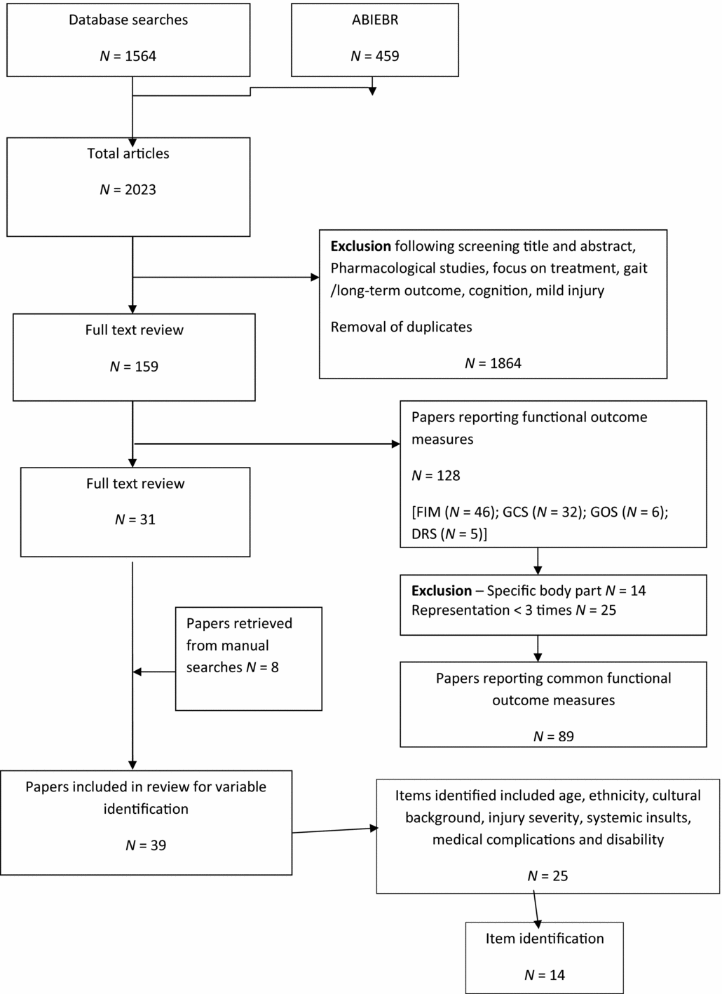
The flow chart for the search strategy is shown in Figure 1. Initial searches yielded 2023 articles. A total of 1564 articles from databases and a further 459 from ABIEBR were retrieved. Excluded, based on title and abstract, were studies such as those dealing with behaviour, cognition, community focus, long-term outcomes, pharmacological studies or mild injury. One hundred and seventeen articles (n = 117) were retrieved for full-text review from the database search and 127 articles from ABIEBR.
Flow diagram for manuscript identification.

Following removal of duplicates, 159 articles (n = 159) were then grouped into manuscripts outlining commonly used outcomes measures (n = 128) and those articles focusing on item identification required for measuring neuro-motor recovery in ABI (n = 31). Of the articles outlining commonly used outcomes measures, those measures that were reported less than three times or were related to a specific body part such as the upper limb (n = 39) were excluded from further analysis. Reference lists of articles focusing on item identification were further examined to ensure any relevant publications were not overlooked, and eight more studies (n = 8) were included; resulting in a total of 39 articles to be included for item identification relevant to measuring neuro-motor recovery.
Data Extraction
Data were extracted from all articles related to commonly used outcome measures, identifying the component variables of the common outcome measures and items identified as important for measurement of neuro-motor recovery in the ABI population. The most commonly reported outcome measures in the retrieved articles were the FIM or Functional Assessment Measure (n=46), GCS (n=32), GOS (n=6) and DRS (n=5). This finding is supported by previous studies reviewing commonly used outcome measures in ABI (Crooks, Zumsteg, & Bell, Reference Crooks, Zumsteg and Bell2007; Haigh et al., Reference Haigh, Tennant, Biering-Sorensen, Grimby, Marincek, Phillips and Thonnard2001; Laxe et al., Reference Laxe, Tschiesner, Zasler, López-Blazquez, Tormos and Bernabeu2012; Pollock, Morris, Wijck, Coupar, & Langhorne, Reference Pollock, Morris, Wijck, Coupar and Langhorne2011; Shukla, Devi, & Agrawal, Reference Shukla, Devi and Agrawal2011). Of these, the GCS, commonly used in the acute care setting, was selected as an accepted validated outcome measure for comparison with the ABIPA. The FIM was not selected due to its use primarily as a rehabilitation measure (Nichol et al., Reference Nichol, Higgins, Gabbe, Murray, Cooper and Cameron2011) and we were more interested in the acute care setting. In addition, well-known physiotherapy assessment scales of motor function, the COVS (Seaby & Torrance, Reference Seaby and Torrance1989) and MAS (Carr et al., Reference Carr, Shepherd and Nordholm1985), were also selected as comparison measures.
In order to identify common items to measure neuro-motor function, the 39 retrieved studies were then reviewed by an expert consensus panel of three experienced clinical physiotherapists working within the Neuroscience Unit, Princess Alexandra Hospital. Their task was to identify the items of neuro-motor recovery appropriate for inclusion in an ABI measurement scale that could be applied to those with a severe injury. Following full review of each manuscript, further studies were removed if the items identified only included age, ethnicity and cultural background, injury severity, systemic insults and medical complications. Studies were also removed if the focus was on level of disability (inability to perform) functional activities, such as transfers. Fourteen studies (n = 14) remained that identified neuro-motor items.
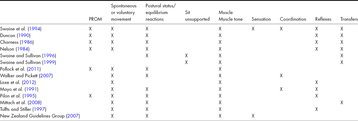
A frequency analysis identified the most important items for inclusion in a measure of neuro-motor impairment following severe ABI. The items were tone (93%), spontaneous and voluntary movements (71%), postural status/equilibrium reactions (64%), passive range of motion (29%) and reflexes (43%) (see Table 1). Passive range of motion, spontaneous movements and postural status were rated as being ‘extremely important’ or ‘very important’ items, requiring evaluation (Mayo, Sullivan, & Swaine, Reference Mayo, Sullivan and Swaine1991; Pilon et al., Reference Pilon, Sullivan and Coulombe1995; Swaine, Sullivan, & Sicotte, Reference Swaine, Sullivan and Sicotte1994; Walker & Pickett, Reference Walker and Pickett2007). Additional items identified as important to measure included the ability to sit unsupported, postural control and ‘tolerance to vertical’ (Pilon et al., Reference Pilon, Sullivan and Coulombe1995), along with muscle tone, voluntary movements, equilibrium reactions, transfers and range of motion (Charness, Reference Charness1986; Duncan, Reference Duncan1990; Laxe et al., Reference Laxe, Tschiesner, Zasler, López-Blazquez, Tormos and Bernabeu2012; Mittrach et al., Reference Mittrach, Grill, Walchner-Bonjean, Scheuringer, Boldt, Huber and Stucki2008; Nelson, Reference Nelson1984; Swaine & Sullivan, Reference Swaine and Sullivan1996, Reference Swaine and Sullivan1999).
Neuro-motor Items Identified from Retrieved Articles
The identified items were grouped under similar categories and became items of muscle power, muscle tone, body alignment and maintaining body position. Piloting of the tool was undertaken with a series of single case studies, with the definitions and procedures for a severe ABI scale explored, developed and refined to enhance the overall clarity of the scale. The final items of the Acquired Brain Injury Physiotherapy Assessment (ABIPA) determined by the expert panel of experienced clinical physiotherapists were: upper limb and lower limb movement, overall muscle tone in each limb, head and trunk alignment in supine, head and trunk alignment in sitting, head and trunk control in sitting and overall presentation.
Scoring
To determine the scoring for the ABIPA scale, the type of data, the evidence supporting scale development, as well as the scoring systems of commonly used validated tools were considered. Scoring these final items of the ABIPA required clinical judgement of the assessor as the data to be scored were observational or qualitative in nature. The best method of scoring qualitative data in a scale format has been suggested as mapping the observational data into measurable dimensions using experienced clinicians’ clinical judgement (Hagerty, Reference Hagerty2002; Guttman, Reference Guttman2004; Guyatt, Krishner, & Jaeschke, Reference Guyatt, Krishner and Jaeschke1992).
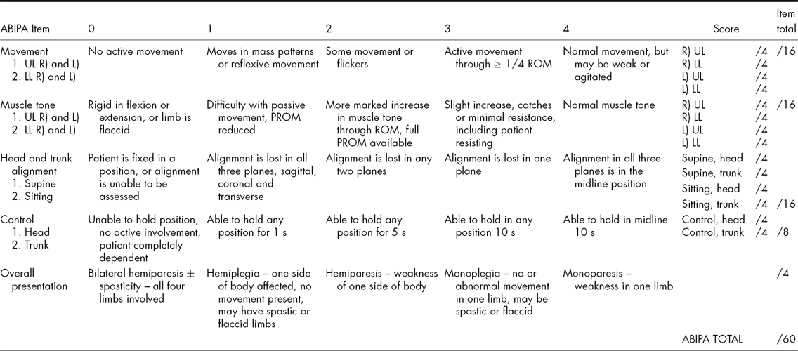
In addition, the retrieved articles relevant to each ABIPA item were further examined to devise a scoring technique relevant to each item. For example, for the first item, upper and lower limb movement, common motor function impairment measures included the manual muscle test (Harms-ringdahl, Reference Harms-ringdahl1993), movement recovery scale (Sodring, Bantz-Holter, Ljunggren, & Wytter, Reference Sodring, Bantz-Holter, Ljunggren and Wytter1995) and the Motoricity Index (Demeurisse, Demol, & Roboye, Reference Demeurisse, Demol and Roboye1980). For these measures, either a five- or six-point scale was used. Two of the more widely used clinical measures for upper and lower limb muscle tone are the Modified Ashworth Scale (Ansari, Haghdi, Moammeri, & Jalaie, Reference Ansari, Haghdi, Moammeri and Jalaie2006; Pomeroy et al., Reference Pomeroy, Dean, Sykes, Faragher, Yates, Tyrrell and Tallis2000) and the Tardieu scale (Tardieu et al., Reference Tardieu, Rondot, Mensch, Dalloz, Monfraix and Tabary1957). Both are rated using a six-point scale. For the remaining items – head and trunk alignment in sitting and supine, and head and trunk control in sitting – consideration was given to the cardinal planes of movement (i.e., sagittal, coronal and horizontal) and whether the head or trunk was fully aligned or not able to be assessed. Considering the range of scales supported by the literature, the experienced clinicians developed the dimensions that were considered clinically significant. A series of single-case pilot studies clarified the dimension and a five-point scale emerged. Scores for each item range from 0 to 4, with low scores representing poorer function and a score of 4 representing best function (Hagerty, Reference Hagerty2002). The ABIPA scale, its items and scoring are set out in Table 2.
Description and Scoring of the ABIPA
PART B: Responsiveness of the ABIPA to Change and Concurrent Validity
Design
Part B investigated the responsiveness of the ABIPA to changes in a patient in the early stages of recovery following an ABI. It also investigated its concurrent validity with other assessment tools currently in use with this population. To achieve this, a prospective cohort study was conducted using a sample of convenience of patients admitted to the neurosurgical unit at Princess Alexandra Hospital. Blinded assessments were conducted on patients throughout their acute hospital stay, until each patient was discharged or showed a change in scores on two other commonly used scales of motor function (COVS and MAS).
Participants
The neurosurgical unit is based in a tertiary referral hospital in Brisbane, Queensland, with state-wide admissions covering Queensland and northern New South Wales. The unit is comprised of 36 beds and is staffed by a multidisciplinary team including physiotherapists, occupational therapists, speech pathologists, social workers, neuropsychologists and a medical team.
Patients were included in the study if they had recently suffered either a moderate (GCS 9–12) or severe (GCS 3–8) ABI or a grade four or five subarachnoid haemorrhage, were medically stable (i.e., had been discharged from intensive care) and were aged between 16 and 70 years. Patients were excluded if they had major musculoskeletal disorders that may impact on movement return (e.g., amputation or fracture) or if there were any residual deficits from previous neurological insult or conditions (e.g., previous stroke or Parkinson's disease). Patients with subarachnoid haemorrhage who were awaiting clipping of an aneurysm or those not deemed medically stable were also excluded.
Ethical clearance was obtained from the Metro South Human Resource Ethics Committee and Griffith University Human Resource Ethics Committee, and the study was supported by the Medical Director of the neurosurgical unit. Informed consent was obtained from the next of kin or legal guardian, as required.
Procedure
Participants were assessed throughout their acute hospital admission. The first assessment took place on the first weekday post admission to the neurosurgical unit. The second assessment occurred on day 3 following admission. Subsequent assessments occurred on day 7 post neurosurgical unit admission then at weekly intervals until the patient showed a change in scores on the two selected scales of motor function, COVS and MAS. Assessments took place at approximately the same time of day.
At each assessment, current GCS, presence of a tracheostomy and weaning status, along with any changes to relevant medications, were recorded. The GCS was included as a measure of consciousness with established responsiveness in the early stages following severe ABI.
Two assessors were involved in each participant assessment and all assessments were completed by one of three physiotherapists, each with 10–20 years of clinical experience in the management of ABI patients. Assessors were randomly allocated to concurrently assess the patients with either the ABIPA assessment or the COVS and MAS assessment, to minimise the time burden for these highly dependent patients at this stage of their rehabilitation. The GCS data were collected from ward assessments. Physiotherapist assessors were blinded to each other's scores, previous scores and were not involved in patient care in the neurosurgical unit. Participants were assessed using the ABIPA (Table 2), which included five items: movement of the limbs, general tone, head and trunk alignment in sitting and supine, head and trunk control in sitting, and overall presentation. ABIPA items were assessed in a consistent order for all participants, commencing with resting alignment in bed (supine) and movement and general tone before assisting the patient into sitting.
Measures
The ABIPA was conducted using a standardised procedure and scored as outlined in Table 2. On initial approach to the bedside the resting alignment of the patient's head and trunk was observed. The patient was then placed in a supine position with a single pillow under the head and allowed to settle. Head alignment was observed, scoring for obvious deviations from the midline, noting rotation, lateral flexion and flexion. Trunk alignment was assessed with observations confirmed by palpation. The therapist observed lateral trunk angle, rib height, iliac crest height and compared equal presentation for both right and left sides. The shoulder girdle, pelvis alignment and lumbar lordosis were also observed, and then overall alignment scored.
Muscle tone and movement were assessed first for the upper limbs and then for the lower limbs. Initially the presence of any spontaneous movement (including reflexive, patterned or selective movement) was observed. Each major muscle group of the upper limb and lower limb was moved through a passive range of motion three times to assess muscle tone and determine a score using the ABIPA scale. The lowest score from the major muscle groups for each limb was recorded as the overall score for that limb.
Active movement was assessed for each of the four limbs individually. The patient was asked to move the limb as able. The patient's limb was then positioned in mid-range and any muscle activity or ability to hold the position recorded. Finally, the limb was moved through range for the major joints, noting any active movement. The highest score was then recorded as movement for that limb.
Head and trunk control was assessed in sitting. This relates to the active movement of the trunk and head and is defined as the ability to maintain a position in space with some muscle activity, normal or abnormal. It was assessed with the patient sitting on a firm surface, with feet supported, and timed using a stopwatch. To assess head control, the trunk was fully supported in the midline while the head was placed in the upright position, head support was then removed. Trunk control was assessed in the same manner, with the trunk placed in the midline and hand support then removed. If the patient was unable to sit (e.g., medical limitations, safety or concomitant injuries), the head and trunk were scored as 0.
Alignment in sitting was assessed using the same scale and procedure as alignment in supine. Head alignment was assessed by positioning the head and trunk in the midline and while fully supporting the trunk, the quality of head alignment in the upright position was assessed. Trunk alignment was assessed in the same manner – positioning the trunk and then removing support. The alignments achieved for both head and trunk were scored. For patients who were constantly moving, the movement was repeated three times and the best alignment achieved was scored. A score of 0 was recorded: if the patient was unable to sit (e.g., medical limitations, safety or concomitant injuries), if the patient did not have any head or trunk control (as per control scale) or if the patient required maximum assistance to sit.
Finally, overall presentation was scored on the completed assessment of tone, movement, alignment and control as per Table 2.
As part of the assessment procedure three comparative measures were conducted: the Glasgow Coma Scale (GCS) (Chieregato et al., Reference Chieregato, Martino, Pransani, Nori, Russo, Nato and Simini2010; McNett, Reference McNett2007), the COVS (Seaby & Torrance, Reference Seaby and Torrance1989) and the MAS (Carr et al., Reference Carr, Shepherd and Nordholm1985). As a well-known measure of consciousness, the GCS evaluates a patient's best verbal response, eye opening and motor response during the early stages of recovery, with scores ranging from 3 to 15.The COVS has established clinometric properties in a range of patient populations (Barker, Amsters, Kendall, Pershouse, & Haines, Reference Barker, Amsters, Kendall, Pershouse and Haines2007; Salter, Jutai, Foley, & Teasell, Reference Salter, Jutai, Foley and Teasell2010) including ABI (Low Choy, Kuys, Richards, & Isles, Reference Low Choy, Kuys, Richards and Isles2002). It is scored using a seven-point scale across 13 domains, including rolling, transfers, sitting balance, wheelchair mobility and gait. Lower scores indicate greater dependence in each domain with total scores ranging from 13 to 91.
The MAS was developed to measure functional progression amongst people following stroke. It comprises a seven-point scale across eight domains: supine to side lying; supine to sitting; balanced sitting; sit to stand; walking; upper arm function; hand movements and advanced hand activities. The MAS has good reliability and validity (Poole & Whitney, Reference Poole and Whitney1988). For the purposes of this study, only the domains of sitting balance and upper arm function (left and right) were adopted, as they were domains represented in the ABIPA. Thus, the possible score range is 0–21.
Data Analysis
Each scale was scored according to standard criteria and the items for each scale were totalled. Descriptive statistics including mean (standard deviation), median (range) and frequency were generated for all scales at each assessment point from admission to discharge.
Standardised response means (SRM) were calculated to determine responsiveness to change for all measures on day 3, day 7 and discharge. The standard response mean is defined as the mean change in score between the first assessment and the comparison assessment, divided by the standard deviation of the individual changes in scores (Portney & Watkins, Reference Portney and Watkins2000). The higher the SRM, the greater the responsiveness to change, whereby a value of >.8 is considered a large effect, >.5 a moderate effect and one of .2 a small effect (Cohen, Reference Cohen1977).
A Spearman's rho correlation coefficient was calculated to investigate the concurrent validity of the ABIPA compared to the GCS, COVS and MAS. Admission and discharge scores were analysed separately. Comparisons were made between day 1 scores and day 3, day 7 and discharge. Discharge data were the last recorded for each participant. Rho coefficients greater than .75 were considered good to excellent, while those between .50 and .75 were considered moderate to good (Portney & Watkins, Reference Portney and Watkins2000).
Results
Participants
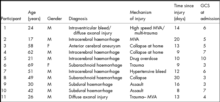
Eleven patients (mean age 41; SD 18 years) were recruited to this study. Participant characteristics are included in Table 3.
Participant Characteristics
MVA, motor vehicle accident.
Three participants were assessed over three data points (days 1, 3 and 7). These patients were discharged from the study at day 7 as they had achieved changes in the scores on the validated motor assessment scales (COVS and MAS). The highest number of assessments that a participant received was nine. In total, 57 assessments were completed for the eleven participants. On three occasions GCS data were not available.
Responsiveness to Change
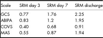
Table 4 illustrates the standardised response means (SRM) from initial assessment for all scales on day 3, day 7 and discharge from the acute ward to other rehabilitation settings or to home. On day 3, the ABIPA showed the greatest responsiveness to change (SRM > .83) compared to the other measures of motor function (SRMs <.55), although the GCS was similar (SRM = .77). By day 7, the GCS demonstrated the greatest responsiveness to change while the ABIPA was higher than the other measures (SRMs < .87). At discharge all scales showed good responsiveness to change (SRMs > .9), with the strongest score demonstrated by the GCS followed by the ABIPA and the selected MAS items. The responsiveness of the MAS and COVS was consistently low to moderate on day 3 of the assessments and continued to be lower than the ABIPA on day 7. The total COVS was also lower at discharge, with the MAS items showing a similar SRM as the ABIPA by discharge.
Standardised Response Means (SRM) from Initial Assessment for all Scales
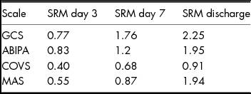
GCS, Glasgow Coma Scale; ABIPA, Acquired Brain Injury Physiotherapy Assessment; COVS, Clinical Outcome Variable Scale; MAS, Motor Assessment Scale.
Concurrent Validity of ABIPA
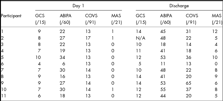
Table 5 illustrates admission and discharge scores on all scales for all participants. Across all assessments (n = 57) the ABIPA demonstrated good to excellent correlations with the GCS (rho > .76, p ≤ .001), COVS (rho > .82, p ≤ .001) and MAS (rho > .66, p ≤ .001). The investigation of concurrent validity at specific assessment points – such as day 1, 3 and 7 – showed that the ABIPA was moderately associated with all scales across the first week at admission to the acute neuroscience ward (rho > .53, p ≤ .001), whereas at discharge the associations were higher (rho > .72, p ≤ .001).
Admission and Discharge Scores for all Scales (n = 11)
N/A, Not available.
Figure 2 shows the mean ABIPA, COVS, GCS and MAS scores for all participants converted to a percentage of the total possible score for each scale over the 7-week period. Three participants had been discharged from the study by day 14 as they had improved sufficiently to be suitable for measurement using the COVS and MAS. Therefore, fewer participants, who demonstrated slower recovery and lower scores, were included at each time point following day 14. Dips at day 14 and day 42 in overall ABIPA scores (Figure 2) represent the loss of those patients who were discharged from the study. By day 42, nine participants demonstrated changes in scores on the other scales and were discharged.
Mean ABIPA, COVS, GCS and MAS scores for all participants, converted to a percentage of the total possible score. ABIPA, Acquired Brain Injury Physiotherapy Assessment; COVS, Clinical Outcome Variable Scale; GCS, Glasgow Coma Scale; MAS, Motor Assessment Scale.
Discussion
We have described the development of the ABIPA, examined its responsiveness to change against other common measures and established its concurrent validity with other common assessment tools. We found that the ABIPA score had a strong positive relationship with the GCS score, the current standard measure of early brain injury, and showed a greater responsiveness to change when compared to the COVS (a measure of motor function) and selected MAS Items (a measure of motor recovery of bed mobility and sitting ability). The results of this study indicate that the ABIPA could be used as a responsive measure of neuro-motor change across the early stages of recovery, providing physiotherapists with a useful tool to use at this stage following ABI.
The ABIPA had the highest level of responsiveness to change when comparing scores on day 1 to day 3 after admission to the neurosurgical ward. Between day 1 and day 7, the GCS and the ABIPA continued to show higher responsiveness to change than the COVS and MAS. The ABIPA was able to detect change earlier than the other motor scales for any given patient. This is an important finding as physiotherapists must make decisions regarding suitability for rehabilitation very early in a patient's acute hospital stay. If such decisions are based on COVS and MAS alone, it would be difficult to advocate objectively for the patient as the existing scales may not detect change during the immediate period after ABI. As the ABIPA continues to show high responsiveness to change during the stages of acute hospital care, it is a promising tool for clinical use during early recovery.
To date, there is no specific scale to monitor early incremental changes in a patient's neuro-motor recovery across the acute period of care, for those with severe brain impairment following ABI. The majority of scales focus on the patient's level of consciousness, cognitive functions, behaviour, social participation and functional limitations (Wright et al., Reference Wright, Bushnik and O’Hare2000). The absence of an appropriate outcome measure for this patient population significantly impacts on clinicians’ ability to objectively assess the effectiveness of interventions, communicate changes in a patient's condition with other team members and advocate for patients. It is also a significant barrier to the advancement of research and evidence-based practice in the early stages of rehabilitation for this complex and challenging clinical population.
Available evidence is often based on retrospective analysis when evaluating long-term outcomes (Chua & Kong, Reference Chua and Kong2002; McNett, Reference McNett2007; Pape et al., Reference Pape, Lundgren, Heinemann, Guernon, Giobbie-Hurder, Wang and Williams2006) and there is little data to determine the impact of different types of acute care intervention on prognosis (New Zealand Guidelines Group, 2007; Teasell et al., Reference Teasell, Bayona, Marshall, Cullen, Bayley, Chundamala and Tu2007). No scales were located that specifically monitored neuro-motor function in the early stages of recovery, which is the focus of physiotherapy management following severe ABI (Canedo et al., Reference Canedo, Grix and Nicoletti2002; O’Dell et al., Reference O’Dell, Jasin, Stivers, Lyons, Schmidt and Moore1996; Pape et al., Reference Pape, Lundgren, Heinemann, Guernon, Giobbie-Hurder, Wang and Williams2006; Teasdale & Jennet, Reference Teasdale and Jennet1974). We observed the ABIPA to be a responsive measure of change in neuro-motor function following severe brain injury.
Limitations
A key challenge was the recruitment of an adequate number of participants for the study. The number of patients admitted with severe brain injuries each year is relatively low and, as motor vehicle accidents account for a large percentage, often patients have concomitant orthopaedic injuries and, thus, have to be excluded. There were only 11 participants in the initial sample and eight participants following the third assessment. Three participants were excluded when they were able to achieve scores above the minimum on either the COVS or MAS and the flooring effect on these established measures no longer existed. In addition, we encountered difficulty in assessing those patients who were agitated and restless, who have reasonable movement but whose language, cognition or behaviour was such that they precluded accurate assessment. While this challenge is not unique to the ABIPA, it further limited our approach.
The participant cohort suffered predominately severe ABI (GCS 3–8), with only one patient representative of the moderate brain injury population (GCS 9–12). This limits the current generalisability of our results and suggests the need for further evaluation of the ABIPA in a broader cohort of participants with ABI. With a larger dataset, the factor structure of the ABIPA could be examined using factor analysis; while investigation of a more refined scoring approach and item generation could be pursued using Rasch analysis and Delphi Survey techniques.
Conclusion
This study has provided preliminary psychometric support for the utility of the ABIPA. It exhibits sound correlations with other measures, and scores during the acute phase show it is more responsive to change than other common measures of neuro-motor function. It is now necessary to test the reliability of assessors using the tool, and to involve multiple assessors to further investigate the test/re-test and intra- and inter-rater reliability of the instrument.