Introduction
The left atrial appendage is a trabeculated, finger-like pouch that projects from the anterolateral wall of the left atrium. Embryologically distinct from the smooth-walled atrial body, it takes four recognised morphological forms—chicken wing, cactus, cauliflower, and windsock—each with its own geometry and mechanical behaviour. Clinical interest in the left atrial appendage has grown steadily, driven largely by its central role as a thrombus reservoir during atrial fibrillation. Reference Vella, Musotto, Cook, Bosi and Burriesci1–Reference Arguelles, Mihalatos, Leung, Colangelo, Jayam and Fujikura3
Inversion of the left atrial appendage, the process by which the appendage folds inward into the atrial cavity, was first described as a surgical complication. The mechanism is mechanical: de-airing manoeuvres at the end of cardiopulmonary bypass generate abrupt negative intra-atrial pressure, and the appendage, whose slender neck and compliant walls offer little resistance, collapses inward. Reference Buttar, Andersen, Poulsen and Thyregod4–Reference Meredith and Kimball7 The result is an intracardiac mass that echocardiography can readily misread as thrombus, myxoma, or vegetation, triggering unnecessary anticoagulation, re-exploration, or reoperation before the true anatomy is recognised. Reference Buttar, Andersen, Poulsen and Thyregod4,Reference Fadia, Vaideeswar and Pandit8
Spontaneous inverted left atrial appendage—occurring without any cardiac operation—is considerably rarer. Only a handful of cases populate the published literature prior to this report. Reference Ankersmit, Mohl and Moser5–Reference Gecmen, Candan and Guler9 The entity is easily missed. Transthoracic echocardiography may show nothing; correct diagnosis depends on knowing the anatomy, recognising the echocardiographic signature, and maintaining a high index of suspicion in settings where the condition is known to arise.
We report an autopsy case of spontaneous inverted left atrial appendage in an 8-month-old infant with mitochondrial cytopathy, in whom a pericardial effusion was identified at post-mortem examination. To our knowledge, this is the first documented spontaneous inverted left atrial appendage in an infant without CHD. The case is placed alongside an updated review of publications from 2020 to 2024 and revisit the evolving understanding of the underlying mechanisms.
Case description
An 8-month-old male infant with no documented cardiac history arrived at the emergency department in acute respiratory distress. He was markedly tachycardic and tachypnoeic. Chest radiography showed bilateral pulmonary congestion, thoracic hyperinflation, and a globular cardiac silhouette pointing to either cardiac enlargement or effusion. The electrocardiogram demonstrated diffuse low voltage. Laboratory findings painted a severe metabolic picture: lactic acidosis, hyperglycaemia, acute kidney injury, and life-threatening hyperkalaemia.
Resuscitation was initiated promptly, but the infant deteriorated rapidly and died within hours of admission. Because no cause of death had been established, the judicial authority ordered a medico-legal autopsy, performed 16 hours post-mortem.
External examination was unremarkable. Internal examination revealed:
-
• A pericardial effusion of 45 mL, with no signs of haemorrhage or exudate.
-
• Marked bilateral pulmonary oedema (right lung 120 g; left lung 184 g), consistent with either cardiogenic or metabolic origin.
-
• Cardiomegaly with global four-chamber dilation and hypertrophy.
-
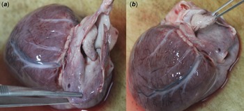
• Absence of the left atrial appendage from its expected anatomical position. On opening the left atrium, the appendage was found inverted into the atrial cavity, taking on a characteristic finger-in-glove configuration (Figure 1 a and 1 b ).
All organ weights exceeded age- and sex-matched reference values, reflecting generalised visceral congestion.
Opening of the left atrium reveals the inverted appendage prolapsing into the atrial cavity, resembling a finger-in-glove structure.

Figure 1. Long description
Two side-by-side autopsy photographs of the same infant heart on a tan surface. The heart is dark reddish-purple, with a pale curved coronary artery running across its surface. In the left photograph (Fig 1a), the wall of the left atrium is opened and metal forceps enter from the lower left. Inside the opened atrium, a pale pink-white, rounded, finger-like structure projects into the cavity, like a finger inside a glove: this is the left atrial appendage turned inside out. In the right photograph (Fig 1b), the heart is seen from a similar angle and forceps enter from the upper right, lifting a pale flap of atrial tissue to expose the same inverted, finger-shaped appendage.
Toxicological screening was entirely negative. Histological examination of multiple organs, most notably cardiac and skeletal muscle, liver, and brain, revealed mitochondrial structural abnormalities consistent with mitochondrial cytopathy. Death was attributed to severe metabolic derangement (lactic acidosis, hyperkalaemia, multi-organ dysfunction) in the setting of underlying mitochondrial disease.
The finding of the inverted left atrial appendage was incidental at autopsy. No thrombus was identified within the inverted appendage.
Discussion
The surgical form of inverted left atrial appendage is mechanically straightforward. De-airing at the end of cardiopulmonary bypass abruptly decompresses the atrium, generating a negative intracardiac pressure spike that the appendage—narrow-necked, thin-walled, and trabeculated—cannot sustain. Reference Buttar, Andersen, Poulsen and Thyregod4,Reference Meredith and Kimball7,Reference Liaqath Ali, Bharucha, Karane and Hayes10,Reference Ekanem, Gattani, Shea, Zhao, Singh and Liam11 The same pressure logic operates after left ventricular assist device implantation, where haemodynamic shifts of comparable magnitude have triggered inversion. Reference Ankersmit, Mohl and Moser5,Reference Fadia, Vaideeswar and Pandit8
The question of why only some appendages invert received its first quantitative answer in 2024. Vella et al. built the field’s first biomechanical computational model of inverted left atrial appendage, simulating pressure conditions across all four morphological subtypes. Reference Vella, Musotto, Cook, Bosi and Burriesci1 The data show that windsock and cauliflower appendages—characterised by longer, narrower necks—bear disproportionately higher wall stress under equivalent pressure loads, making them meaningfully more susceptible to inversion. In spontaneous cases, where no surgical trigger intervenes, appendage geometry may itself be the decisive variable.
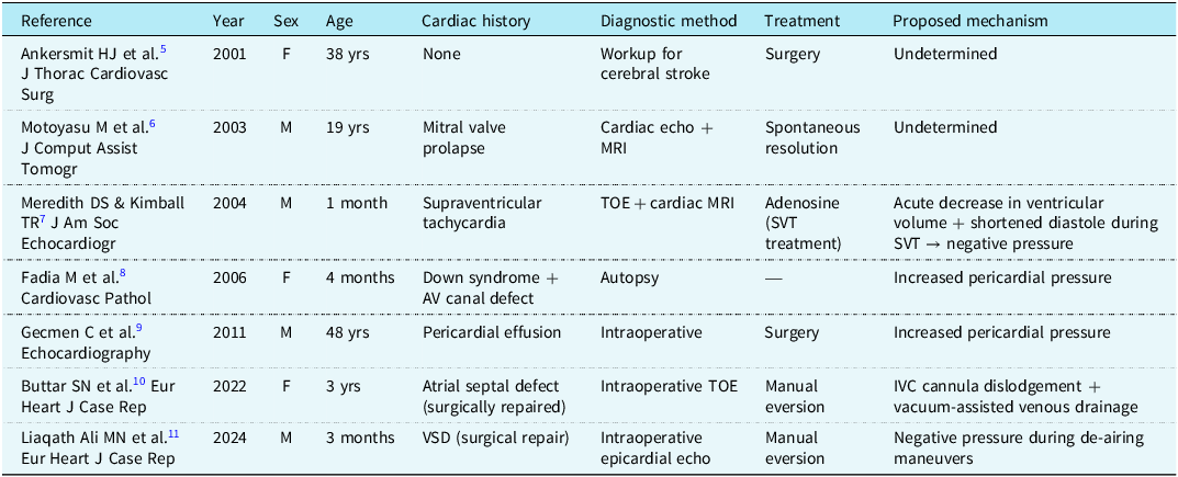
Spontaneous inverted left atrial appendage stands apart. The entire published record, including the present case, amounts to fewer than ten patients worldwide. Table 1 makes the pattern legible: the common denominator across spontaneous cases is a transmural pressure imbalance rather than a surgical event. In three cases including ours, Reference Fadia, Vaideeswar and Pandit8,Reference Gecmen, Candan and Guler9 pericardial effusion drove the process—accumulated fluid exerted external compression on the appendage wall and forced it inward. A fourth case followed a different but equally coherent mechanism: supraventricular tachycardia curtailed diastolic filling so sharply that transient negative pressure developed within the left atrium. Reference Meredith and Kimball7 Two distinct triggers, one shared result.
Summary of published cases of spontaneous left atrial appendage inversion (updated literature review 2001–2024)

Table 1. Long description
Fields: year, sex, age, history, diagnosis, treatment, mechanism. 1) Ankersmit 2001, female, 38 years, none, stroke work-up, surgery, undetermined. 2) Motoyasu 2003, male, 19 years, mitral valve prolapse, echocardiography and magnetic resonance imaging (MRI), resolved spontaneously, undetermined. 3) Meredith 2004, male, 1 month, supraventricular tachycardia, transoesophageal echocardiography and MRI, adenosine, shortened diastole, negative pressure. 4) Fadia 2006, female, 4 months, Down syndrome with atrioventricular canal defect, autopsy, none, raised pericardial pressure. 5) Gecmen 2011, male, 48 years, pericardial effusion, intraoperative, surgery, raised pericardial pressure. 6) Buttar 2022, female, 3 years, repaired atrial septal defect, intraoperative transoesophageal echocardiography, manual eversion, cannula dislodgement. 7) Liaqath Ali 2024, male, 3 months, repaired ventricular septal defect, epicardial echocardiography, manual eversion, negative pressure at de-airing.
The clinical course of inverted left atrial appendage is not uniform. Most spontaneous cases follow a benign trajectory, discovered incidentally and never causing symptoms. Several complications, however, are serious enough to warrant explicit attention:
-
• Thromboembolism: The most clinically consequential complication. The earliest published spontaneous case Reference Ankersmit, Mohl and Moser5 presented as cryptogenic cerebral stroke, with inverted left atrial appendage later identified as the embolic source. The irregular trabecular surface of the inverted appendage likely creates a prothrombotic microenvironment.
-
• Mitral inflow obstruction: A progressively inverted appendage may bulge towards the mitral annulus during diastole, producing functional mitral stenosis, pulmonary hypertension, and right ventricular failure. Reference Fadia, Vaideeswar and Pandit8,Reference Ekanem, Gattani, Shea, Zhao, Singh and Liam11
-
• Arrhythmias: The mass effect of the inverted appendage against the atrial wall can mechanically irritate the myocardium and trigger atrial arrhythmias.
-
• Appendiceal incarceration and necrosis: Rare but potentially fatal, requiring urgent surgery. Reference Ekanem, Gattani, Shea, Zhao, Singh and Liam11
Transoesophageal echocardiography is the diagnostic modality of choice. The characteristic picture is recognisable: a mobile, finger-like mass at the anterolateral left atrial wall, with no colour-flow Doppler signal traversing it and a diastolic excursion towards the mitral annulus. Reference Ekanem, Gattani, Shea, Zhao, Singh and Liam11 When these features cluster together, the diagnosis is essentially secured. Cardiac MRI resolves residual ambiguity. In the operative setting, routine post-bypass TOE—or direct visual inspection of the left atrium before chest closure—constitutes a straightforward safeguard that surgical teams should apply whenever the anatomy raises any doubt. Reference Liaqath Ali, Bharucha, Karane and Hayes10,Reference Ekanem, Gattani, Shea, Zhao, Singh and Liam11
This case advances the literature on two specific points. First, spontaneous inverted left atrial appendage in an infant without CHD has not, to our knowledge, been previously documented. Its occurrence in the setting of mitochondrial cytopathy raises a question the data cannot yet answer: does impaired myocardial energy metabolism alter the biomechanical properties of the appendage wall, reducing its resistance to pressure-driven inversion? The hypothesis is speculative, but the case places it on the record for future investigation. Second, the diagnosis was reached exclusively at autopsy. The finding was not subtle; it was invisible because the treating team’s attention was, understandably, directed entirely at a life-threatening metabolic emergency.
Management of inverted left atrial appendage in a living patient follows the clinical context. Manual eversion—intraoperatively or via catheter-based techniques—is the preferred first step when anatomically feasible. Where the appendage has already served as an embolic source, surgical ligation or device-based closure (AtriClip, WATCHMAN) addresses the risk of recurrence. Reference Ankersmit, Mohl and Moser5,Reference Gecmen, Candan and Guler9 Anticoagulation functions as a bridge when thrombogenic risk is high and surgical intervention cannot be arranged promptly.
Conclusion
Spontaneous left atrial appendage inversion is rare. Rare enough to be missed, yet clinically consequential enough that missing it matters. The differential diagnosis of any left atrial mass—regardless of the patient’s age and regardless of whether cardiac surgery has ever taken place—should include inverted left atrial appendage. The data consistently point to a transmural pressure gradient as the operative trigger, most often generated by pericardial effusion or acute hemodynamic instability. Natural history is generally benign, but the condition carries real risks of thromboembolism, mitral obstruction, and arrhythmia. Clinicians who recognise its echocardiographic signature will not mistake it for something that requires a very different intervention.
Computational biomechanics is beginning to explain why certain appendage morphologies invert while others do not—a line of inquiry with direct implications for risk stratification. The next step requires prospective data: studies that can establish true incidence, map the risk factors with precision, and generate the evidence base needed to guide management of an entity that remains, for now, systematically under-recognised.
Financial support
This research received no specific grant from any funding agency, commercial, or not-for-profit sectors.
Competing interests
None.