Introduction
Research over the past 15 years suggests that depression occurring in later-life has a complex and multifactoral etiology. In particular, the roles of stress, hypothalamic–pituitary–adrenal (HPA) axis dysfunction, neurotrophic factors, neurodegenerative changes, vascular, genetic and epigenetic factors, and the contribution of estrogen and other hormones have been studied (Belmaker and Agam, Reference Belmaker and Agam2008). In the study of major depressive disorder (MDD) generally, there has also been less focus on the reductionist monoamine deficiency hypothesis that assumes depressive symptoms are caused by a deficiency of serotonin, noradrenaline, or dopamine alone.
These developments have been summarized in a number of reviews (Nestler et al., Reference Nestler, Barrot, DiLeone, Eisch, Gold and Monteggia2002; Belmaker and Agam, Reference Belmaker and Agam2008; Krishnan and Nestler, Reference Krishnan and Nestler2010) and in general suggest that differential pathophysiological mechanisms contribute to depression in younger people compared to that occurring in later-life (Krishnan et al., Reference Krishnan, Hays, Tupler, George and Blazer1995; Alexopoulos et al., Reference Alexopoulos, Meyers, Young, Campbell, Silbersweig and Charlson1997). Data suggest that late-life depression (LLD) is particularly associated with volume loss in key frontal and subcortical areas (Schweitzer et al., Reference Schweitzer, Tuckwell, Ames and O'Brien2001; Hickie et al., Reference Hickie2005), white-matter hyperintensities (WMH) on magnetic resonance imaging (MRI) scans (Hickie et al., Reference Hickie, Scott, Wilhelm and Brodaty1997), and neuropsychological impairments in the domains of processing speed, memory, and executive functioning (Naismith et al., Reference Naismith2003). A body of evidence suggests that such changes are also more pronounced in those older people who have later ages of onset of depression and/or vascular risk factors (Hickie and Scott, Reference Hickie and Scott1998). Overall, the neurobiological profile suggests that key frontal-subcortical neuronal networks are implicated, as described below (Mayberg, Reference Mayberg2003; Drevets et al., Reference Drevets, Price and Furey2008a).
In addition to explicating the neurobiological correlates of LLD, there is growing interest in elucidating how glia, acting alone or as part of a glial–neuronal network, may contribute to the pathophysiology of depression. In this paper we explore this literature, particularly with respect to how glia may modulate neurotoxicity (defined here as neuronal dysfunction and possible degeneration) and how they may contribute to the volume loss reported in key frontal and sub-cortical structures.
It is now widely accepted that synapses consist of a pre- and post-synaptic neuronal component as well as a functionally apposed glial cell process or even sheath. Consequently, interactions between neurons and glial cells need to be considered when evaluating normal or pathological CNS activity. The active interconnections between glia and neurons are what we refer to as glial–neuronal networks (GNN). These interactions can be metabolic, electrochemical, neurohumoral, and immunological, and also allow for superimposition of the effects of different GNNs. The resulting level of complexity has to be taken into account when diagnostic results (e.g. from neurophysiology or neuroimaging) are interpreted and when pharmacological interventions are devised.
Further exploration of the complex role of GNN will facilitate our understanding of the pathophysiology of LLD, help to identify biomarkers, and may lead to the development of preventative treatments.
Neural circuitry of depression
As mentioned above, evidence from neuroimaging, neuropsychological, neuropathological, and lesion analysis studies has established that disruptions of fronto-subcortical circuitry most likely underpin the symptoms of MDD (Nestler et al., Reference Nestler, Barrot, DiLeone, Eisch, Gold and Monteggia2002; Mayberg, Reference Mayberg2003; Naismith et al., Reference Naismith2003; Drevets et al., Reference Drevets, Price and Furey2008a; Krishnan and Nestler, Reference Krishnan and Nestler2010). Numerous cortical and subcortical areas have been implicated, as befits a disorder that involves not only emotive changes, but also cognitive, endocrine, autonomic, and psychomotor activity manifestations. Five segregated and parallel striatal–thalamocortical pathways have been described, of which damage to dorsolateral prefrontal cortex (DLPFC), lateral orbitofrontal cortex (OFC), and anterior cingulate cortex (ACC) loops may be implicated in depression (Alexander et al., Reference Alexander, DeLong and Strick1986). Limbic structures, particularly the hippocampus and amygdala, play an important role, as can be demonstrated by the dense interconnections within these key circuits; the amygdala, for example, is critical for the assessment of the emotional significance of sensory input, and modulating monoamine and steroid secretion in response to aversive or novel stimuli (Drevets et al., Reference Drevets, Savitz and Trimble2008b). At the subcortical level, due to anatomical proximity, lesions may affect several circuits and produce a varied array of symptoms. Lesions to, or functional impairment of, the striatum, prefrontal cortex (PFC), OFC, or their associated white matter tracts may cause disinhibition of the amygdala and downstream structures. This may result not only in mood changes via modulation of structures such as the locus ceruleus and raphe nucleus, but also disruption in emotion-mediated memory via the hippocampus and alteration in interoceptive states, including autonomic and metabolic changes via the hypothalamus.
Late-life depression
LLD presents additional challenges to etiological models since it incorporates those forms of depression that emerge earlier in life (early-onset depression; EOD) as well as depression that occurs for the first time late in life (commonly defined as occurring after age 50 or 60 (late-onset depression; LOD)). Evidence now suggests that these two groups have differing environmental and genetic risks, and importantly, that the age-of-onset differentiation may be useful when considering treatment decisions (Hickie et al., Reference Hickie, Naismith, Norrie and Scott2009).
LOD, in particular, is associated with cerebrovascular disease and increased rates of progression to dementia (Byers and Yaffe, Reference Byers and Yaffe2011). MRI studies in LLD, and particularly LOD, tend to show an increase in the prevalence and severity of white matter lesions in the deep white matter (Hickie et al., Reference Hickie, Scott, Wilhelm and Brodaty1997), and periventricular and subcortical regions. This is an important component of the “vascular depression” hypothesis, which postulates that cerebrovascular disease precipitates and perpetuates depression in later life (Alexopoulos et al., Reference Alexopoulos, Meyers, Young, Campbell, Silbersweig and Charlson1997). These WMH, thought to be ischemic in origin (Thomas et al., Reference Thomas2002), may represent damage to the key fronto-subcortical circuits that mediate the emotive, movement, and cognitive symptoms of LLD.
There have been a number of reviews regarding the specific structural neuroimaging findings in LLD (Schweitzer et al., Reference Schweitzer, Tuckwell, Ames and O'Brien2001; Vaishnavi and Taylor, Reference Vaishnavi and Taylor2006). Several groups have reported decreased frontal lobe volumes as well as decreased volumes of subcortical structures that are critical components of the limbic and cognitive circuitry. Quantitative MRI studies have demonstrated that volumetric loss is apparent in the PFC, including the OFC, ACC, and gyrus rectus, as well as subcortical regions including the caudate nucleus (mainly restricted to the head of the caudate), putamen, amygdala, and hippocampus (Husain et al., Reference Husain1991; Naismith et al., Reference Naismith2002; Lee et al., Reference Lee2003; Ballmaier et al., Reference Ballmaier2004; Hickie et al., Reference Hickie2005; Elderkin-Thompson et al., Reference Elderkin-Thompson, Hellemann, Pham and Kumar2009; Burke et al., Reference Burke, McQuoid, Payne, Steffens, Krishnan and Taylor2011). The hippocampus appears to be particularly sensitive to volume loss. It has been reported to be reduced in younger people with depression, and there appears to be a consistent relationship with duration of depression, with more pronounced volume loss being associated with longer durations of untreated illness in particular (Sheline et al., Reference Sheline, Sanghavi, Mintun and Gado1999). This body of research has been supported by a wealth of functional imaging studies showing reductions in glucose metabolism (Smith et al., Reference Smith2009), regional cerebral blood flow (Hickie et al., Reference Hickie1999; Reference Hickie2007), and blood oxygen level dependent activation (Naismith et al., Reference Naismith, Lagopoulos, Ward, Davey, Little and Hickie2010) in the striatum and prefrontal regions. Accordingly, the neuropsychological profile of LLD is one of fronto-subcortical dysfunction, with impaired performance evident on tests of executive functioning, processing speed, and, to a lesser extent, learning and memory (Naismith et al., Reference Naismith2003; Reference Naismith, Hickie, Ward, Scott and Little2006).
While the above findings regarding structural and functional imaging changes have certainly been informative regarding the underlying neurobiology of LLD, these technologies are less informative regarding the underlying cellular, neuronal, and/or glial mechanisms that may underpin the disease. In this regard, multiple pathological processes may be responsible, including cerebrovascular disease, glucocorticoid excess, and other inflammatory, neuro-chemical, and illness-specific factors. While many of these pathways have been considered, few studies have attempted to integrate findings within the broader context of glial pathology and perturbations of GNN.
Glia
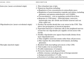
Glia are non-neuronal cells that function to support and collaborate with neurons to maintain homeostasis, and are crucial for synaptic plasticity (Allen and Barres, Reference Allen and Barres2009). The importance and complexity of glial cells are being increasingly recognized. Table 1 outlines the main functions of microglia, astrocytes, and oligodendrocytes. Astrocytes were long considered to have a simple supportive role for neurons, providing structure to the neuropil and adequate environmental conditions for neuronal function. This view was altered by the discovery that astrocytes use variations in cytoplasmic Ca2+ as a form of cellular excitability, allowing signaling to other glia, neurons, and blood vessels (Schummers et al., Reference Schummers, Yu and Sur2008). There is now a reconceptualization of neuronal signaling, with astrocytes forming an integral part of “the tripartite synapse” (Araque and Navarrete, Reference Araque and Navarrete2010), along with the pre- and post-synaptic neurons. Astrocyte excitability can be triggered by various neurotransmitter receptors expressed on glia and, in turn, these cells can release a wide variety of gliotransmitters, including glutamate, adenosine triphosphate (ATP), and D-Serine, which regulate synaptic transmission and plasticity. Although much of the detail of glial–neuronal signaling is unknown, astrocytes may be in contact with many thousands of synapses and there is increasing evidence that astrocytes have an integral role in the wider GNN as well as in information processing (Araque and Navarrete, Reference Araque and Navarrete2010).
CNS glial cell types and their functions

Microglia are also recognized as having more diverse functions than initially thought. Arising from myeloid, pre-hematopoietic precursor cells during development (Ginhoux et al., Reference Ginhoux2010), they are the first line of parenchymal CNS defense in their role as macrophages, and they form an immune network of antigen presenting cells (Graeber and Streit, Reference Graeber and Streit1990). Activated microglia can produce cytokines and stimulate astrocytes. They may also have a role in neurogenesis and post-lesional synaptic plasticity as well as in the maintenance of synaptic integrity (Graeber, Reference Graeber2010; Tremblay et al., Reference Tremblay, Lowery and Majewska2010). In addition, synaptic pruning by microglia appears necessary for normal brain development (Paolicelli et al., Reference Paolicelli2011).
Glial and neuronal pathology in depression
There is a burgeoning literature on phenotypic and functional changes in glial and neuronal cell populations in key brain regions implicated in major depression. Generally, the evidence from histological studies of patients diagnosed with MDD suggests regionally specific decreases in the density and number of glial cells (Rajkowska and Miguel-Hidalgo, Reference Rajkowska and Miguel-Hidalgo2007) across several aspects of fronto-striatal circuitry. However, these studies have generally investigated either mixed or younger samples and few have examined LLD specifically.
While studies examining the anterior cingulate gyrus (subgenual area and supragenual areas) in mixed age samples have demonstrated reduced glial cell density (Ongur et al., Reference Ongur, Drevets and Price1998; Cotter et al., Reference Cotter, Mackay, Landau, Kerwin and Everall2001; Gittins and Harrison, Reference Gittins and Harrison2011), the evidence for changes in neuronal density or size is more equivocal. A similar pattern was found for the DLPFC (Rajkowska et al., Reference Rajkowska1999), with one study also finding a reduction in neuronal size (Cotter et al., Reference Cotter, Mackay, Chana, Beasley, Landau and Everall2002). There have been mixed findings in the OFC, with both reductions in glial density (Rajkowska et al., Reference Rajkowska1999) and no reductions reported (Cotter et al., Reference Cotter, Hudson and Landau2005), accompanying a reduction in neuronal size in cortical layers 2 and 3. Glial reduction in mood disorders is not uniform throughout the cortex, with no reduction seen in the somatosensory cortex (Ongur et al., Reference Ongur, Drevets and Price1998).
Few studies have examined in detail other areas of the limbic system or striatum. However, in the hippocampus, Stockmeier et al. (Reference Stockmeier2004) reported a 35% increase in both the glial and neuronal densities in the brains of those with MDD, of mixed ages of depression onset, when compared to controls. There was a significant decrease in neuronal soma size and the authors suggested that the different pattern seen in the hippocampus compared to other frontal structures may reflect reduced neuropil in the hippocampus, including a loss of dendritic branching and glial processes. It was postulated that these changes may have been related to the reduction in neurotrophic factors seen in MDD and that the loss of neuropil was responsible for the volume reduction seen in the hippocampus of those with MDD (Hickie et al., Reference Hickie2005). This study however, used frozen tissue, which is highly susceptible to ice crystal formation and other artifacts, which could lead to alterations in cell density and which might explain the finding of increased neuronal and glial densities, in contrast to studies in other cortical areas.
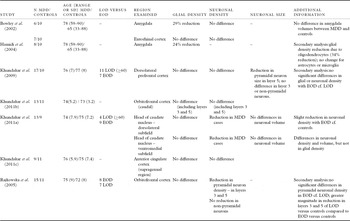
In patients with LLD specifically, a relative dearth of neuropathological studies have examined the cytoarchitecture of the key areas demonstrating volume loss described above. Table 2 summarizes the findings from the seven known studies of LLD that examined patients older than 50 years at the time of death and included an assessment of glial density. These findings suggest that the neuropathology differs from that reported in younger patients (Khundakar and Thomas, Reference Khundakar and Thomas2009). Of the seven studies, only one set of MDD cases (reported in two papers) was found to have a reduction in glial density, occurring in the amygdala (Bowley et al., Reference Bowley, Drevets, Ongur and Price2002; Hamidi et al., Reference Hamidi, Drevets and Price2004). The other five studies, however, reported no significant differences in glial density in the other regions examined: the entorhinal cortex, the DLPFC, the OFC, the ACC, or the head of caudate nucleus (Bowley et al., Reference Bowley, Drevets, Ongur and Price2002; Rajkowska et al., Reference Rajkowska, Miguel-Hidalgo, Dubey, Stockmeier and Krishnan2005; Khundakar et al., Reference Khundakar, Morris, Oakley, McMeekin and Thomas2009; Reference Khundakar, Morris, Oakley and Thomas2011a; Reference Khundakar, Morris, Oakley and Thomas2011b; Reference Khundakar, Morris, Oakley and Thomas2011c). This is in contrast to the previous studies involving a younger sample described above (Rajkowska et al., Reference Rajkowska1999; Cotter et al., Reference Cotter, Hudson and Landau2005).
Summary of post-mortem studies examining glial and neuronal pathology in patients with late-life depression

Note: LOD = late onset depression; EOD = early onset depression; MDD = major depressive disorder. Hamidi et al. (Reference Hamidi, Drevets and Price2004) used predominantly the same cases and controls as the Bowley et al. (Reference Bowley, Drevets, Ongur and Price2002) study. The four Khundakar et al. studies (Khundakar et al., Reference Khundakar, Morris, Oakley, McMeekin and Thomas2009; Reference Khundakar, Morris, Oakley and Thomas2011a; Reference Khundakar, Morris, Oakley and Thomas2011b; Reference Khundakar, Morris, Oakley and Thomas2011c) used predominantly the same group of cases and controls.
In those with LLD, there may, however, be age-dependent changes in neuronal pathology. For instance, Rajkowska et al. (Reference Rajkowska, Miguel-Hidalgo, Dubey, Stockmeier and Krishnan2005) reported a reduction in pyramidal neuronal density in the OFC, mainly due to significant reductions in layers 3 and 5, and found an association between increasing age and decreased neuronal density in their subjects. A similar pattern was reported by Khundakar et al. (Reference Khundakar, Morris, Oakley, McMeekin and Thomas2009), who examined the DLPFC. They found no overall reduction in neuronal density, but a reduction in neuronal volume in cortical layer 3 and most markedly, layer 5. These layers contain pyramidal neurons, projecting to the striatum from the DLPFC and OPC, regions implicated in the etiology of depression. En-route to the striatum, the axons of these neurons traverse white matter, where they may be susceptible to ischemic damage associated with white-matter lesions (Thomas et al., Reference Thomas2002; Khundakar et al., Reference Khundakar, Morris, Oakley, McMeekin and Thomas2009), as discussed above. Van Otterloo et al. (Reference Van Otterloo, O'Dwyer, Stockmeier, Steffens, Krishnan and Rajkowska2009) examined a different but overlapping area of the DLPFC and reported the conflicting finding of no overall or layer-specific differences in neuronal density between an LLD and control group. Neurons may also be damaged by elevated cortisol from excessive HPA system activation, which may be more severe with age (Khundakar et al., Reference Khundakar, Morris, Oakley, McMeekin and Thomas2009), or a function of repeated episodes of depression throughout life (Sheline et al., Reference Sheline, Sanghavi, Mintun and Gado1999). Accordingly, a recent epidemiological study of neuropathology associated with depression in an elderly community sample found that LLD was significantly associated with hippocampal neuronal loss, measured by a semi-quantitative approach (Tsopelas et al., Reference Tsopelas2011). This was postulated to reflect the chronic effects of hypercortisolemia.
Given the association between cerebrovascular disease and later age of illness onset, two papers investigated whether cases of LOD demonstrated distinct neuropathological features compared to cases where depression arose earlier in life. In both studies, subjects were divided into those with EOD versus those with LOD using a cut-off age of 60 years. Khundakar et al. (Reference Khundakar, Morris, Oakley, McMeekin and Thomas2009) examined the DLPFC in six patients with EOD and 11 patients with LOD. They found no overall correlations between age of depression onset and glial and neuronal densities. Rajkowska et al. (Reference Rajkowska, Miguel-Hidalgo, Dubey, Stockmeier and Krishnan2005) included eight EOD and seven LOD subjects, and reported no major difference in overall or laminar density of pyramidal neurons between EOD and LOD.
The lack of significant associations between age of onset and glial–neuronal pathology could be explained by a number of factors. First, different mechanisms may underpin the reductions in neuronal density seen in EOD and LOD. In EOD, genetic factors are likely to play a critical role in illness onset, and with progression, the effects of hypercortisolemia, sleep and circadian changes, reduction in key neurotrophins, and the inflammatory and immunological responses may predominate (Hickie et al., Reference Hickie, Naismith, Norrie and Scott2009; Hickie and Rogers, Reference Hickie and Rogers2011). Vascular factors and neurodegenerative changes, by comparison, may be more pertinent to the neurobiological changes observed in later life (Rajkowska et al., Reference Rajkowska, Miguel-Hidalgo, Dubey, Stockmeier and Krishnan2005). Second, the sample sizes of all studies have been small, with a maximum of 17 subjects with MDD, and may have been underpowered to detect differences between the two groups of patients. Third, the studies have examined different cortical and subcortical regions, which make comparisons between studies limited. Finally, although the two papers discussed above dichotomized their group into EOD and LOD, other studies did not make this division. That is, in the former studies, although age was noted, it is unclear if participants with LLD had LOD or were “graduates” from EOD.
There are several methodological difficulties that arise when investigating glial pathology. Reports of changes in glial density are complicated by the difficulties in sub-typing glial cells, partly due to the limitations of Nissl staining. This technique does not allow for easy discrimination of astrocytes, oligodendrocytes, and microglia (Hamidi et al., Reference Hamidi, Drevets and Price2004). It may be that increases or decreases in sub-types partially cancel each other out when looking at overall glial density. Further, unlike neurons, volumetric changes in glia cannot be assessed (using stereological tools such as the “nucleator” or “rotator”) and so morphological changes may occur in glial cells in LLD, which cannot be detected using the prevailing methods. Finally, the estimation of glial density is based on cell counts within the neuropil and is therefore susceptible to the effects of tissue processing, such as shrinkage, particularly when frozen tissue is used, as in the Stockmeier et al. (Reference Stockmeier2004) study. Using additional staining with S-100β and human leukocyte antigen, Hamidi et al. (Reference Hamidi, Drevets and Price2004) reported that the glial reduction in the amygdala of elderly depressed subjects was primarily due to oligodendrocyte reduction. Uranova et al. (Reference Uranova, Vostrikov, Orlovskaya and Rachmanova2004) have previously reported oligodendrocyte reductions in the PFC of adults with depression and further research is needed to replicate this finding in LLD and to explore the extent to which oligodendrocytes are responsible for reductions in glial density and the mechanisms underlying this process.
A further limitation to the pathology studies is the imprecise use of cognitive impairment as an exclusion criterion for those with MDD. The studies by Khundakar et al. (Reference Khundakar, Morris, Oakley, McMeekin and Thomas2009; Reference Khundakar, Morris, Oakley and Thomas2011a; Reference Khundakar, Morris, Oakley and Thomas2011b; Reference Khundakar, Morris, Oakley and Thomas2011c) exclude those with any evidence of “significant cognitive impairment” during life; the study by Rajkowska et al. (Reference Rajkowska, Miguel-Hidalgo, Dubey, Stockmeier and Krishnan2005) excludes those with neurologic disease including Alzheimer's disease; and the Bowley et al. (Reference Bowley, Drevets, Ongur and Price2002) and Hamidi et al. (Reference Hamidi, Drevets and Price2004) studies do not report exclusion of participants on the basis of cognition but use a staining technique to exclude histologically diagnosed dementia. Although excluding participants with dementia is necessary, approximately 50% of those with LLD demonstrate mild cognitive impairment (Lee et al., Reference Lee, Potter, Wagner, Welsh-Bohmer and Steffens2007) and so exclusion of some of these participants may represent a systematic bias and limit the validity of the conclusions.
Rajkowska (Reference Rajkowska2000) has proposed a model to explain the age-related differences in glial pathology. In early MDD, a combination of stress-related glucocorticoid toxicity, glutamate toxicity (McDonald et al., Reference McDonald, Levine and Qu1998), and/or a deficiency of trophic and angiogenic factors may lead to a reduction of astrocytes and oligodendrocytes. Over time, this may contribute to neuronal damage and destruction via excess glutamate as discussed below. Later, in the disease course, the neuronal injury may lead to a compensatory gliosis and an increase in glial density. This model is supported by the discovery of increased glial fibrillary acidic protein (GFAP), an astrocytic marker, in cortical layer 1 of the DLPFC in subjects with LLD (Davis et al., Reference Davis, Thomas, Perry, Oakley, Kalaria and O'Brien2002). By contrast, in younger patients with depression, levels of GFAP in the PFC were significantly lower compared to controls (Si et al., Reference Si, Miguel-Hidalgo, O'Dwyer, Stockmeier and Rajkowska2004), while there was no significant difference in the level of GFAP between depressed and control subjects older than 60 years at time of death. This perhaps reflects a compensatory increase in astrocytes seen in older people – levels of GFAP becoming comparable with older subjects without a history of depression (Rajkowska, Reference Rajkowska2000).
Alternatively, differences in GFAP expression may result from differing underlying pathophysiologies in EOD and LOD. Associations between LLD, WMHs, and vascular risk factors suggest a role for ischemic vascular disease and a subsequent inflammatory response in underpinning LLD. Deep WMHs have been demonstrated to be frequently due to cerebral ischemia (Thomas et al., Reference Thomas2002), and upregulation of the intracellular adhesion molecule-1 (ICAM-1), a vascular marker of inflammation, has been reported in the gray and white matters of the DLPFC in patients with LLD (Thomas et al., Reference Thomas2000), providing direct support for the involvement of vascular pathology in LLD. Additionally, microglial activation has been demonstrated in periventricular and deep subcortical WMHs in aging brains (Simpson et al., Reference Simpson2007), with the response associated with periventricular lesions in particular, possibly reflecting activation related to disruption of the blood brain barrier. However, despite these subtle indicators of vascular pathology, no association between macrovascular pathology (as defined by the presence of hemorrhages, infarcts, lacunes, or small vessel disease) and LLD was found in a recent post-mortem study (Tsopelas et al., Reference Tsopelas2011).
Glial mediation and modulation of neurotoxicity
The mechanisms by which glia are associated with the symptom complex of MDD, and more particularly LLD, remain unclear. An inflammatory basis for depression has been postulated for many years; the evidence has been reviewed by both Miller et al. (Reference Miller, Maletic and Raison2009) and Leonard (Reference Leonard2001). Studies in patients with depression have demonstrated increased level of the pro-inflammatory markers C-reactive protein (CRP), tumor necrosis factor (TNF), interleukin-1 (IL-1), interleukin-2 (IL-2), and interleukin-6 (IL-6; Zorrilla et al., Reference Zorrilla2001; Dowlati et al., Reference Dowlati2010), and a disordered ratio of the pro-inflammatory cytokine interferon gamma (IFN-γ) to the anti-inflammatory cytokine interleukin-4 (IL-4). Aging has also been suggested to be associated with a shift towards a pro-inflammatory state, leading to activated microglia and increased production of cytokines (Sparkman and Johnson, Reference Sparkman and Johnson2008).
Surprisingly, there has been a relative lack of studies examining inflammatory mediators in LLD. Overall, the results are also less consistent compared to changes in inflammatory markers in younger or mixed samples, possibly due to the smaller sample sizes and inconsistent definitions of depression. Of the 14 studies found, the only inflammatory mediator consistently reported to be higher in LLD than controls was IL-1β, examined in two studies (Thomas et al., Reference Thomas, Davis, Morris, Jackson, Harrison and O'Brien2005; Diniz et al., Reference Diniz, Teixeira, Talib, Gattaz and Forlenza2010). Other commonly examined mediators included CRP, IL-6, and TNF-α, for which there appears to be no consistent difference between patients with LLD and controls (Dentino et al., Reference Dentino1999; Brown and Bauman, Reference Brown and Bauman2000; Penninx et al., Reference Penninx2003; Tiemeier et al., Reference Tiemeier, Hofman, van Tuijl, Kiliaan, Meijer and Breteler2003; Empana et al., Reference Empana2005; Thomas et al., Reference Thomas, Davis, Morris, Jackson, Harrison and O'Brien2005; Almeida et al., Reference Almeida, Norman, Hankey, Jamrozik and Flicker2007; Bremmer et al., Reference Bremmer2008; Lee et al., Reference Lee2009; Diniz et al., Reference Diniz, Teixeira, Talib, Gattaz and Forlenza2010; Forti et al., Reference Forti2010).
There are several mechanisms by which glia may induce depressive symptomatology. Indoleamine 2,3-dioxygenase (IDO) is an enzyme that converts tryptophan to kynurenic acid and quinolinic acid through the kynurenine pathway. IDO is expressed by microglia and macrophages in response to pro-inflammatory cytokines (IFN-γ, TNF, IL-1) and is inhibited by anti-inflammatory cytokines (IL-4, IL-10). Excess release of pro-inflammatory cytokines therefore increases IDO expression, which is associated with reduced plasma tryptophan levels (Capuron et al., Reference Capuron, Ravaud, Neveu, Miller, Maes and Dantzer2002). Tryptophan is an essential amino acid and is converted to serotonin via tryptophan hydroxylase. Tryptophan depletion may therefore reduce serotonin, and has been shown experimentally to induce depressive symptoms in susceptible individuals (Delgado et al., Reference Delgado1994).
“Inflammation” associated changes (Graeber et al., Reference Graeber, Li and Rodriguez2011) in glutamate metabolism may also play a key role in the regulation of mood. Kynurenic acid is an N-methyl-D-aspartate (NMDA) glutamate receptor antagonist and quinolinic acid a NMDA receptor agonist. Microglia preferentially express quinolinic acid and astrocytes kynurenic acid, so in the presence of pro-inflammatory cytokines, there may be an imbalance in the kynurenine pathway, with more quinolinic acid produced. In addition to glutamate receptor agonism, quinolinic acid also causes direct release of glutamate and induces oxidative stress, all of which may contribute to CNS excitotoxicity (Miller et al., Reference Miller, Maletic and Raison2009). Clinical studies have reported elevated quinolinic acid levels in the blood and cerebrospinal fluid of patients with depression and a recent study found severe depression was associated with increased microglial quinolinic acid in subregions of the anterior cingulate gyrus (Steiner et al., Reference Steiner2011).
An important function of astrocytes is the removal of glutamate from the synapse via the excitatory amino acid transporter (EAAT). This converts glutamate to glutamine through the action of the enzyme glutamine synthetase. The glutamine is later transported back to neurons where it can be converted back to glutamate. Several mediators released by activated microglia could impair astroglial expression of the EAAT and reduce glutamate reuptake (Vercellino et al., Reference Vercellino2007; McNally et al., Reference McNally, Bhagwagar and Hannestad2008).
It is unclear exactly how glutamate is neurotoxic. Excess glutamate with NMDA receptor stimulation may lead to a rise in intracellular calcium, which can depolarize mitochondria, producing free radicals such as nitric oxide that can directly damage the cell. Animal models have shown that cytokines may also have a role in these processes, as blocking the enzyme required for IL-1 synthesis inhibits glutamate neurotoxicity and activation of microglia (Tikka and Koistinaho, Reference Tikka and Koistinaho2001). In response to peripheral inflammatory mediators, activated microglia produce a variety of neurotoxic substances including reactive oxygen and nitrogen species, IL-1β, and TNFα (Block et al., Reference Block, Zecca and Hong2007). Neurons and oligodendrocytes are sensitive to the toxic effects of the inflammatory cascade, in particular TNF-α, which could lead to demyelination and apoptosis.
Several lines of evidence support the role for glutamate in depression and the biological mechanisms described previously. Post-mortem studies have shown elevated glutamate in the frontal lobe of younger patients with MDD (Hashimoto et al., Reference Hashimoto, Sawa and Iyo2007) and proton magnetic resonance spectroscopy (MRS) has also demonstrated increased glutamate concentrations in the occipital cortex of younger MDD patients (Sanacora et al., Reference Sanacora2004). Indirect evidence also comes from findings showing that Ketamine, an NMDA receptor antagonist (Berman et al., Reference Berman2000), and Riluzole, a glutamate modulator, may also have antidepressant effects (Zarate et al., Reference Zarate2004).
In addition to their role in neurotoxicity, glia also produce neuroprotective factors. Microglia have a neuroprotective role (Li et al., Reference Li, Lu, Tay, Moochhala and He2007) and, when non-activated, produce both IL-1ra, which protects neurons from the harmful effects of IL-1, and brain-derived neurotrophic factor (BDNF). BDNF has an important role in neuronal survival and neuroplasticity. Reductions may be associated with the atrophy seen in the limbic areas of patients with LLD (Duman and Monteggia, Reference Duman and Monteggia2006) and serum levels are elevated by chronic antidepressant treatment (Dimopoulos et al., Reference Dimopoulos2006). It therefore may represent a mechanism of antidepressant treatment. Other systems also have a role in modulating neurotoxicity, with the endogenous cannabinoid anandamine having been shown to limit microglial associated neurotoxicity following CNS injury (Eljaschewitsch et al., Reference Eljaschewitsch2006). Astroglia (and oligodendrocytes) produce S-100β. This regulates cell size and shape, has a role in cell-to-cell signaling, and can be either responsible for growth and differentiation or apoptosis, depending on its concentration (Schroeter et al., Reference Schroeter, Abdul-Khaliq, Krebs, Diefenbacher and Blasig2008). Schroeter et al. (Reference Schroeter, Abdul-Khaliq, Krebs, Diefenbacher and Blasig2008) demonstrated elevated serum S-100β levels in their hospitalized patients with MDD, both at admission and discharge, compared to control subjects. They also measured levels of the neuron marker neuron-specific enolase and found no difference between patients and controls, further emphasizing the importance of glia in depression.
There is further direct evidence for the importance of glia from a recent animal model study, which showed that inducing glial loss in the PFC, through administration of a astrocyte-specific gliotoxin, produces depressive-like behaviors (Banasr and Duman, Reference Banasr and Duman2008). The behaviors observed in such models mimicked the effects of chronic stress on the animals, and supports a role for glia (Hinwood et al., Reference Hinwood, Morandini, Day and Walker2011) in the symptom complex of MDD.
A novel technique for examining the vascular and inflammatory mechanisms that may contribute to the etiology of LLD is PET scanning, using the [11C](R)-PK11195 ligand. This ligand displays upregulation of peripheral benzodiazepine binding sites, which provides a cellular marker for microglial activation (Banati and Hickie, Reference Banati and Hickie2009). Initial studies utilizing this ligand in multiple sclerosis displayed increased binding in association with focal white matter pathology as well as in gray matter (Banati et al., Reference Banati2000). The PK11195 ligand has since been utilized across a range of neurodegenerative disorders including Alzheimer's disease, but to date, no known studies have utilized this ligand to measure microglial activation in LLD, or more specifically, in LOD.
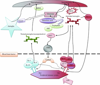
Figure 1 presents a summary of the mechanisms of glia-associated neurotoxicity and neuroprotection described above.
Summary of the glia associated neurotoxic and neuroprotective mechanisms involved in the pathophysiology of major depressive disorder.
Note: Dotted line indicates inhibitory process. BDNF = brain-derived neurotrophic factor; ROS = reactive oxygen species; IL = interleukin; EAAT = excitatory amino acid transporters; 5-HT = serotonin; PGE2 = prostaglandin E2; CRH = corticotropin-releasing hormone; IFN = interferon; TNF = tumor necrosis factor; Th = T helper cell; LPS = lipopolysaccharide. Peripheral inflammatory mediators induce microglial activation, which release a variety of neurotoxic substances, including ROS and pro-inflammatory cytokines. Normal microglia are also neuroprotective, releasing protective factors, such as BDNF. Astrocytes release neurotrophic factors and recycle glutamate. Pro-inflammatory mediators and activated microglia impair the ability of astrocytes to remove glutamate, which can be neurotoxic and limit the synthesis of serotonin as more tryptophan is converted to kynurenic acid and quinolinic acid. Quinolinic acid is preferentially expressed over kynurenic acid in the pro-inflammatory state, resulting in further excitotoxicity.
Source: Adapted with permission from McNally et al. (Reference McNally, Bhagwagar and Hannestad2008).

Conclusions
The literature to date points to a complex and important role for glial cells, particularly astrocytes, in mediating and modulating neurotransmitter levels, and in the regulation of neuroprotective and toxic elements, particularly when associated with what is commonly referred to as inflammation (Graeber et al., Reference Graeber, Li and Rodriguez2011). The extent to which their function changes with age is less clear however, and there is less evidence for peripheral cytokine changes in LLD.
LLD is associated with volume reduction in a number of key frontal-subcortical regions. When these regions are analyzed histologically, there is however, no clear evidence that glial cell reduction is responsible for the gray matter loss seen in LLD. This is in contrast to data obtained from younger people with MDD, where a reduction in glial density is commonly seen. Gliosis has been postulated to account for the discrepancy, suggesting that subtle vascular or inflammatory changes may be important in LLD. Furthermore, neuropathological studies have demonstrated neuronal volume reduction in the cortical layers 3 and 5 of the PFC where pyramidal neurons project to the striatum, routes that would be susceptible to ischemic damage associated with white matter or vascular lesions. This lends some support to the role of vascular pathology in the etiology of LLD, but as discussed above there are limitations to the pathology studies, including the inability of Nissl staining to allow easy discrimination of glial subtypes, the small number of studies in different cortical and subcortical areas, and the lack of differentiation of LLD by age of onset.
Further studies are now required to explore the complex relationship between WMHs, ischemic damage, the role of pro-inflammatory cytokines both in the CNS and periphery (Nomura et al., Reference Nomura2011), and the contribution of glial pathology to these processes. These should differentiate individuals with LLD by age of onset to examine more clearly the potential differences in pathophysiology associated with depression occurring for the first time later in life. These different pathophysiologies may be confounding studies of glial–neuronal pathology. Other directions for future research include analysis of specific glial-cell types and outputs (e.g. Il-1β, S-100β, BDNF), as well as the implications of glial–neuronal dysfunction for cognitive dysfunction. The deficits in executive functioning and processing speed seen in LOD are consistent with disruption to the fronto-subcortical circuit, and there is some preliminary evidence for an association between raised inflammatory cytokine levels with cognitive impairment (Weaver et al., Reference Weaver, Huang, Albert, Harris, Rowe and Seeman2002).
Novel techniques such as PK11195-ligand PET scanning may allow in vivo visualization of microglial activation in LLD, as an early biomarker to better guide treatment. There are also the tantalizing possibilities of using glia as a new therapeutic direction in the treatment of depression, either as a vehicle for CNS gene delivery of neuroprotective factors (Polazzi and Monti, Reference Polazzi and Monti2010), or as a target for therapy. Minocycline, an inhibitor of microglial activation and proliferation, for example, has been shown in animal models to reduce the neurobiological effects of chronic psychological stress (Hinwood et al., Reference Hinwood, Morandini, Day and Walker2011), and to have anti-depressant properties, using the common model of the forced swim test (Pae et al., Reference Pae, Marks, Han and Patkar2008). Exploration of the role of glia in LLD may therefore both improve our understanding of the etiology of the disorder, and allow us to develop potential new therapeutic avenues.
Conflict of interest
None.
Description of authors’ roles
IH conceived the idea for the paper; MP wrote the first draft; and the other authors (SN, LN, MG) contributed equally to editing the final paper.