The Occlutech Figulla Flex II (FFII) Occluder (Occlutech GmbH, Jena, Germany) is a commonly used percutaneous transcatheter device for closing atrial septal defects. As percutaneous device closure is a less invasive alternative to surgical repair, it has been commonly used for anatomically suitable atrial septal defects in recent years. The success rates of the percutaneous approach are comparable to that of the surgical approach for atrial septal defects anatomically suitable for this procedure. Reference Du, Hijazi, Kleinman, Silverman and Larntz1 Many patients do not experience severe complications following percutaneous treatment. Infective endocarditis is a relatively rare but very severe complication associated with atrial septal defects closure devices. Reference Masura, Gavora and Podnar2 Despite advances in diagnostic and therapeutic techniques, morbidity and mortality rates in IE cases remain high. Therefore, the current guidelines recommend 6 months of antibiotic prophylaxis after device implantation, based on animal study results. Reference Walter, Taubert and Gewitz3 In particular, IE following atrial septal defect closure with an occluding device in a child is rare. We report a case of late bacterial endocarditis in an 11-year-old girl with incomplete endothelialisation after percutaneous device closure of atrial septal defects.
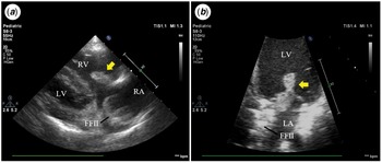
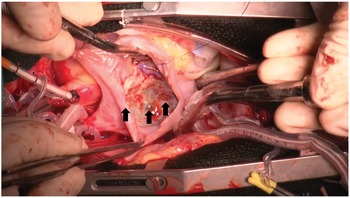
An 11-year-old girl with a secundum atrial septal defect (17.7 mm) underwent percutaneous repair with a 21-mm FFII occluder device in July 2017. She presented with a 2-week history of persistent fever, ankle pain, and coxalgia in June 2021. Additionally, she developed mucosal bleeding from the palpebral conjunctiva and oral mucosa, Janeway lesion, and Osler’s node. She had no notable medical history except for the treatment of atrial septal defects. Her vital signs were stable, except for the body temperature (40°C). She did not experience septic shock. While she had not received dental treatment, she had lost her deciduous tooth 1 month before presentation. Sepsis was not clearly associated with the loss of the deciduous tooth. Laboratory results indicated bacterial infection but no signs of pancytopenia, abnormal blood clotting, and multiple organ failure. Blood culture revealed methicillin-resistant Staphylococcus aureus. Cardiomegaly and lung consolidation were barely noticeable on chest radiography. Electrocardiogram findings were normal. Fundoscopy revealed the presence of Roth spots in both eyes. Transthoracic echocardiography revealed large hypermobile masses on her tricuspid and mitral valves. The mass on the tricuspid valve and on the mitral valve was 18 mm and 17 mm, respectively (Fig 1). No residual atrial septal defect shunt, valve regurgitation, and destruction were observed. Because several cerebral microemboli were already detected on brain magnetic resonance imaging during hospitalisation, we decided to perform an urgent surgery to resect the vegetations and infected device, followed by patch closure for the atrial septal defect using fresh pericardium. During surgery, we found that neo-endothelialisation of the occluder device was partially insufficient (especially at the hub) and that the metallic mesh was exposed (Fig 2), although adhesions around the disc rim were very strong. The vegetation extended to the surface of the disc. Postoperatively, the first-degree atrioventricular block persisted for several weeks. However, sinus rhythm recovered spontaneously. She received approximately 6 weeks of vancomycin therapy (10–30 mg/kg/day) after blood culture was negative for infection, followed by 5 months of Bactrim (sulfamethoxazole and trimethoprim) at 10 mg/kg/day. We temporarily substituted vancomycin for daptomycin for 1 week because of drug-associated granulocytopenia and drug eruption. She was discharged without any neurological deficits due to effective exercise therapy. At the most recent follow-up echocardiography, we observed mild tricuspid regurgitation, no mitral regurgitation, and normal cardiac function.
Transthoracic echocardiography images at hospitalisation. Large vegetations are on the tricuspid and mitral valve (arrows). They are hypermobile and extended to the occluding device.
RA = right atrium, LA = left atrium, RV = right ventricle, LV = left ventricle, FFII = The Occlutech Figulla Flex II.

Neo-endothelialisation of the occluder device is partially insufficient, and the metallic mesh is exposed (arrows), although the very strong adhesion around the disc rims.

In many cases, transcatheter repair of atrial septal defect is safe, non-invasive, and effective. Percutaneous closure of atrial septal defect has been reported since the 1970s. Reference King, Thompson, Steiner and Mills4 Compared with the early counterparts, current occlusion devices have improved markedly; therefore, percutaneous device closure is performed widely instead of surgical repair in many hospitals. The percutaneous approach is not complicated. Device malposition, fracture, atrio-aortic erosion, residual shunt, cardiac arrhythmias, cardiac tamponade, valve regurgitation, thrombosis, and infective endocarditis is the known significant complications of occluding device deployment. However, the incidence of late infective endocarditis is significantly low. The European Society of Cardiology, American College of Cardiology, and American Heart Association recommend 6 months of antibiotic prophylaxis following percutaneous device closure of atrial septal defects, as devices for atrial septal defects are fully endothelialised within 1–3 months after deployment in several animal studies. Reference Sharafuddin, Gu, Titus, Urness, Cervera-Ceballos and Amplatz5,Reference Lock, Rome and Davis6 However, the same may not be applicable to humans. It is unclear whether complete endothelialisation occurs within 6 months in humans after deployment of closure devices for atrial septal defects. Insufficient endothelialisation of the disc may increase the risk of infective endocarditis. Some cases of late infective endocarditis associated with closure devices for atrial septal defects involved implanted devices that were not completely endothelialised, although over 6 months had elapsed since deployment. Reference Amedro, Soulatges and Fraisse7–Reference Jha, Kiraly and Murala9 Tanabe et al. reported neo-endothelialisation in humans in vivo. They evaluated neo-endothelialisation on the surface of discs with angioscopy 6 months after device implantation. Reference Tanabe, Suzuki and Kuwata10 They described that the neo-endothelialisation of the devices varied among the study patients and that the patients treated with a large device might show poor endothelialisation. Moreover, the endothelialisation of the central areas was insufficient. They considered that incomplete endothelialisation was related to the device size, right ventricular systolic pressure, and enlargement of the right ventricle volume overload. Reference Tanabe, Suzuki and Kuwata10 The period until complete endothelialisation in humans may differ substantially among individuals. The current guidelines are primarily focused on appropriate antibiotic prophylaxis for infective endocarditis. Our report suggests that clinicians should decide the need for prolonged antibiotic administration after large occluder device deployment flexibly to prevent severe complications, such as late infective endocarditis.
Acknowledgements
We thank the members of the cardiac team and the infection control team for their attentive medical care.
Financial support
This research received no specific grant from any funding agency, commercial, or not-for-profit sectors.
Conflicts of interest
The authors declare that they have no competing interests.
Patient consent
The patient’s mother provided written consent for the publication of this case report.
Social media synopsis
We report a rare case of late bacterial endocarditis in an 11-year-old girl with incomplete endothelialisation after percutaneous device closure of ASD.