Introduction
The key to the survival of adult fishes in the freezing waters of Antarctica lies in the production of antifreeze molecules that are distributed throughout the body (DeVries & Cheng Reference DeVries and Cheng2005). The major antifreezes found in notothenioid fishes (Perciformes, Notothenioidei), which dominate the high latitudinal Antarctic fish fauna, belong to a single family of glycoproteins, the antifreeze glycoproteins (AFGPs). Members of the AFGP family vary in size from about 2.6 to 33.1 kDa, and are usually grouped into eight size clusters (AFGPs 1–8) where AFGP 8 is the smallest (DeVries Reference DeVries1971, Reference DeVries1988, DeVries & Cheng Reference DeVries and Cheng2005). AFGPs are synthesized in cells of the exocrine pancreas, oesophagus and anterior stomach as large molecular weight polyproteins that are subsequently processed enzymatically to yield the different members of the AFGP family (Cheng et al. Reference Cheng, Cziko and Evans2006). Since all these sites of AFGP synthesis secrete into the gastrointestinal tract, a key question relates to how AFGPs are distributed to other parts of the body where protection from freezing is paramount.
Antarctic fish AFGPs essentially consist of different lengths of a repetitive (N = 4–50) alanine-alanine-threonine (AAT) backbone, with a disaccharide (β-D-galactosyl-(1,3)-α-D-N-acetylgalactosamine) O-linked through the hydroxyl of each threonine. Some of the smallest antifreezes, however, may substitute an alanine with a proline in the amino acid backbone. The antifreezes function through an adsorption-inhibition mechanism, most probably hydrogen binding through their sugar moieties to specific planes in the ice and inhibiting crystal growth through the Kelvin effect (Raymond & DeVries Reference Raymond and DeVries1977). This mechanism of action requires that ice be present for the AFGPs to work i.e. AFGPs do not function by inhibiting ice crystal nucleation. The manner by which AFGPs operate presents a significant problem for the survival of Antarctic fishes, since the accumulation of internal ice is potentially lethal. This raises another question, which relates to how fishes deal with internalized, microscopic ice crystals.
The primary barrier to freezing in most poikilothermic vertebrates is an intact epithelium (Valerio et al. Reference Valerio, Kao and Fletcher1992) as shown in the larvae of certain Antarctic fish species, which lack adequate levels of antifreeze and yet survive exposure to external ice as long as the epithelium is intact (Cziko et al. Reference Cziko, Evans, Cheng and DeVries2006). When the epithelium is compromised (through an injury to the skin, for example) ice will propagate in these larval fish since their equilibrium melting point (due to the colligative properties of their plasma) is well above the freezing point of sea water (c. -1.91°C). Larval (and adult) fish with adequate levels of antifreeze are protected from freezing at temperatures at least as low as their blood non-equilibrium freezing point (determined experimentally using serum), which typically confers 1–2°C thermal protection below their equilibrium melting point. This separation of the equilibrium melting point and the non-equilibrium freezing point is known as thermal hysteresis, and is a defining property of biological antifreezes (DeVries Reference DeVries1988, Peltier et al. Reference Peltier, Brimble, Wojnar, Williams, Evans and DeVries2010).
Fish collected from freezing Antarctic waters have ice associated with their integument, gill epithelium and gastrointestinal tract, but not their body fluids, muscle or major organs, except the spleen (DeVries & Cheng Reference DeVries and Cheng1992, Praebel et al. Reference Praebel, Hunt, Hunt and DeVries2009). The association of ice with the integument and gills is not surprising since these tissues form the barrier between the internal milieu and the ice-laden environment. Ice adherent to the body surface is a potential problem, however, since it could grow and penetrate the otherwise protective epithelium, which may be compromised by some form of damage of physical (e.g. injury) or biological (e.g. pathogenic) origin. This raises the question as to whether antifreeze might also be present in the mucus on the body surface, preventing potential injury to the epithelium from the growth of adherent crystals. Here, using the naked dragonfish Gymnodraco acuticeps as an example, we explore the possibility that mucus secreted on the scaleless body surface might contain AFGPs capable of limiting ice crystal growth.
The discovery of ice in the gastrointestinal tract is also not unexpected, since Antarctic fish imbibe ice-laden food and water. AFGPs secreted directly into the gastro-intestinal tract have the capacity to protect the fish from any potential damage (Cheng et al. Reference Cheng, Cziko and Evans2006). The presence of ice in the spleen, however, indicates that ice can penetrate the epithelial barrier and reach the circulation. Whilst AFGPs confer protection by stopping ice crystal growth the fish is still left with the problem of how to deal with the offending ice crystals, which in the circulation have the potential to damage organs and tissues if lodged in small diameter vessels. The key issue now is to address exactly how Antarctic fish survive internal ice. We hypothesize that circulating AFGPs bind to internal ice crystals and that this complex is then phagocytosed by cells in the spleen. Our approach has involved the synthesis of AFGP-coated silica nanoparticles, small enough to pass relatively unimpeded through the fish vascular bed, and then utilizing them as a proxy for AFGP-coated ice crystals.
Methods
Preparation of antifreeze
Blood collected from the Antarctic toothfish Dissostichus mawsoni Norman was allowed to clot for 4 h in the cold (4°C) and centrifuged (3000 x g, 10 min, 4°C) to prepare the serum, which was then stored at -80°C until required. The trichloroacetic acid (TCA) soluble AFGPs were separated from Antarctic toothfish serum by precipitating the other serum proteins with the addition of 5% TCA (1:1). Following centrifugation (10 000 x g, 10 min, 4°C) the supernatant was dialysed for 24 h against three changes of distilled water using Spectra/Por 3 dialysis tubing (MW cut-off 3500Da). The dialysed AFGPs were lyophilized, dissolved in 50 mM ammonium bicarbonate, and separated into large (AFGP 1–5) and small (AFGP 7,8) size classes on a Sephacryl HR-100 column (2.5 x 150 cm). The column fractions were then lyophilized and the size classes verified using non-denaturing polyacrylamide gel electrophoresis after labelling with fluorescamine (O’Grady et al. Reference O’Grady, Schrag, Raymond and DeVries1982, Ahlgren et al. Reference Ahlgren, Cheng, Schrag and DeVries1988). The different AFGP size classes were subsequently coupled to FSE-NPs (AFGP 1–5) or complexed with FITC (AFGP 1–5 and AFGP 7,8).
Complexing of fluorescein isothiocynate with AFGP
Ten mg of dry AFGP 1–5 or AFGP 7,8 was dissolved in 2 ml sodium borate buffer (50 mM, pH 8.6). One ml of fluorescein isothiocyanate (FITC) dissolved in anhydrous dimethyl sulphoxide (20 mg ml-1) was added and the mixture incubated at room temperature for 2 h in the dark. Unbound or hydrolyzed FITC was then separated from the fluorescein-AFGP conjugates using a Sephadex G-25 column (2.5 x 30 cm) equilibrated with water. The separation was followed by illuminating the column with an ultraviolet lamp, and after elution the fluorescent conjugates were lyophilized and stored at 4°C. Prior to injection the fluorescein-AFGP conjugates were dissolved in notothenioid PBS (Cziko et al. Reference Cziko, Evans, Cheng and DeVries2006).
Synthesis of labelled nanoparticles
Fluorescein-labelled silica nanoparticles were prepared using a standard microemulsion method (Bagwe et al. Reference Bagwe, Yang, Hilliard and Tan2004, Yao et al. Reference Yao, Wang, Wu, Smith, Xu, Zhao, Lee and Tan2006). First, 5(6)-carboxyfluorescein diacetate N-succinimidyl ester (FSE) was conjugated to aminopropyltriethoxysilane (APTES) by dissolving 1.6 mg of FSE (or 2.4 mg of NIR-664) in 1.2 ml of anhydrous n-hexanol. Pure APTES (3.44 μl) was then added under a nitrogen atmosphere and the solution stirred for 4 h at room temperature. The FSE-APTES conjugate was then added to a mix of cyclohexane oil phase (7.5 ml), n-hexanol co-solvent (0.6 ml) and Triton® X-100 surfactant (1.77 g) followed by 230 ml of deionized water. The solution was stirred for 5 min and then 100 ml of tetraethylorthosilica (TEOS) was added. After 30 min, 40 ml of ammonium hydroxide was added and the microemulsion stirred for 24 h. To stop aggregation and prepare the nanoparticles for bioconjugation, 50 ml of TEOS was then added with rapid stirring, followed 30 min later by the addition of 40 ml of 42 wt% in water 3-(trihydroxysilyl)propyl methyl phosphonate, monosodium salt and 10 ml of aminopropyltrimethoxysilane. The nanoparticles were separated from the solution with the addition of excess ethanol and centrifuged three times in ethanol, with resuspension between the washing steps by sonication. The dye-doped nanoparticles were stored in the dark, in ethanol, at 0.5 mg ml-1 and at 4°C.
Antifreeze - nanoparticle coupling
Generation 4.5 (G4.5) poly(amidoamine) dendrimers (PAMAM) bearing -COOH surface groups (Gubala et al. Reference Gubala, Le Guevel, Nooney, Williams and MacCraith2010) were first activated with a 1-ethyl-3-[3-dimethylaminopropyl] carbodiimide hydrochloride (EDC)/sulpho-N-hydroxysulphosuccinimide (sulpho-NHS) mixture in 0.1 M 2-(N-morpholino)ethanesulphonic acid (MES) buffer, pH 4.5 before reacting with purified AFGP 1–5. Briefly, PAMAM G4.5 (1 μmol, 128 x -COOH) was dissolved in 0.5 ml of MES buffer. To this solution, sulpho-NHS (0.192 mmol, 1.5 equiv. per one -COOH group) and EDC (0.384 mmol, 3.0 equiv. per one -COOH group) were added. The final volume was then adjusted to 1 ml with MES buffer and the reaction allowed to proceed for 15 min. The sulpho-NHS-activated dendrimers were then added directly into the FSE-NPs (2 mg ml-1), while keeping the total volume at 1 ml. This mixture was allowed to react for 20 min and excess, unreacted dendrimers were then removed by centrifugation (17 000 x g, 5 min). The dendrimer-modified nanoparticles were re-dissolved in MES buffer, pH 4.5 and purified AFGP 1–5 (300 μg) was added. The reaction mixture was gently shaken for 4 h and 100 μl of Tris (2-amino-2-hydroxymethyl-propane-1,3-diol) buffer (pH 8.0) was then added to hydrolyze the remaining NHS esters. The mixture was then purified by centrifugation (4x, 17 000 x g, 5 min) and the AFGP-NP bioconjugate was re-dissolved in 0.1 M PBS buffer, pH 7.4, with 0.01% (w/v) sodium azide. All reactions involving FSE-NPs were performed under reduced light conditions to prevent photo-bleaching and all final samples were freeze dried and stored in the dark at 4°C before they were used. An equivalent protocol was used to prepare pegylated NPs on the same scale.
Mass spectrometry analysis (LC-MS/MS)
Fish mucus was stripped from the external epithelium of an adult Gymnodraco acuticeps Boulenger by running a gloved hand over the surface of the fish in the mid-trunk region and transferring the exudate to a cryovial for storage in liquid nitrogen and subsequent analysis by mass spectrometry. For analysis, the sample was treated with 0.1% formic acid (final volume 1.0 ml) and subjected to solid phase extraction on a 10 mg Oasis HLB SPE cartridge (eluted with 0.3 ml of 10% acetonitrile in water). The eluate was concentrated to a few microlitres and then made up to 10 μl with 0.1% formic acid before injection into a LC Packings Pepmap C18 (0.3 mm x 5 mm) trap column (2% acetonitrile in 0.1% formic acid as the mobile phase) to concentrate the sample after injection. The sample was then passed through an Agilent Zorbax SB300 C18 (0.3 mm x 10 mm) column with 0.1% formic acid in water and 0.1% formic acid in acetonitrile as mobile phases A and B respectively. For the first three minutes the column was flushed (6 μl min-1) with 2%B, then the trap column was switched in-line and B increased to 35% by 23 min. The column was then washed by ramping up to 95%B by 20 min and holding for a few minutes before re-equilibrating at 2%B for 35 min. A QSTAR XL was set to monitor all species with m/z 300–1600, followed by MS/MS on the most abundant multiply charged species.
Manipulative experiments
Fish (Pagothenia borchgrevinki (Boulenger)) were caught using baited hooks cast through holes drilled in the summer sea ice in McMurdo Sound, near the location of the 2009 sea ice runway (77°50.959′S, 166°33.643′E), and returned to the aquarium facilities at Scott Base. Experimental fish were treated with the fish aesthetic MS222 (250 mg l-1 in the swimming water) until they were rendered unconscious. Anaesthetized fish were subsequently injected intraperitoneally (i.p.) through the ventral body wall or intravascularly (i.v.) via the caudal vein with a) fluorescein-AFGP 1–5,7,8 (0.5 mg i.v., 2.0 mg i.p.), b) AFGP-NP (0.7 mg), or c) PEG-NP (0.78 mg).
At different time intervals up to 72 h, injected fish were lethally exposed to MS222 and autopsied. Samples of different organs and tissues were fixed in 2% (w/v) freshly dissolved paraformaldehyde in phosphate buffer (pH 7.3) and prepared for microscopy as frozen sections (5–10 μm thick) with or without 300 nM 4,6′-diamidino-2-phenylindole (DAPI) counterstaining of nuclei. Some sections were counterstained with haemotoxylin and eosin or Masson’s trichrome according to established procedures. Sectioned tissues were mounted in FluoroGuard antifade (Bio-Rad Labs) and examined in either an Olympus BHS or a Leica DMR microscope with fluorescence, bright field, phase and differential interference contrast (DIC) capacity. Images were collected using either a Leica DC500 or an Infinity 2.1C digital camera (Lumenera Corp) and processed using either Spot Imaging Software (Diagnostic Instruments, Inc) or Adobe Photoshop (Adobe Systems, Inc).
Results
Synthesis of nanoparticles
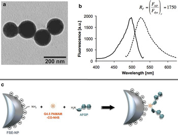
Fluorescein-labelled nanoparticles (FSE-NPs) were synthesized as described and coupled to generation 4.5 (G4.5) poly(amidoamine) dendrimers (PAMAM) derivatized to crosslink to antifreeze glycoproteins (AFGP 1–5) as illustrated (Fig. 1). The average diameter of the nanoparticles could be controlled within limits, and nanoparticles averaging 208 ± 34.9 nm (as measured by dynamic light scattering) were employed in this study. FSE-NPs were also coupled directly to a branched polyethylene glycol derivative (TMS(PEG)12, MW 2420.8) to serve as an independent control.
Preparation of conjugated nanoparticles. a. TEM micrograph of unfunctionalized FSE-NPs approximately 143 ± 41 nm in average diameter, b. Excitation (solid) and emission (dashed) fluorescence spectrum of FSE-NPs with fluorescence relative to a single luminophore, c. Cartoon illustrating the bioconjugation reaction between the NP and AFGP 1–5.

Fish mucus contains AFGPs
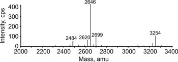
LC-MS/MS of mucus from the surface epithelium of Gymnodraco acuticeps yielded molecules that eluted around 12.5–15 min as triple-charged species with m/z (mass to charge) ratios of 882.724 and 1085.482. The MS/MS spectra of these molecules showed the distinctive oxonium ions for Hex-HexNac, reflecting the presence of the disaccharide β-D-galactosyl-(1,3)-α-D-N-acetylgalactosamine characteristic of the AFGPs, as well as backbone fragment ions consistent with the AFGP peptide sequence. The deconvoluted molecular weights of the identified molecules indicated the presence of AFGP 8 (2646Da) and AFGP 7 (3254Da) with traces of other species corresponding to sugar loss or exchange of proline for alanine (Fig. 2). Detection of high molecular weight AFGP species was not undertaken in this analysis.
Fish mucus contains AFGPs. Mass reconstruction from an MS analysis of mucus from Gymnodraco acuticeps shows peaks at 2646 Da and 3254 Da eluting at about 12.5–15 min and representing AFGP 8 and AFGP 7 respectively.

Free fluorescein-AFGP conjugates are widely dispersed via the lymph and blood
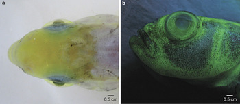
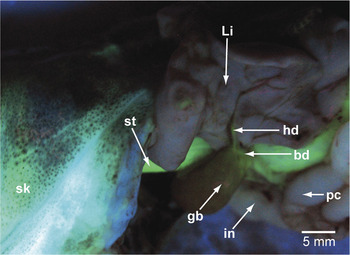
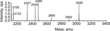
Free fluorescein-labelled AFGP 1–5,7,8 (i.e. not conjugated with beads) was not endocytosed by spleen cells following either i.p. or i.v. injection (results not shown). Instead, the fluorescein-AFGPs were widely dispersed though the lymphatic and blood vascular beds, regardless of the route of injection, such that under UV light the living fish took on a distinct green appearance (Fig. 3a & b). Free fluorescent AFGP reached the bile after i.v. injection as shown by the presence of fluorescence in the common bile duct (Fig. 4) and LC-MS/MS analysis in which a peak representing fluorescein-labelled AFGP 8 was detectable at 3036 Da (Fig. 5). This peak eluted considerably later than unlabelled AFGP 8, reflecting its less hydrophilic nature. Externally, fluorescence was particularly evident in non-pigmented regions of the skin, and it was also seen in both the anterior and posterior chambers of the eye. On dissection, the tongue, buccal cavity, oesophagus and stomach were brightly fluorescent, whereas the more posterior parts of the gut (from and including the pyloric caeca) were less so. The gills, liver and spleen appeared non-fluorescent, as did the brain, the urinary bladder and gonads. The gall bladder had a fluorescent tinge to it, but near its neck fluorescence was unmistakable in the hepatic and common bile ducts (Fig. 4).
Free fluorescein-AFGP is widely dispersed. Fluorescein-labelled AFGP 1–5,7,8 (injected i.p.) is visible through the skin and in the anterior chamber of the eye in these images taken under incandescent (a. dorsal) and UV light (b. lateral). Heavily pigmented regions in the skin obscure the fluorescent signal. Scale bar: 0.5 cm.

Free fluorescein-AFGP reaches the bile via the liver. Fluorescein-labelled AFGP 1–5,7,8 is visible in the skin (sk), stomach (st), hepatic duct (hd) and bile duct (bd) under UV light. Pigment in the skin, liver and bile obscures the fluorescent signal to different degrees. The pyloric caeca (pc) and intestine (in) are non-fluorescent. Li = liver, gb = gall bladder. Scale bar: 5 mm.

Free fluorescein-AFGP traverses the liver to enter the bile. Mass reconstruction from an LC-MSMS analysis of bile from Pagothenia borchgrevinki injected i.v. with fluorescein-labelled AFGP 1–5,7,8. The peak at 3036 Da represents fluorescein-conjugated AFGP 8, which eluted at 38 min.

Phagocytic cells in the spleen engulf AFGP-coated nanoparticles
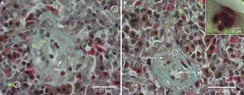
AFGP-coated fluorescent nanoparticles were found within splenic phagocytes 4–6 h after intravenous injection (Fig. 6a & b). Some of these cells were identifiable as foamy macrophages that surround blood vessels (arterioles and sinusoids) within the spleen, whereas other cells were more distant from the vasculature, presumably representing a motile phagocytic cell population. AFGP-coated nanoparticles were not found within endothelial cells lining blood vessels and nor were they found in the liver, which contains phagocytic Kupffer cells (results not shown). In contrast to AFGP-coated nanoparticles, those coated with PEG were poorly phagocytosed by spleen or liver cells (results not shown).
Phagocytosis of AFGP-coated nanoparticles. AFGP-coated FSE-NPs (green) are shown in the reticulendothelial cells that surround splenic ellipsoids in P. borchgrevinki. Note single AFGP-coated nanoparticles within the spleen remote from the ellipsoid (a. circles). A single AFGP-coated FSE-NP is shown in a foamy cell of the reticuloendothelial system that contributes to splenic ellipsoids (b. circle). Fluorescence microscopy with Masson trichrome counterstaining. Reticulin blue-green, erythroid cells red. Nu = nucleus of macrophage in ellipsoid, en = endothelial cell of ellipsoid. Scale bar: 20 μm. Insert shows a single phagocytosed AFGP-coated nanoparticle. Fluorescence microscopy with haemotoxylin and eosin counterstaining. Scale bar: 5 μm.

Discussion
The Antarctic notothenioid Pagothenia borchgrevinki is a pelagic fish found in McMurdo Sound at depths from immediately below the sea ice to at least 500 m (Gon & Heemstra Reference Gon and Heemstra1990). It is characteristically described as a cryopelagic species, since near the surface it is often found inhabiting the platelet ice layer (immediately below the solid sea ice) where it can both obtain food and hide from predators. This distribution brings it into intimate contact with ice, some of it in the form of minute ice crystals, which contribute to the layer of platelet ice. To survive in this habitat, P. borchgrevinki requires an appropriate freeze-avoidance system. Indeed, it has the highest level of AFGPs and antifreeze activity of all Antarctic fishes, commensurate with the severity of its habitat (DeVries & Cheng Reference DeVries and Cheng2005).
Ice crystals within the freezing environment inhabited by P. borchgrevinki are imbibed along with food and seawater (drunk to maintain the internal milieu) and are consequently found within the gastrointestinal tract. Here, they are inhibited from growing further through interaction with AFGPs secreted by cells in the exocrine pancreas (which discharges into the anterior of the small intestine), oesophagus and stomach wall (Cheng et al. Reference Cheng, Cziko and Evans2006). Ice that remains associated with external surface structures is probably embedded in the protective mucus coat (Praebel et al. Reference Praebel, Hunt, Hunt and DeVries2009) which begs the question as to what limits its crystalline growth in this location. One possible explanation is that when mucus-embedded ice crystals reach a certain size they detach through shear effects, clearing the surface of potentially lethal ice. However, even minute external ice crystals have the potential to initiate lethal internal ice growth if the protective epithelium is damaged in some way, such as through lesions opened by bacterial pathogens or physical abrasion. We have tested the possibility that notothenioid mucus might contain AFGPs by using a scaleless species that produces abundant mucus (the naked dragonfish, Gymnodraco acuticeps) as a model system for proof in principle. Using mass spectrometry, we have shown that mucus from the body surface of this notothenioid species contains AFGPs, which have the potential to inhibit ice crystal growth at this location. How these AFGPs are incorporated into the mucus is unclear since none of the currently identified sources of AFGP production provides a direct route to the surface for incorporation into mucus, and whether they are present at functionally effective concentrations in the mucus remains to be tested.
Although P. borchgrevinki collected from freezing Antarctic waters have ice associated with their integument, gill epithelium and gastrointestinal tract, there is none detectable in their body fluids, muscle or major organs, other than the spleen (Praebel et al. Reference Praebel, Hunt, Hunt and DeVries2009). As in other vertebrates, the spleen represents an important defence system that clears foreign particulates from the circulation, particularly microorganisms. However, the observation of ice specifically in this organ within P. borchgrevinki suggests that it also has a protective role by removing ice from the circulation of Antarctic fishes.
The presence of ice within the vertebrate circulation is not a common observation, even within cold environment poikilotherms. When present, however, internal ice may be derived by either endogenous nucleation or from external sources. Under experimental conditions, larval Antarctic fish can survive to temperatures well below their serum non-equilibrium freezing point in the presence of external ice, a phenomenon attributable to the protective properties of the epithelium (Cziko et al. Reference Cziko, Evans, Cheng and DeVries2006) and the lack of endogenous nucleators. Adult fish freeze at temperatures close to their non-equilibrium freezing point in the presence of external ice (DeVries & Cheng Reference DeVries and Cheng2005) indicating the epithelium is no longer a perfect protective barrier. Adult fish that freeze at temperatures close to their non-equilibrium freezing point in the absence of external ice probably have ice in their spleens or intestinal fluid whereas those that do not must be ice free. Taken together, these observations suggest that endogenous ice nucleators are not present in notothenioid fishes; thus when ice is present it has an external origin, most probably deriving from damage to the epithelium in ice-laden water.
Although ice represents a foreign body there is only one report (in two northern hemisphere fish species, the Atlantic herring Clupea harengus L. and the ocean pout Zoarces (Macrozoarces) americanus (Bloch & Sneider)) that it is antigenic (Verdier et al. Reference Verdier, Ewart, Griffith and Hew1996). This result is unexpected in antifreeze-bearing fish since anti-ice antibodies would compete with endogenous antifreeze and there is no evidence that antibodies have protective antifreeze effects. Instead of initiating an immune response leading to the production of anti-ice antibodies that offer no protection against freezing, the major body of evidence indicates that crystals of ice in the circulation of Antarctic notothenioid fishes react essentially instantaneously with endogenous AFGPs, which then act to inhibit crystal growth. The problem now is how these ice crystals, bound to adsorbed antifreeze, are removed from the circulation. To resolve this problem we propose a novel second function for ice-adsorbed AFGPs within the circulation, namely the promotion of uptake by phagocytic cells, primarily in the spleen.
The spleen is arguably the most phagocytically active organ in the vertebrate body in response to foreign particles (living or dead) in the bloodstream. It contains abundant phagocytic cells, especially macrophages, which are distributed throughout the stroma. In the spleens of many species phagocytes are also found as condensations in the form of ellipsoids ensheathing the endothelial cells of the narrow arterioles, as well as in association with the more dilated sinusoids. In fishes, the splenic ellipsoids may be indistinct or lacking, but the macrophages typically display a vacuolated or foamy cytoplasm (Fange & Nilsson Reference Fange and Nilsson1985). Phagocytosed material in fishes is typically transported via the macrophages to melanomacrophage centres, which vary in abundance in different species and under different pathological conditions (Agius & Roberts Reference Agius and Roberts2003).
The spleen of P. borchgrevinki contains intermingled lymphoid and erythroid areas, similar to those described in related notothenioids (Romano et al. Reference Romano, Ceccariglia, Abelli, Baldassini, Picchietti, Mazzini and Mastrolia2004), although the ellipsoids are not always distinct. After intravenous injection, fluorescent nanoparticles coated with AFGP are found in phagocytic cells associated with blood vessels but not in the lining endothelial cells, suggesting that the nanoparticles escape from the circulation through a paracellular route. AFGP-coated nanoparticles are also found in phagocytic cells not intimately associated with the vasculature, suggesting that migration of phagocytic cells occurs within the spleen, presumably leading to the formation of melanomacrophage centres.
Importantly, our results show that free fluorescent-labelled AFGPs are not significantly endocytosed by spleen cells whereas AFGP-coated nanoparticles are. This distribution of free fluorescent-labelled AFGP concurs with the results of other studies using anti-AFGP antibodies, which show that intracellular AFGP is found only in cells that secrete it, namely within the exocrine pancreas, oesophagus, and anterior stomach (Cheng et al. Reference Cheng, Cziko and Evans2006). This difference in uptake between free- and bound-AFGP suggests that AFGP-coated particles are engulfed in a process akin to Fc receptor-mediated phagocytosis, in which clustering of multiple receptors induced by immunoglobulin-bound foreign particles initiates the phagocytic response (Swanson & Hoppe Reference Swanson and Hoppe2004). Immunoglobulins not bound to particles do not activate the phagocytic response and are displaced from the cell surface receptors in the process. In a similar way, we envisage that AFGP-coated nanoparticles, acting as a proxy for AFGP-coated ice crystals, are distinguished from single endogenous AFGP molecules and actively phagocytosed.
To function effectively, AFGPs need to be widely distributed through the entire extracellular space (both lymph and blood) so that they can immediately bind to and inhibit the otherwise fatal endogenous growth of ice. The nature of the fish lymphatic system is the subject of current research, with a case being made for direct blood-lymphatic conduits connecting arterial vessels to the lymphatic system in at least some species, such as the zebrafish Danio rerio (Hamilton) (Jensen et al. Reference Jensen, Cao, Hedlund, Söll, Lundberg, Hauptmann, Steffensen and Cao2009). Our results using free fluorescent-labelled AFGPs injected i.v or i.p. show AFGPs are indeed distributed widely throughout the body, reflecting transport between the lymphatic and blood vascular compartments.
Whole body fluorescence following injection of fluorescently labelled AFGPs is quenched to some degree by tissue pigments, including melanin in the skin, haemoglobin in erythrocytes, and bile components. In our study, fluorescence in the gills, liver and spleen was quenched by haemoglobin, and bile pigments presumably accounted for the poorly reactive gall bladder, although LC-MS/MS confirmed the presence of labelled AFGPs in the bile. Fluorescent AFGPs were visible in the hepatic and common bile ducts, indicating that AFGPs reach the bile from the blood via the liver hepatocytes, to be discharged ultimately into the gut. Since all known sources of AFGP discharge into the gastrointestinal tract, how high levels (c. 35 mg ml-1) are maintained in the blood and lymph remains a challenge for further research. The lack of fluorescence from free AFGPs in the brain and the urinary bladder is consistent with results from other studies (Ahlgren et al. Reference Ahlgren, Cheng, Schrag and DeVries1988), and is attributable to the efficiency of the blood-brain barrier and the presence of aglomerular nephrons in the kidneys of AFGP-bearing notothenioid species respectively (DeVries Reference DeVries1988).
Ice is known to be trapped in the spleens of Antarctic fishes (DeVries & Cheng Reference DeVries and Cheng1992, Praebel et al. Reference Praebel, Hunt, Hunt and DeVries2009) and we have shown using AFGP-coated nanoparticles, as a proxy for ice, that it is likely to be engulfed by phagocytic cells within this organ. Our observations that pegylated nanoparticles are not significantly trapped or phagocytosed in the spleen are significant in that they show that it is not the presence of nanoparticles per se but the nature of their surface moieties (provided by a coating of AFGP) that is significant in trapping and phagocytosis.
We hypothesize that the removal of ice crystals from the circulation confers a protective advantage to the fish, reducing the possibility of tissue damage from entrapment in small blood vessels. The question now is what might happen to ice engulfed by splenic phagocytes. Evidence suggests that it would accumulate during the lifetime of the fish until some event (such as seasonal warming of the environment) occurs that enables the ice to melt (Hunt et al. Reference Hunt, Hoefling and Cheng2003). Fish that do not experience such a warming event will continue to carry an increasing burden of ice, possibly leading to their death unless there is a mechanism other than melting to enable its disposal.
Acknowledgements
This research was supported by grants from the Human Frontier Science Programme (RGP003/2009-C) to MAB and ALD, the Science Foundation Ireland (05/CE3/B754) to VG and RN, and the Faculty Research Development Fund (University of Auckland) to CWE. We thank Martin Middleditch and Natasha Lucas for mass spectrometry analysis, Jennifer Chen for histological preparation, Antarctica New Zealand for logistical support, and staff and colleagues at Scott Base (Antarctica) for their assistance in the field. We are grateful to Vivian Ward for assistance with the images. The constructive comments of the reviewers are also gratefully acknowledged.