INTRODUCTION
Lymphatic filariasis is an ancient mosquito-borne parasitic disease caused by Wuchereria bancrofti and Brugia sp. World-wide, 120 million people are affected and more than 1·1 billion are at risk of infection, living in the endemic countries. The disease is identified as the second leading cause of permanent and long-term disability with morbidity estimated at 5·5 million disability-adjusted life years (DALYs) (Melrose, Reference Melrose2004). Annual doses of diethylcarbamazine (DEC) plus albendazole, albendazole plus ivermectin or use of DEC fortified salt are the currently available drugs for the treatment of lymphatic filariasis (Pokharel et al. Reference Pokharel, Rai, Kodumudi, Reddy and Rathaur2006). Still, these drugs remained ineffective on adult parasites and are known to cause side effects. Further, wider usage of these drugs in endemic areas may pose a possibility of drug resistance making the population more vulnerable to the infection. The World Health Organization also affirms the necessity of alternative modes of treatment such as vaccine or adulticides as an ancillary to the existing treatment for the successful elimination of lymphatic filariasis. Therefore, it is essential to identify a novel filarial target that can be exploited for the discovery of a vaccine or drug against this disease.
Acid phosphatase (EC.3.1.3.2) catalyses the hydrolysis of a wide variety of phosphomonoesterase substrates as well as transphosphorylation reactions at an acidic pH. In higher eukaryotes, reversible phosphorylation of proteins on serine, threonine, and tyrosine residues plays a central role in the integration of signals involved in cellular proliferation and differentiation. The dephosphorylation of phosphoproteins is universally catalysed by protein phosphatases that are classified into 2 major functional groups: protein tyrosine phosphatase (PTP) and protein serine/threonine phosphatase (PP). Two types of PTPs, conserved in sequence and structure can be distinguished: classical PTPs, which are specific for phosphotyrosine residues and dual-specificity phosphatases (DSPs) which can additionally dephosphorylate phosphoserine/threonine residues (Zhang, Reference Zhang2001). The DSPs from the PTP superfamily have the same general 3-dimensional structure and catalytic mechanism as PTP's but differ in their size of active-site pocket. These enzymes have been implicated to play a role in cell cycle control.
Phosphorylation of proteins is a key element of signalling pathways induced by environmental stimuli that regulate cell growth, cell-cell communication, differentiation, metabolism, mitogenesis, immune response, survival and cell migration (Zhang, Reference Zhang2001). It is reported that protein phosphatases also influence survival, internalization and replication of pathogens by interfering with many host signal transduction pathways (DeVinney et al. Reference DeVinney, Steele-Mortimer and Finlay2000).
PTPs have been identified in parasites such as Ascaris suum, Trypanosoma brucei, Leishmania sp. and Entamoeba histolytica (Bakalara et al. Reference Bakalara, Seyfang, Davis and Baltz1995a, Reference Bakalara, Seyfang, Baltz and Davisb; Wimmer et al. Reference Wimmer, Schimid, Tag and Hofer1998; Aguirre-Garcia et al. Reference Aguirre-Garcia, Escalona-Montano, Bakalara, Perez-Torres, Gutierrez-Kobeh and Becker2006; Rodriguez et al. Reference Rodriguez, Pacheco, Rohana and Encina2006) and were used as a virulence factor by many pathogens like Yersinia virus (yop-H) (Guan and Dixon, Reference Guan and Dixon1990) and Coxiella sp. (Baca et al. Reference Baca, Roman, Glew, Christner, Buhler and Aragon1993). SDF-9, a PTP is reported to involve in developmental regulation in C. elegans (Ohkura et al. Reference Ohkura, Suzuki, Ishihara and Katsura2003).
Despite these many predominant reports available, PTP activity in filarial helminths remained unexplored to date. Preliminary studies on AcP have been done in filarial parasites Dirofilaria immitis (Maki and Yanagisawa, Reference Maki and Yanagisawa1980) and Wucheraria bancrofti (Kharat and Harinath, Reference Kharat and Harinath1985). To our knowledge, this is the first report on the identification and biochemical characterization of AcP having dual specific phosphatase activity from a bovine filarial parasite, Setaria cervi.
MATERIALS AND METHODS
Collection of parasitic material and preparation of homogenates
Adult, motile S. cervi worms were procured from the peritoneal folds of freshly slaughtered Indian water buffaloes. They were brought to the laboratory from local abattoir in Krebs Ringer solution supplemented with streptomycin, penicillin, glutamine and 1% glucose. Worms were washed with phosphate-buffered saline and maintained in supplemented Ringer solution or frozen at −70°C until further use. Microfilariae were obtained by dissecting the uterus of gravid females.
Adult parasites and mf were maintained in supplemented Ringer at 37°C for 4 h under sterile conditions. Maintenance medium was centrifuged and clear supernatant was treated as excretory–secretory (ES) product.
Adult female S. cervi extract was prepared as described by Singh and Rathaur (Reference Singh and Rathaur2005). Briefly, worms were dried on filter paper folds, weighed and homogenized at 4°C in 20 mM Tris-HCl buffer, pH 7·0, using a motor-driven REMI homogenizer type RQ127A and then centrifuged at 16 000 g for 1 h. The resulting supernatant was used for purification and further characterization. Soluble extract of mf was prepared by sonicating the mf using an ultra- sonicator (MSE 150 W) for 10 min with a cycle of 30 sec followed by a 30 sec interval at 20 KC in cold 20 mM Tris-HCl buffer, pH 7·0, on ice. The extract was centrifuged at 12 000 g for 30 min. The clear supernatant was collected and stored at −70°C until further use.
Acid phosphatase assay
Acid phosphatase activity was measured according to the method of Chen et al. (Reference Chen, Ansai, Awano, Iida, Barik and Takehara1999) using para-nitrophenyl phosphate (pNPP) as substrate. Briefly, crude extract or purified acid phosphatase was incubated in 0·1 M sodium acetate buffer, pH 4·8, containing 50% glycerol, 10 mM NaCl and 10 mM pNPP in a final reaction volume of 0·5 ml for 30 min at 37°C. The reaction was terminated by the addition of equal volumes of 1·0 M NaOH and liberated para-nitrophenol was measured spectrophotometricaly at 405 nm. Activity was calculated using an extinction coefficient of 17·5 M−1cm−1. Hydrolysis of other substrates (phospho-L-tyrosine, phospho-L-serine, sodium-α-naphthyl phosphate, fructose-6-phosphate, adenosine-di-phosphate and phosphoenol pyruvate) was studied by the method as described by Taga and Etten (Reference Taga and Etten1982). Activity was determined by the quantification of reduced phosphomolybdic acid at 700 nm using a molar extinction coefficient of 4×10³ M−1cm−1. Purified enzyme was incubated at 37°C in 1 ml of solution containing 50 mM sodium acetate buffer, pH 5·0, 10 mM substrate and 100 mM NaCl. The reaction was stopped after 20 min by the addition of 1 ml of 3% ammonium molybdate in 200 mM sodium acetate, pH 4·0. Then, 0·1 ml of 1% ascorbic acid in 200 mM sodium acetate, pH 4·0, was added and allowed to develop colour for 30 min.
Detection of AcP in native PAGE
AcP activity staining in native PAGE was performed according to the method of Gomez (Reference Gomez1978), with some modifications. Briefly, adult female extract was loaded on 7·5% native PAGE, electrophoresed in Tris-boric acid, pH 8·9, and then stained for acid phosphatase in a 0·1 M sodium acetate buffer, pH 5·0, containing 1 mM MnCl2, 1 mg/ml of sodium-naphthyl phosphate and 1 mg/ml of Fast Blue BB salt.
Purification of ScDSP
ScDSP was purified from adult female S. cervi using cation exchange, gel filtration and concavalin affinity sequential chromatography. In the first step, somatic extract was loaded on a CM-Sephadex C-50 column pre-equilibrated with 50 mM sodium acetate buffer, pH 6·0, and protein was eluted with 0–0·5 M NaCl gradient. AcP activity was eluted in unbound as well as at 0·2 M in the gradient. Protein peaks having AcP activity in unbound fractions were concentrated and loaded on Sephadex G-200 column pre-equilibrated with 0·1 M Tris buffer, pH 7·0. Fractions having enzyme activity were pooled and further loaded on Concavalin-Sepharose affinity matrix and ScDSP was eluted with 1 M mannose.
Protein estimation and SDS-PAGE
Protein concentration was measured according to dye-binding method using bovine serum albumin as standard protein (Bradford, Reference Bradford1976). SDS-PAGE was performed under non-reducing conditions (Laemmli, Reference Laemmli1970) and protein bands were visualized by silver stain. Pre-stained molecular weight markers (20–97·4 kDa) were used for the determination of subunit weight of the purified ScDSP.
pH and temperature optima
Optimum pH for ScDSP activity was determined with 0·1 M sodium acetate buffer at a pH range of 3·6–7·5 using pNPP as substrate. Optimum temperature was determined by incubating the routine assay mixture at 10, 20, 30, 37, 40, 50 and 60°C respectively.
Effect of inhibitors and metal ion
Purified ScDSP was pre-incubated with inhibitors/activators at 37°C for 15 min in the reaction buffer (0·1 M sodium acetate). pNPP was then added and the tubes were further incubated for 30 min. Reaction was terminated by the addition of 1 M NaOH and O.D. was measured at 405 nm. Inhibitors included ethylenediamine tetraacetate (EDTA, 2·5 mM), sodium fluoride (2·5 mM), ammonium molybdate (2·5 mM), sodium orthovanadate (50 μM), L (+) tartrate (2·5 and 5·0 mM), DTT (2·0 mM), para-hydroxy mercuric benzoate (pHMB, 1 mM), silver nitrate (2·5 mM), zinc chloride (2·5 mM) and manganese chloride (2·5 mM).
Kinetic studies
Km and Vmax values for purified ScDSP were determined with pNPP, phospho-L-tyrosine and phospho-L-serine as substrates at a concentration range of 2·0 mM to 20·0 mM. The values were obtained by plotting a Lineweaver-Burk plot using Origin 6·0 software. Sodium orthovanadate was used for inhibition studies at a concentration range of 10 μM to 50 μM.
Modification of histidine and carboxylate residues
Purified ScDSP was incubated with diethyl pyrocarbonate (DEPC) (1 mM) at 37°C for different time-intervals in 0·1 M sodium acetate buffer, pH 5·5. AcP activity was routinely assayed using pNPP. The effect of 1-ethyl-3-dimethylaminopropyl carbodiimide (EDAC) on AcP activity was studied according to the method as described by Amutha et al. (Reference Amutha, Khire and Khan1999) with some modifications. Briefly, purified ScDSP was incubated with various concentrations of EDAC in the presence of 30 mM alanine ethyl ester in 100 mM Tris-HCl, pH 6·0. After incubation at 37°C for 45 min, reaction was quenched by the addition of equal volumes of 1 M Na-acetate buffer, pH 5·5, and residual activity was determined under standard assay conditions.
ELISA
IgE response of purified ScDSP against various categories of W. bancrofti-infected human sera i.e., endemic normal (EN), asymptomatic microfilaraemic, chronic (CH) and tissue fluid (from a chronic patient) was assessed using ELISA (Rathaur et al. Reference Rathaur, Robertson, Selkirk and Maizels1987). Briefly, 2 μg/ml solution of purified ScDSP in 60 mM carbonate buffer, pH 9·6, was coated onto polystyrene microplate wells (Nunc, USA) and followed by blocking with 5% skimmed milk in PBS, addition of different categories of filarial sera as primary antibodies (1:100 dilution) and detection of bound antibodies with alkaline phosphatase-conjugated rabbit anti-human IgE solution (1:5000 dilution) (Sigma–Aldrich). Colour was developed using p-nitrophenyl phosphate as substrate and absorbance was measured at 405 nm using an ELISA plate reader (Bio-Rad).
Western blotting
Western blotting was performed according to the method described by Lunde et al. (Reference Lunde, Paranjape, Lawley and Ottesen1988). Purified protein (15 μg) run on 10% SDS-PAGE under non-reducing conditions was electro-transferred on to polyvinylidene fluoride (PVDF) membrane by the semi-dry method. This membrane was cut into 0·3 cm wide strips and incubated in blocking buffer (5% skimmed milk in PBS) for 1 h at room temperature. After 3 washes of 5 min each with wash solution (2·5% skimmed milk in PBS–0·05% Tween 20), the membranes were incubated in different categories of filarial-infected human sera and other helminth-infected human sera (diluted 1:100 in wash solution) for 1 h at room temperature. Following the incubation period, the membranes were washed as described above and then incubated with peroxidase-conjugated goat anti-human IgG (Bangalore Genei, India) (1:5000 in dilution solution). After 3 consecutive washes, the blots were developed using the substrate 2′,2′ diaminobenzidine (DAB) and H2O2.
Effect of ScDSP on eosinophil effector function
Isolation of eosinophils
Eosinophils were separated from the normal human blood according to the method described by Koenderman et al. (Reference Koenderman, Kok, Hamelink, Verhoeven and Bruijnzeel1988). Briefly, the blood was diluted twice with PBS and then centrifuged (100 g, 10 min, RT). Buffy coats were collected and again diluted with PBS and centrifuged over Histopaque (1000 g, 20 min, RT). The upper layer of mononuclear cells was removed and granulocytes with contaminating erythrocytes were collected from the bottom of the tubes. Erythrocytes were lysed with isotonic ammonium-chloride solution (containing 10 mM KHCO3 and 0·1 mM EDTA) at 0°C for 15 min. After centrifugation (400 g, 10 min, 4°C) the sedimented cells were washed once with PBS, resuspended in 10 ml of sterile FBS (pH 7·4) and incubated for 30 min at 37°C, then again centrifuged (400 g, RT) and resuspended in PBS. The mixed granulocyte preparation was incubated with fMLP (formyl-methionyl-leucyl-phenylalanine) (10 nM) in PBS for 10 min at 37°C. Thereafter, 1 ml of cell suspension was layered carefully onto a Percoll layer of density 1·082 g/ml, under-layered by Percoll of density 1:100 g/ml and centrifuged (1000 g, 15 min, RT). Pure eosinophils obtained from the interface between the two Percoll layers were collected, washed with PBS and resuspended in a suitable buffer. Eosinophils, thus obtained were stained with eosin and purity was found to be approximately 91%.
Superoxide anion generation
Superoxide anion generation by the human eosinophils stimulated by IgG in the absence and presence of purified ScDSP was studied according to the method described by Shin et al. (Reference Shin, Kita, Park and Seoh2001). Briefly, tissue-culture plates were coated with human IgG (50 μl/well) diluted in PBS at 2 different concentrations (10 and 30 μg/ml) in the absence/presence of ScDSP for 2 h at 37°C. After incubation, the wells were aspirated and washed with PBS and 200 μl of eosinophil suspension (2·0×104cells/ml) in HBSS with 10 mM HEPES, 0·03% gelatin and 100 μM cytochrome C were dispensed in to the wells. Immediately after the addition, absorbance was measured at 550 nm at different time-intervals.
Statistical analysis
Statistical analysis of data obtained (mean and standard deviation) was performed using the PC SPSS application package and Origin 6.0 software. Experiments were performed in triplicate and all results are mean values unless otherwise stated.
RESULTS
Identification and purification of ScDSP
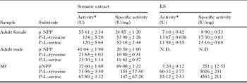
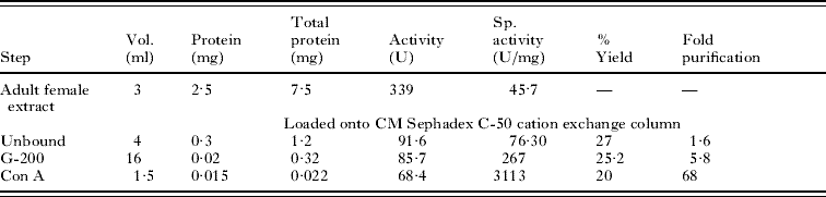
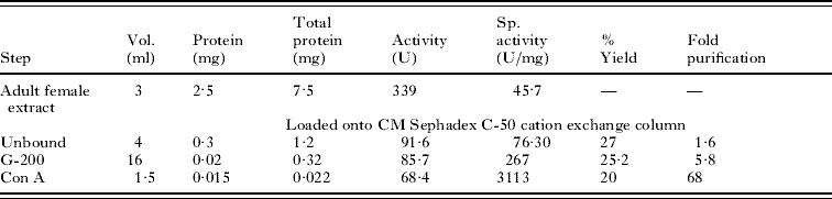
A significant amount of AcP activity was detected in the adult female extract and ES of different life stages of S. cervi using general substrates pNPP as well as phospho-L-tyrosine and phospho-L-serine. The activity was highest in adult female extract followed by mf and adult male. The ES product of mf showed higher activity than the adult female worm. However, no activity was detected in male ES (Table 1). ScDSP from adult female extract was purified up to homogeneity by 3 subsequent column chromatographic procedures. Two forms were separated by CM-Sephadex C-50. ScDSP eluted in the unbound fraction was further purified up to 68-fold with 20% yield after a 2-step purification by G-200 gel filtration and Concavalin-A affinity matrix. Table 2 summarizes the overall purification steps involved in ScDSP purification. Subunit molecular mass and other properties of both AcPs separated during this purification procedure differ markedly (data not shown).
AcP activity in excretory-secretory products (ES) and somatic extracts of microfilariae and adult worms of Setaria cervi

* One unit of enzyme activity is defined as micromoles of p-nitrophenol produced ml−1 min−1 with pNPP and amount of free phosphate produced using P-L-tyrosine and P-L-serine as substrates. Values are mean±s.d. of 3 different determinations.
N.D., Not detectable.
Purification steps of ScDSP from adult female somatic extract
Characterization of ScDSP
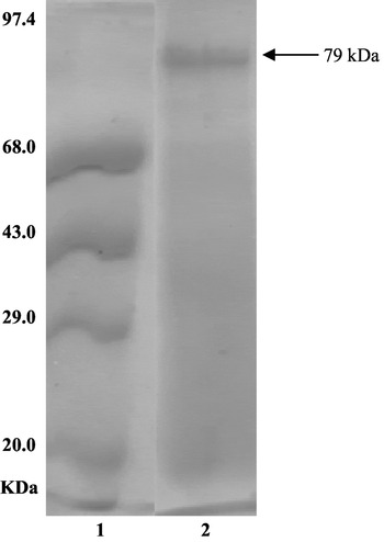
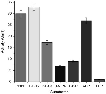
SDS-PAGE analysis of purified ScDSP (Fig. 1) revealed a subunit molecular mass of approximately 79 kDa. It was active over a broad pH range having maximum activity at pH 4·8 and almost negligible activity at pH 6·0 and above. Enzyme activity was found to be highest at 40°C in a temperature range of 0 to 60°C. Activity at 60°C was almost 91% less than that at 40°C. Along with pNPP, the enzyme showed its affinity towards phospho-L-tyrosine, ADP, phospho-L-serine, fructose-6-phosphate and sodium-napthyl-phosphate. However, phosphoenolpyruvate was not hydrolysed by the enzyme (Fig. 2). The enzyme followed Michaelis-Menten kinetics with pNPP as substrate. Km and Vmax values using pNPP/phospho-L-tyrosine/phospho-L-serine as substrates were calculated from a double reciprocal plot and were found to be 16·66 mM, 25·0 μM/ml/min; 20·0 mM, 40·0 μM/ml/min and 27·0 mM, 25·0 μM/ml/min respectively. Sodium orthovanadate, a specific inhibitor for the PTP class of phosphatases showed a competitive type of inhibition. KI with pNPP and sodium orthovanadate (IC50 33·0 μM) was calculated to be 50·0 mM.
SDS – PAGE analysis of purified ScDSP on 12·5% polyacrylamide gel. Lane 1, Molecular weight markers. Lane 2, Purified ScDSP.

Substrate specificity for ScDSP.

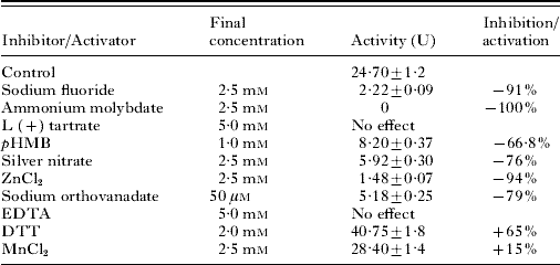
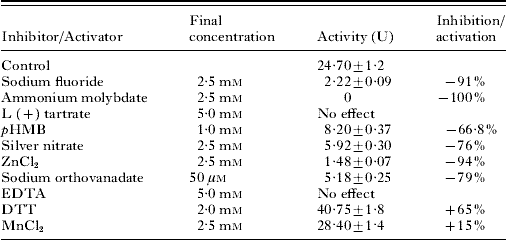
Table 3 summarizes the effect of various inhibitors and metal ions on the enzymatic activity of purified ScDSP. The enzyme was found to be sensitive with known PTP inhibitors such as fluoride (91%), molybdate (100%), zinc chloride (94%) and silver nitrate (76%) at 2·5 mM; sodium orthovanadate at 50 μM, (79%), and pHMB at 1·0 mM (66·8%). ScDSP was found to be resistant for L (+) tartrate (2·5 mM and 5·0 mM) and EDTA (2·5 mM). The presence of DTT has strongly enhanced the enzymatic activity (65%) whereas Mn2+ showed a slight increase (15%).
Effect of inhibitors and metal ions on purified ScDSP
(The concentration of inhibitors and metal ions indicated are those exhibiting highest inhibition tested so far. Activity was measured using pNPP (10 mM) as substrate. Results indicated are mean±S.D. of 4 different determinations.)
DEPC, a histidine-group specific inhibitor, showed time-dependent inhibition on ScDSP activity. It accounted for 86% inhibition at 1·0 mM concentration after 30 min. Similarly, EDAC, a specific inhibitor for carboxylate residue significantly inhibited ScDSP activity in a concentration-dependent manner accounting for 66·4% inhibition at 25 mM concentration.
ELISA, Western blotting and superoxide anion generation
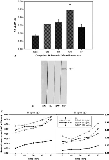
In ELISA, purified ScDSP showed significant levels of IgE response in all the categories of W. bancrofti-infected human sera (Fig. 3A). An increasing trend in O.D. was observed from EN to CH. Considerable cross-reactivity was also observed with tissue fluid collected from chronic patients. Western blotting of purified ScDSP further confirms its cross-reactivity with W. bancrofti-infected microfilaraemic sera (Fig. 3B). IgG-induced eosinophil degranulation, in the presence of ScDSP, showed an increase in superoxide anion generation on addition of cytochrome C. However, superoxide generation was suppressed with increasing concentration of ScDSP (Fig. 3C).
(A) IgE ELISA of ScDSP against Wuchereria bancrofti-infected human sera. With ScDSP as antigen maximum IgE level was detected in chronic filarial pathology. NEN, non-endemic normal; EN, endemic normal (n=17); MF, microfilaraemic (n=18); Ch, chronic (n=10); TF, tissue fluid. (B) Western blot of purified ScDSP revealing significant cross-reactivity with W. bancrofti-infected microfilarial sera. EN, endemic normal; Ch, chronic; RW, round worm; MF, microfilaraemic. (C) Kinetics of superoxide anion production by eosinophils incubated in microplate wells coated with IgG in the presence or absence of ScDSP. Wells were coated with IgG (10 μg/ml and 30 μg/ml) in the absence or presence of purified enzyme, at the doses indicated, for 2h at 37°C. After washing, eosinophils were added to the wells, and superoxide production from eosinophils was measured by reduction of cytochrome c, as described in the Materials and Methods section.

DISCUSSION
A stage-specific screening for AcP having PTP activity in secretions and somatic extracts of S. cervi clearly demonstrated its presence both in mf and adult stages of parasite. Highest specific activity was observed in microfilariae, a developing stage of the parasite. Significant activity was also detected in the ES of adult females and microfilariae suggesting an extra cellular form of PTP. S. cervi AcP utilized pNPP as well as protein phosphatase substrates phospho-L-tyrosine and phospho-L-serine. A marked difference in enzyme activity with different substrates at various life stages suggested that AcP activity might be developmentally regulated. Earlier, it has been reported in T. cruzi and T. brucei too that PTP activity varied in different developmental stages (Bakalara et al. Reference Bakalara, Seyfang, Baltz and Davis1995b). Significant PTP activity in excretory-secretory products suggested that the parasitic enzyme could modify the physiology of host cell (Kutuzov and Andreeva, Reference Kutuzov and Andreeva2008). The role of PTPs in regulating the immune response has been well reported (Dolton et al. Reference Dolton, Sathish and Mathews2006). ScDSP was purified to homogeneity using cation exchange, gel filtration and concavalin-A affinity chromatography. Binding of AcP to concavalin matrix indicates its glycosylated nature. Earlier, 3 distinct AcPs have been separated, purified and characterized from L. donovani using similar protocol (Remaley et al. Reference Remaley, Das, Campbell, LaRocca, Pope and Glew1985).
The estimated molecular mass of ScDSP was 79 kDa which was close to a membrane-bound AcP purified and characterized in L. mexicana (70–72 kDa) (Menz et al. Reference Menz, Winter, Ilg, Lottspeich and Overath1991) and PTP from E. histolytica (97 kDa) (Anaya-Ruiz et al. Reference Anaya Ruiz, Perez Santos and Talamas Rohana2003). However, it was higher than the DSP reported from Vaccinia virus (43 kDa) and from humans (20 and 21 kDa) (Guan et al. Reference Guan, Broyles and Dixon1991; Hood et al. Reference Hood, Tobin and Yoon2002). Purified enzyme was most active at pH 4·8 which is in accordance with the usual pH range 3·8 to 6·0 of parasitic helminth acid phosphatases (Maki and Yanagisawa, Reference Maki and Yanagisawa1980; Fetterer and Rhoads, Reference Fetterer and Rhoads1980). A marked difference has been reported in the case of the human enzyme where the pH range is 6·0 to 7·0 (Hood et al. Reference Hood, Tobin and Yoon2002).
Substrate specificity studies revealed that purified ScDSP has maximum affinity towards phospho-L-tyrosine followed by pNPP and phospho-L-serine and also towards two physiological substrates i.e., ADP and fructose-6-phosphate. DSP capable of dephosphorylating P-Tyr as well as P-Ser were also reported from P. falciparum (PfYVH1) and Vaccinia virus (VH1) (Kumar et al. Reference Kumar, Musiyenko, Cioffi, Oldenburg, Adams, Bitko, Sri Krishna and Barik2004; Guan et al. Reference Guan, Broyles and Dixon1991). Dependence of initial enzyme activity on substrate concentrations and appearance of the hyperbolic curve suggest that the purified enzyme follows Michaelis–Menten kinetics (data not shown). The Km value of S. cervi DSP obtained with pNPP, phospho-L-tyrosine and phospho-L-serine was very high when compared with other parasitic PTPs for respective substrates. In the case of filarial parasites preliminary kinetic studies were performed in D. immitis using β-glycerophosphate as substrate in crude extract that showed a relatively high Km value of 32 mM (Maki and Yanagisawa, Reference Maki and Yanagisawa1980). The affinity of this phosphatase towards ADP and fructose-6-phosphate suggested that it might be playing an essential role in providing nutrition and energy metabolism as described earlier by Remaley et al. (Reference Remaley, Das, Campbell, LaRocca, Pope and Glew1985).
A study on the effect of specific inhibitors and metal ions on AcP activity proved crucial in confirming the mechanistic class of this enzyme. Inhibition of enzyme activity with known PTP inhibitors such as flouride, molybdate, orthovanadate and resistance to tartrate and EDTA confirmed its identity as PTP.
Sodium orthovanadate showed marked inhibition of ScDSP activity and the IC50 value was found to be 33 μM. This value is very low in comparison to other parasites, for example, 162 μM for AcP purified from F. tularensis (Almeida Amaral et al. Reference Almeida Amaral, Firpo, Vannier-Santos and Meyer Fernandes2006), 75·8 μM for ecto-PTP in E. histolytica (Anaya Ruiz et al. Reference Anaya Ruiz, Perez Santos and Talamas Rohana2003) and 200 μM for PTP in a crude cell preparation of T. brucei (Bakalara et al. Reference Bakalara, Seyfang, Davis and Baltz1995a). It has been reported that sodium orthovanadate is a competitive inhibitor of PTP and DSP (Almeida Amaral et al. Reference Almeida Amaral, Firpo, Vannier-Santos and Meyer Fernandes2006). In our study too, orthovanadate showed competitive inhibition of ScDSP. The apparent Km in the presence of orthovanadate was found to be 50 mM. It was stated that orthovanadate acts as a phosphate analogue and is generally thought to bind as a transition state analogue to the phosphoryl transfer enzymes (Almeida Amaral et al. Reference Almeida Amaral, Firpo, Vannier-Santos and Meyer Fernandes2006). EDTA showed no effect on enzyme activity, thus excluding the possibility of involvement of metal ion in enzyme catalysis. Activation of enzyme by DTT, a thiol group-protecting reagent, and inhibition with pHMB indicates the involvement of cysteine residue in enzyme catalysis. The presence of a cysteine residue at the active site is a key for the catalytic function of PTPs. Furthermore, inhibition with zinc also confirms the presence of cysteine as it binds tightly to sulphydryl groups and has the same redox properties (Fernandes et al. Reference Fernandes, Granjeiro, Taga, Meyer-Fernandes and Aoyama2003; Almeida Amaral et al. Reference Almeida Amaral, Firpo, Vannier-Santos and Meyer Fernandes2006).
It has been reported that PTP and DSP employ an identical mechanism for phosphate monoester hydrolysis. Both families of enzymes share an identical active site having cysteine and aspartic acid residues (Denu and Dixon, Reference Denu and Dixon1995). ScDSP appears to have similar amino acids at the active site as characterized by using pHMB and EDAC. Inhibition of our enzyme with DEPC suggests the presence of a histidine residue at its active site which is in accordance with some earlier studies (Chen et al. Reference Chen, Ansai, Awano, Iida, Barik and Takehara1999; Taga and Etten, Reference Taga and Etten1982; Denu and Dixon, Reference Denu and Dixon1998). Cysteine and aspartate were reported to be involved in key steps of DSP-mediated catalysis during the formation of thiol-phosphate intermediate (Denu and Dixon, Reference Denu and Dixon1995). These results confirm that S. cervi AcP has a dual specific PTP activity.
Cross-reactivity of ScDSP with W. bancrofti-infected microfilarial cases was confirmed by Western blotting. Antigenic cross-reactivity between S. cervi and W. bancrofti has previously been established (Kaushal et al. Reference Kaushal, Kaushal and Ghatak1987; Sharma et al. Reference Sharma, Mishra and Rathaur1998). These observations suggest that a protein homologous to ScDSP is expressed by W. bancrofti and thus it could be used for the screening of possible vaccine antigen, drug target and diagnostic marker against human lymphatic filariasis. Moreover, this analysis also indicated the potential of ScDSP as a possible diagnostic marker for the detection of active infection. Human lymphatic filariasis produces a range of host immune responses that have been implicated in the pathogenesis of different clinical manifestations of the disease. Filarial parasites are known to highly modulate the immune system during their long-term co-evolutionary interaction with the host (Allen and Maizels, Reference Allen and Maizels1996). The persistence of the parasites was attributed to the modulation of the immune system, leading to inhibition of inflammatory responses required for the elimination of parasite and also implies highly effective immune strategies (Maizels et al. Reference Maizels, Bundy, Selkirk, Smith and Anderson1993). Manipulation of the host cellular phosphorylation state has been used for evading host immune reactions by several pathogens because reversible phosphorylation of tyrosine residues has been shown to represent a key mechanism for the transduction of signals of immune reactions as well as other physiological processes (Mustelin et al. Reference Mustelin, Alonso, Bottini, Huynh, Rahmouni, Nika, Louis-dit-Sully, Tautz, Togo, Bruckner, Mena-Duran and Maria al-Khouri2004). The role of IgE and eosinophils in combination has been reported in filarial infections. It was shown that eosinophils are recruited at the site of helminth infection following IgE-induced mast cell degranulation. Parasites opsonized with IgG, IgE permits eosinophil adhesion to the parasite surface and facilitates eosinophil degranulation, albeit the mechanism by which eosinophils mediate protection against helminth infection remains poorly understood (Klion et al. Reference Klion and Nutman2003). Elevated levels of IgE against ScDSP have been observed in all categories of W. bancrofti-infected human sera with highest in chronic pathology. We have also studied the in vitro effect of purified parasitic DSP on superoxide anion generation by eosinophils stimulated by IgG, as they are known to be important effector cells in the host defence against helminth parasites. ScDSP showed an increased effector function of eosinophils as observed by the increase in superoxide anion generation suggesting that it could play important role in host protection. However, there was suppression in release of superoxide ion when the concentration of ScDSP increased. The way in which eosinophils are activated by ScDSP is not clear, but involvement of a signal transduction mechanism cannot be ruled out. Such a type of enhancement of eosinophil effector function has already been reported for a protease purified from S. mansoni (Auriault et al. Reference Auriault, Caprom, Cesari and Capron1983). Degranulation of adhering eosinophils could be the underlying mechanism of eosinophil-mediated parasite killing (Shin et al. Reference Shin, Kita, Park and Seoh2001). After migration into inflamed tissue eosinophils become activated and release various mediators such as reactive oxygen intermediates, lipid mediators and cytotoxic granular proteins. Tissue-invading helminths may have an immune escape mechanism of down regulation of eosinophil effector function, thus enabling the worm to pass through host immune defences undisturbed (Shin et al. Reference Shin, Kita, Park and Seoh2001).
In summary, we report that S. cervi AcP having dual specific phosphatase activity is unique and differs from other parasitic and mammalian counterparts. Identification of this class of phosphatases in filarial helminths opens new dimensions for parasitologists to study the host-parasite interaction and mechanism of survival of parasite within the host.
The authors are grateful to CSIR, New Delhi, scheme no. 37 (1189) EMR II for financial assistance. The authors are also grateful to UGC, India for providing the financial support under the RGNF scheme.