There are several paediatric cardiologists and cardiac surgeons from the former Children's Memorial Hospital who have made significant contributions to the care of children with transposition of the great arteries and common arterial trunk – truncus arteriosus. The purpose of this review is to highlight those contributions. Several of these contributions are now a standard part of practice of most physicians caring for children with these congenital cardiac anomalies. The individuals include Thomas G. Baffes, Milton H. Paul, Farouk S. Idriss, Constantine Mavroudis, Robert H. Anderson, Hyde M. Russell, and myself.
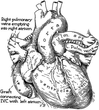
Thomas G. Baffes was born in New Orleans in 1923. He attended medical school and received his training in general surgery at the Charity Hospital in New Orleans. He was a member of the hospital staff at the Children's Memorial Hospital from 1952 to 1997. During that time, he published 89 peer-reviewed papers. Dr Baffes’ sentinel contribution was a new operation for the surgical palliation of transposition of the great arteries (Fig 1). This operation was originally published in Surgery, Gynecology & Obstetrics in 1956.Reference Baffes 1 The Baffes operation was described at a time when there was no effective palliation for children with transposition of the great arteries. The only described operation was the Blalock–HanlonReference Blalock and Hanlon 2 atrial septectomy. The operation that came to be known as the “Baffes procedure” was performed through a right thoracotomy. Cardiopulmonary bypass was not used. The operation was performed with partial occlusion clamps and consisted of a homograft conduit directing the inferior caval vein blood to the left atrium. The right pulmonary veins were transected from their entrance into the left atrium and were anastomosed directly to the right atrium. Surgical lore has it that the key to this operation was occluding the right pulmonary artery during the procedure so that the right lung did not become engorged with blood.
This illustration shows a completed Baffes operation. A homograft connects the inferior caval vein to the left atrium. The right pulmonary veins have been anastomosed to the right atrium. This effectively was a 50% correction of transposition of the great arteries.Reference Baffes 1 IVC = inferior vena cava.

This operation was first performed at the Children's Memorial Hospital on May 6, 1955. The “Baffes procedure” was used for the next 10 years as the primary procedure of choice for infants with transposition of the great arteries. Baffes performed a total of 202 cases employing this technique. Several patients were later converted to a full atrial level repair and survived into adulthood.
Milton H. Paul (b. 1928) was an attending cardiologist at the Children's Memorial Hospital between 1957 and 1989. He trained at the Children's Hospital Boston with the late Alexander Nadas. He was Chief of Cardiology at the Children's Memorial Hospital from 1963 to 1982. Dr Paul published over 30 peer-reviewed articles on transposition of the great arteries between 1960 and 1999. He was the author of the chapter, “Transposition of the Great Arteries” in the classic text, Moss’ Heart Disease in Infants, Children, and Adolescents, edited by Adams and Emmanouilides.Reference Paul 3 Dr Paul was involved in the first balloon atrial septostomy performed at the Children's Memorial Hospital (1967) with the assistance of Dr RashkindReference Rashkind and Miller 4 who happened to be visiting Chicago during a meeting when a child with transposition of the great arteries presented to our hospital.
Farouk S. Idriss was Chief of Pediatric Cardiovascular Surgery at the Children's Memorial Hospital between 1967 and 1990. Dr Idriss was born in Beirut, Lebanon, in 1928. He studied at the American University in Beirut where he received his Bachelor of Arts degree in 1949 and his Doctor of Medicine degree in 1953. Dr Idriss did his residency in general surgery at Wesley Memorial Hospital. He was a paediatric surgery fellow at the Children's Memorial Hospital between 1958 and 1959. He became director of the surgical laboratory at the Children's Memorial Hospital in 1960 and Division Head of Cardiovascular-Thoracic Surgery (the first Division Head) in 1967. Idriss was a Professor of Surgery at Northwestern University Medical School. He published over 120 peer-reviewed articles and book chapters. He was involved in the training of 125 surgical fellows from Northwestern, Loyola, and Rush Universities. He founded the heart transplant programme at the Children's Memorial Hospital. He was married to Lorraine Idriss for 34 years and had four sons.
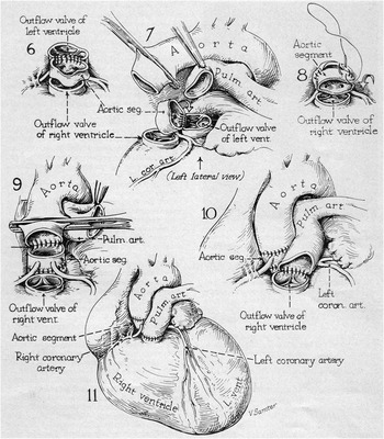
Dr Idriss became interested in transposition of the great arteries and, in fact, attempted a version of the arterial switch operation in 1960 (Fig 2). This was published in Circulation in 1961.Reference Idriss, Goldstein, Grana, French and Potts 5 This was 15 years before Jatene et al'sReference Jatene, Fontes and Paulista 6 report of the arterial switch operation. After performing trials in the surgical laboratory with cardiopulmonary bypass, Idriss embarked on a clinical trial in the summer of 1960. He operated on four patients; two died in the operating room; two actually survived for 24 hours and then died.
The “Idriss switch” is demonstrated in this drawing. Panel 6 shows how a ring of aorta is harvested with the coronary artery orifices. Panel 7 illustrates how this aortic segment is flipped 180° and brought into the left ventricular outflow tract. In Panel 9, the ascending aorta is being anastomosed to this aortic segment. Panel 10 illustrates the pulmonary artery being anastomosed to the outflow tract of the left ventricle. Panel 11 shows the completed switch operation.Reference Idriss, Goldstein, Grana, French and Potts 5

The steps of this operation are illustrated in Figure 2. The essence of the operation was to harvest a circular segment of the aorta that also contained the orifices of the coronary arteries. The aortic segment was then flipped 180° and sutured into the outflow tract of the left ventricle. The transected great vessels were then anastomosed to their appropriate outflow tracts. The completed operation shows the pulmonary artery anastomosed to the right ventricular outflow tract and the ascending aorta anastomosed to the small aortic segment, which was then anastomosed to the ventricular outflow tract.
The anatomic specimen from the first “switch” has been saved in the Farouk S. Idriss Cardiac Registry. This examination of the specimen shows patent coronary orifices but a rather “thinned out” left ventricle. This patient in retrospect was not haemodynamically prepared; the left ventricle was not robust enough to support the systemic circulation. Idriss et alReference Idriss, Goldstein, Grana, French and Potts 5 actually suggested in the manuscript that, “young infants with equally developed right and left ventricles and older children with associated ventricular septal defects and high left ventricular pressure would probably be the best candidates for arterial switching procedure”. I am certain this manuscript stimulated other surgeons such as Jatene to continue the quest for the ideal operation for transposition of the great arteries, which is now the arterial switch procedure as we know it.
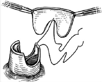
Early on in the experience at the Children's Memorial Hospital with the arterial switch operation polytetrafluoroethylene patches were used to augment the neopulmonary valve sinuses after the coronary buttons had been removed. Idriss noted a high incidence of post-operative supravalvar pulmonary artery stenosis in this patient group. He thought of a solution to this problem, which was to use an autologous patch of pericardium cut in a pantaloon shape for the pulmonary artery reconstruction (Fig 3). He reported using this technique in 23 patients.Reference Idriss, Ilbawi and DeLeon 7 Long-term follow-up for these patients demonstrated a very low incidence of recurrent supravalvar pulmonary stenosis. We have now adopted this technique as our standard operation for the arterial switch operation. Worldwide, many other surgeons now also use this technique for the neopulmonary artery reconstruction.
This is an illustration of the pantaloon-shaped autologous pericardial patch, which is used to reconstruct the neopulmonary artery valve sinuses following harvesting of the coronary artery buttons.Reference Idriss, Ilbawi and DeLeon 7

Our current standard approach to the patient with transposition of the great arteries is to use continuous cardiopulmonary bypass at 24°C. The patient receives two doses of del Nido cardioplegia solution. The coronary buttons are anastomosed to the neoaorta in a “Best Lie” configuration. All patients receive the manoeuvre of LeCompte et al.Reference Lecompte, Zannini and Hazan 8 The pulmonary artery is then reconstructed with a pantaloon-shaped autologous pericardial patch. Using this technique, our mortality rate for the past 10 years in over 90 cases is <2%. In 1991, Dr Mavroudis and myself published a monograph entitled, “The Arterial Switch Operation”.Reference Backer, Idriss and Mavroudis 9 This 200-page text was dedicated to Farouk S. Idriss and Milton H. Paul. The two of them worked together at the Children's Memorial Hospital for over 25 years, achieving new levels in patient care and scientific research.
We recently published two articles updating the experience at the Children's Memorial Hospital with the arterial switch operation. First was a 25-year experience with 258 patients.Reference Rudra, Mavroudis and Backer 10 The conclusion in that article was that the arterial switch operative mortality has decreased over time. The use of the redundant pantaloon-shaped pericardial patch has reduced neopulmonary reintervention. Our current rate of reintervention is <4%. Complex coronary anatomy increases the incidence of need for coronary and/or aortic valve reintervention. Prior banding of the pulmonary artery increases the incidence of the need for aortic reintervention. We also reported our reoperative techniques for complications after the arterial switch operation.Reference Mavroudis, Stewart, Backer, Rudra, Vargo and Jacobs 11 In this manuscript, we reviewed the outcomes of 27 reoperations in 23 patients. These reoperations were performed for lesions relating to the coronary arteries, that is, nine procedures in seven patients; the neoaortic root, that is, 12 procedures in 10 patients; and right ventricular outflow tract, six procedures in six patients. We concluded that although the incidence of late reintervention is low, a subset of patients will require operations for myocardial revascularisation, left ventricular outflow tract reconstruction, and relief of pulmonary stenosis.
Common arterial trunk
Dr Idriss also made significant contributions to the management of infants with common arterial trunk. He established our Cardiac Registry in 1960, which was named after him following his death in 1992. That registry now contains over 2000 cardiac specimens. Within that collection are 17 hearts with a diagnosis of common arterial trunk. That registry has been used by Professor Robert Anderson and Dr Hyde M. Russell to develop a new classification scheme for patients with common arterial trunk.Reference Russell, Jacobs and Anderson 12 Dr Russell used that investigation as a springboard for a Society of Thoracic Surgery registry inquiry to evaluate the outcomes of common arterial trunk with truncal valve surgery.Reference Russell, Pasquali and Jacobs 13
Perhaps our most important contribution to the management of infants with common arterial trunk is from Dr Constantine Mavroudis. Dr Mavroudis trained in general and cardiac surgery at the University of California, San Francisco, between the years 1973 and 1981. His mentor and preceptor at that time was Dr Paul Ebert who went on to become President of the American College of Surgeons. Dr Ebert is well known as the “father” of infant common arterial trunk surgery. In fact, Ebert et al'sReference Ebert, Turley, Stanger, Hoffman, Heymann and Rudolph 14 sentinel paper, published in 1984, described the first series of patients who underwent routine repair of common arterial trunk within the first 6 months of life. The 106 infants reported in that paper were operated on between 1974 and 1981, which was exactly the time period that Mavroudis was a resident at the University of California, San Francisco. That article reported an operative mortality of 11%, which was the lowest reported up to that time.
As his first job out of fellowship training, Mavroudis became Chief of Pediatric Cardiovascular Surgery at Kosair Children's Hospital in Louisville, Kentucky. He held this position from 1981 to 1989. He then accepted the position as Chief of Cardiovascular-Thoracic Surgery at the Children's Memorial Hospital in 1989. He held this position until 2008. He was Chairman of the Department of Congenital and Pediatric Cardiac Surgery at the Cleveland Clinic from 2008 to 2012. He is currently the Director of the Congenital Heart Institute at the Walt Disney Pavilion, Florida Hospital for Children. Mavroudis moved to the Children's Memorial Hospital in Chicago at a time when Paul Ebert was retiring to become President of the American College of Surgeons. Dr Ebert referred many of his patients to the Children's Memorial Hospital so that Dr Mavroudis could perform their conduit change. During Dr Mavroudis’ tenure as Chief at the Children's Memorial Hospital, 76 conduit changes for common arterial trunk were performed. In addition, during that time period Mavroudis and I participated in 16 truncal valve operations.
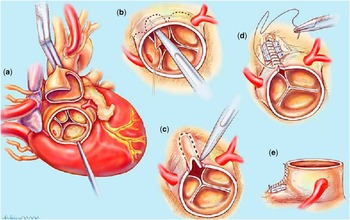
The technique for truncal valve repair is a significant contribution to the care of children with common arterial trunk. We utilised the technique originally reported by Imamura et alReference Imamura, Drummond-Webb, Sarris and Mee 15 and reported our results with that strategy in the Annals of Thoracic Surgery in 2001.Reference Mavroudis and Backer 16 At that time, we reported eight patients who had an operation on the truncal valve either at the time of primary repair – three patients – or in conjunction with conduit replacement – five patients. The essence of the repair is illustrated in Figure 4. After placing the patient on cardiopulmonary bypass, transecting the common trunk, and detaching the pulmonary artery, the truncal valve is evaluated. As shown in panel B, the dimunitive or rudimentary cusp of the quadricuspid valve is excised. In addition, a portion of the truncal valve sinus of Valsalva is excised. This is then reapproximated with pledgeted sutures to make the annulus smaller, but appropriately sized for the size of the now trileaflet valve. A variation of this operation is required when the coronary artery arises from the rudimentary cusp. In this case, that same article also reported successful aortic reimplantation of a transferred coronary artery for this same diagnosis. The long-term results of our truncal valve repair are reported in this same issue authored by Russell et al.Reference Russell, Mavroudis, Backer and Mavroudis 17
This drawing demonstrates repair of a quadricuspid truncal valve that had severe truncal valve insufficiency. In Panel ( a ), the common arterial trunk has been transected and the pulmonary artery excised from the aorta. The quadricuspid valve is exposed. In Panel ( b ), the most dimunitive cusp is being excised. Panel ( c ) shows removal of a portion of the sinus of Valsalva. Panel ( d ) shows the reconstruction of the annulus and sinus so that this is now a trileaflet aortic valve. Panel ( e ) shows the completed procedure with annuloplasty sutures.Reference Mavroudis and Backer 16

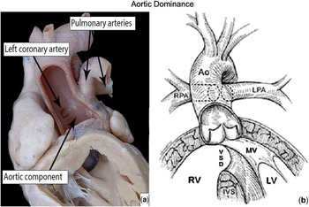
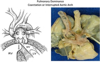
Professor Robert H. Anderson has been visiting our institution since 2007 to catalogue, study, and report on the specimens in our Idriss Cardiac Registry. Anderson is one of the world's leading authorities on congenital heart morphology and pathology. He and Dr Hyde Russell have used our collection and the specimens from two other institutions to examine 28 autopsied hearts with common arterial trunk.Reference Russell, Jacobs and Anderson 12 That same review was used to categorise 42 patients who had undergone surgical correction of common arterial trunk at the Children's Memorial Hospital. All autopsied hearts could be assigned to either aortic or pulmonary dominance of the common arterial trunk. There were 20 aortic dominant and eight pulmonary dominant specimens. Pulmonary dominance was found only when the aortic component of the trunk was hypoplastic and the arterial trunk supplied the majority of flow to the descending aorta. This simplified classification of common arterial trunk into aortic dominance (Fig 5) and pulmonary dominance with coarctation or interrupted aortic arch (Fig 6) is now becoming the standard classification scheme for common arterial trunk. During the years between 1979 and 2012, 56 children have undergone surgical repair for common arterial trunk at the Children's Memorial Hospital. The age ranged from 1 week to 5 years, with a median age of 10 days. There were 48 patients with aortic dominance and eight patients with pulmonary dominance. In all, 12 patients had interventions on the truncal valve. The overall operative mortality was only 5%.
This picture demonstrates the aortic dominant common arterial trunk. Ao = aorta; IVS = intraventricular septum; LPA = left pulmonary artery; LV = left ventricle; MV = mitral valve; RPA = right pulmonary artery; RV = right ventricle; VSD = ventricular septal defect.

This picture shows the pulmonary dominant common arterial trunk typically associated with coarctation of the aorta or interrupted aortic arch. This shows an example of a type B interrupted aortic arch with the ductus arteriosus supplying flow to the lower body and left subclavian artery. Ao = aorta; IVS = intraventricular septum; LPA = left pulmonary artery; LV = left ventricle; MV = mitral valve; RPA = right pulmonary artery; RV = right ventricle; VSD = ventricular septal defect.

The interest in common arterial trunk generated by this review of the collection led to an interrogation of the Society of Thoracic Surgeons Congenital Heart Surgery Database to evaluate on a large-scale basis the outcomes of repair of common arterial trunk with truncal valve surgery.Reference Russell, Pasquali and Jacobs 13 Of 572 patients, the median age at surgery was 12 days; 23 patients underwent common truncal valve surgery and 39 patients underwent interrupted aortic arch repair at the time of common arterial trunk, five of whom had concomitant tricuspid valve surgery. The mortality rate for common arterial trunk repair with truncal valve surgery versus isolated common arterial trunk repair was 30% versus 10% (p = 0.0002). All four patients who required truncal valve surgery later during the admission died. Truncal valve surgery was associated with increasing mortality among patients with common arterial trunk both with and without interrupted aortic arch repair, with the highest mortality (60%) among common arterial trunk patients undergoing interrupted aortic arch repair and truncal valve surgery (n = 5). The conclusion of this review was that truncal valve surgery in patients undergoing common arterial trunk repair is still associated with significant mortality. The repair of interrupted aortic arch and truncal valve surgery at the time of common arterial trunk repair carried particularly high risk.
In summary, the Children's Memorial Hospital (now Ann & Robert H. Lurie Children's Hospital of Chicago) has made multiple contributions to the care of infants with transposition of the great arteries and common arterial trunk. The primary contributions in patients with transposition of the great arteries were the Baffes operation from the 1950s, the attempted arterial switch by Dr Idriss in the 1960s, and the pericardial pantaloon patch, which is now the standard for pulmonary artery reconstruction. For the common arterial trunk, our contribution is truncal valve repair and a new classification scheme. It is our hope that the surgeons and cardiologists at the Ann & Robert H. Lurie Children's Hospital of Chicago will continue to make contributions to the infants with common arterial trunk and transposition of the great arteries.