Introduction
Cyclosporin has been employed as an immunosuppressive agent in patients with psoriasis for 20 years.Reference Krupp and Monka1 An increased risk of developing lymphoproliferative disorders, mostly in association with Epstein–Barr virus (EBV) infection, has been well documented in the context of cyclosporin therapy.Reference Starzl, Nalesnik, Porter, Ho, Iwatsuki and Griffith2 However, most cases have been reported in post-transplantation patients, and rarely in the non-transplant setting of rheumatological disorders.Reference Tao and Wasik3
We present a novel case of non-EBV-associated lymphoproliferative disease following long-standing cyclosporin therapy for psoriatic arthritis, which initially presented as a nasopharyngeal mass.
Case report
A 53-year-old Caucasian man presented with a six-month history of bilateral nasal congestion, with a left-sided conductive hearing loss of 40 dB. He had taken 100 mg cyclosporin daily for eight years to treat psoriatic arthritis affecting both knees. He did not suffer from any skin lesions characteristic of psoriasis, and was a non-smoker.
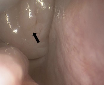
Clinical examination revealed a large lymphoid mass in the post-nasal space (Figure 1), with complete obstruction of both posterior choanae, as well as left-sided otitis media with effusion.
Large soft tissue mass in the nasopharynx, obstructing the right posterior choana (arrow).

The patient's full blood count showed a haemoglobin concentration of 12.5 g/dl, white blood cell count (WBC) of 9.2 × 10⁹/l (neutrophils 3.5 × 10⁹/l and lymphocytes 4.4 × 10⁹/l) and platelet count of 310 × 109/l. His electrolyte concentrations and renal function test results were normal. The C-reactive protein (CRP) concentration was 4 mg/l.
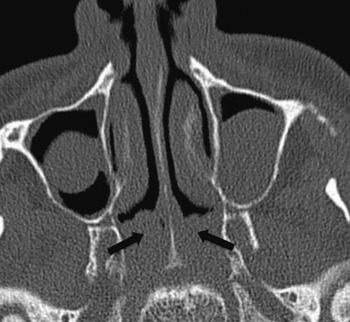
Computed tomography (CT) scanning revealed a soft tissue mass in the nasopharynx, without any abnormality in the sinuses or cranial cavity (Figure 2).
Axial computed tomography scan of the paranasal sinuses, showing a soft tissue mass within the nasopharynx (arrows), abutting the soft palate.

The mass was subsequently biopsied under general anaesthesia and macroscopically cleared by suction diathermy, with insertion of a left tympanostomy tube.
Histological examination revealed reactive lymphoid hyperplasia only.
Four months post-operatively, the patient suffered recurrent nasal congestion. Examination confirmed recurrence of the post-nasal mass, again obstructing both posterior choanae. This time, the full blood count showed a haemoglobin concentration of 11.7 g/dl, WBC of 9.8 × 10⁹/l (neutrophils 3.6 × 10⁹/l and lymphocytes 4.8 × 10⁹/l) and platelet count of 370 × 109/l. The electrolytes and renal function were again normal, while the CRP was 6 mg/l.
Biopsy and complete clearance with suction diathermy were repeated. Histological examination again showed reactive lymphoid hyperplasia.
Four months after the second biopsy, the patient re-presented with similar nasal symptoms due to recurrence of the post-nasal mass. He had no night sweats nor weight loss, and remained extremely well. As the patient was reluctant to undergo further surgery, a three-week course of intranasal and oral steroids (Nasonex® and prednisolone 20 mg daily) was prescribed.
However, this failed to alleviate the patient's symptoms, so he underwent a third operation. The recurrent post-nasal mass was re-biopsied and removed with suction diathermy.
Histological analysis again showed reactive lymphoid hyperplasia, with no suggestion of dysplasia or malignancy. Immunohistochemical staining showed that the lymphocyte population comprised mixed B and T cells, with the T-cell component extensively involving the overlying epithelium, a feature of reactive hyperplasia. CD 21 glycoprotein staining highlighted follicular dendritic cells with residual expanded germinal centres, favouring a diagnosis of reactive lymphoid hyperplasia. Serological tests for cytomegalovirus, human immunodeficiency virus and Epstein–Barr virus, including polymerase chain reaction, were negative.
Coincidentally, just before the third operation, the patient's cyclosporin therapy was stopped by his rheumatologist at another hospital, as his renal function was slightly deranged (as evidenced by the following concentrations: sodium 139 mmol/l, potassium 4.4 mmol/l, urea 12.3 mmol/l and creatinine 132 mmol/l). The patient was also referred for a haematological opinion as his blood film revealed atypical lymphocytosis (haemoglobin concentration 12.6 g/dl, WBC 11.7 × 10⁹/ l (neutrophils 4.3 × 10⁹/l and lymphocytes 6.6 × 10⁹/l) and platelet count 274 × 109/l). The liver function and bone profile were normal, while the CRP was 10 mg/l. A bone marrow biopsy demonstrated several lymphoid aggregates containing mostly reactive lymphocytes. There was a low level increase in plasma cells but these were not light-chain restricted. Serial paraprotein quantitations were normal. A CT chest scan showed a small volume of axillary lymphadenopathy and a small area of consolidation in the right lower zone of the lungs, which on repeated serial scans one, two and three months after cessation of cyclosporin revealed progressive reduction in size to almost complete resolution.
A multidisciplinary team discussion concluded that the patient was likely to be suffering from low-level lymphoproliferative disorder as a result of his prolonged cyclosporin therapy, and reactive change secondary to his psoriatic arthritis.
Three months after his third operation (and five months after cyclosporin cessation), the patient remained asymptomatic, without any clinical evidence of recurrence of the nasopharyngeal mass.
Discussion
The occurrence of lymphoproliferative disorders, mainly linked to Epstein–Barr virus (EBV) infection, is well documented in patients receiving cyclosporin-based immunosuppression. However, cases usually occur following bone marrow or organ transplantation (being termed post-transplant lymphoproliferative disorder) associated with significant immunosuppression involving cyclosporin dosages upwards of 8 mg/kg/day.Reference Leblond, Sutton, Dorent, Davi, Bitker and Gabarre4 Post-transplant lymphoproliferative disorder represents a spectrum of unregulated lymphoid expansion, ranging from polyclonal lymphoid hyperplasia to monoclonal lymphoma; adenotonsillar hypertrophy is the most common presentation in paediatric cardiac transplant patients.Reference Herrman, Sweet, Hayashi, Canter, White and Lieu5 Approximately 80 per cent of post-transplant lymphoproliferative disorder cases are EBV-positive, and commonly involve polymorphic B-cell proliferations.Reference Leblond, Sutton, Dorent, Davi, Bitker and Gabarre4 The aetiology of EBV-negative post-transplant lymphoproliferative disorder remains unknown. Post-transplant lymphoproliferative disorder is extremely rare in non-transplant settings; with cases of PTLD-like disease clustered around immunomodulatory therapy in rheumatoid arthritis.Reference Kamel, Van de Rijn, Weiss, Del Zoppo, Hench and Robbins6
Our patient developed non-EBV-associated lymphoproliferative disease whilst taking low-dose cyclosporin therapy for psoriatic arthritis, presenting initially with recurrent lymphoid hyperplasia in the nasopharynx, a phenomenon not previously described. The overall incidence of lymphoproliferative disorders in patients with psoriasis is extremely low. Psoriatic arthritis affects 10–30 per cent of psoriasis patients. It remains unclear whether the broad immune activation of the underlying disease itself, independent of any therapy administered, is related to an increased risk of developing post-transplant lymphoproliferative disorder like disease.Reference Krupp and Monka1 More aggressive forms of post-transplant lymphoproliferative disorder-like disease, such as non-Hodgkin lymphoma, hairy cell leukaemia and Waldenstrom macroglobulinaemia, have previously been reported in psoriatic patients receiving cyclosporin treatment.Reference Fozza, Dore, Bonfigli, Podda and Longinotti7, Reference Lelievre, Sacre, Adle-Biassette, Molinier-Frenkel, Gaulard and Papo8
In our patient, a seven-year delay was observed between cyclosporin treatment commencement and lymphoproliferation; this suggests that low-dose cyclosporin therapy carries a risk of post-transplant lymphoproliferative disorder like disease, which persists for as long as the immunosuppressive drug is administered. Reversibility and spontaneous regression of lymphoproliferation upon cyclosporin cessation has also been reported in a patient with aggressive EBV-associated lymphoproliferative disease and psoriasis.Reference Lelievre, Sacre, Adle-Biassette, Molinier-Frenkel, Gaulard and Papo8 Classical post-transplant lymphoproliferative disorder usually occurs during the first year of transplantation, correlating with the strength of immunosuppression required to prevent organ rejection. In early post-transplant lymphoproliferative disorder, withdrawal of iatrogenic immunosuppression may lead to clinical and histological improvement, whereas late post-transplant lymphoproliferative disorder tends to resemble lymphoma in immunocompetent patients and does not respond to immunosuppressive therapy dose reduction, ultimately requiring cytotoxic chemotherapy. The reactive lymphoid hyperplasia seen in our patient may correspond to early post-transplant lymphoproliferative disorder like disease, in spite of the extended duration of cyclosporin therapy.
• Lymphoproliferative disorders, mainly linked to Epstein–Barr virus (EBV) infection, are well documented in patients receiving cyclosporin-based immunosuppression
• This paper describes the first reported case of non-EBV-associated lymphoproliferative disease in a patient receiving long-standing cyclosporin therapy for psoriatic arthritis, with the sole initial presentation of a recurrent nasopharyngeal mass
• The otolaryngologist should have a high index of clinical suspicion when repeated biopsies reveal lymphoid hyperplasia, with subsequent aggressive recurrence
In conclusion, otolaryngologists should be aware of this extremely rare complication of cyclosporin therapy in non-transplant patients, especially those with rheumatological disorders such as rheumatoid and psoriatic arthritis, as the sole initial presentation may be a recurrent nasopharyngeal mass. In such patients, repeated biopsies showing lymphoid hyperplasia, together with subsequent aggressive recurrence, should prompt immediate withdrawal of the drug to reduce immunosuppression and promote spontaneous regression. Joint follow up with the haematologists is encouraged, together with careful monitoring for tumour regression after discontinuation of cyclosporin; this should certainly be trialled before cytotoxic chemotherapy is considered.