Recently, it has been shown that leukaemia-initiating cells can be generated in vitro by introducing one molecular hit, such as forced expression of a fusion gene in normal human haematopoietic stem and early progenitor cells. Such fusions have been created from genes encoding acute myeloid leukaemia 1 protein (AML1) and ENL (YEATS-domain-containing protein 1) (Ref. Reference Barabe1) or TEL (transcription factor ETV6) and AML1 (Ref. Reference Hong2). The latency period in vivo from the injection of leukaemia-initiating cells into mice until overt disease is manifest varies, and might reflect a requirement for additional molecular events to cooperate with the initial genetic change for development of disease. Numerous molecular alterations that promote cancer cell initiation and progression have been described, and include mutations in tumour-suppressor genes, oncogenes and DNA-stability genes (Refs Reference Vogelstein and Kinzler3, Reference Garber and Offit4). In addition, the cancer microenvironment might play a crucial role in supporting the selection and propagation of cancer stem cells (Ref. Reference Anderson5). These include increased angiogenesis (Refs Reference Folkman6, Reference Hicklin and Ellis7, Reference Aguayo8), response to ligands secreted by other signalling cells (Refs Reference Sloand9, Reference Liu10) and ineffectively competing nonmalignant cells (Refs Reference Li11, Reference Li12).
Such alterations increase the adaptability of transformed haematopoietic stem cells to the microenvironment and the probability of their selection from the stem cell pool (Refs Reference Gatenby13, Reference Vineis and Berwick14). The potential implications of such mechanisms to the development of leukaemia in inherited bone marrow failure syndromes (IBMFSs) have recently been reviewed (Ref. Reference Bagby and Meyers15). Studies of both human IBMFSs and their animal models suggest that the underlying genetic mutations that cause the marrow failure phenotype also lead either directly or indirectly to the development of cancer (Refs Reference Dror16, Reference Alter17, Reference Houghtaling18, Reference Ruggero19, Reference Putz20). Shwachman–Diamond syndrome (SDS) is one of the most common pre-leukaemic IBMFSs (Refs Reference Steele21, Reference Dror22). Here I describe the general and malignant features of the syndrome and discuss the potential molecular mechanisms that may lead to leukaemia in SDS.
Shwachman–Diamond syndrome
SDS is an autosomal recessive multi-system disorder (Ref. Reference Shwachman23). It usually presents in childhood, and commonly includes a triad of bone marrow failure, pancreatic insufficiency and skeletal abnormalities (Ref. Reference Aggett24). The clinical diagnosis of SDS relies on evidence of bone marrow dysfunction and exocrine pancreatic dysfunction. Other conditions that affect both systems, such as Pearson syndrome, should be excluded. The only gene known to be associated with SDS is SBDS (Shwachman–Bodian–Diamond syndrome, named after the three main investigators who described the disease in 1964), which encodes a ribosome maturation protein. Mutations in SBDS can be identified in 90% of patients with SDS (Ref. Reference Boocock25). The identification of biallelic mutations in the SBDS gene might help to establish a diagnosis in patients with partial clinical phenotype.
Neutropaenia is the most common haematological abnormality that occurs in virtually all patients, followed by reticulocytopaenic anaemia and thrombocytopaenia. Trilineage cytopaenias occur in 10–65% of patients. Severe aplastic pancytopaenia has occasionally been reported (Refs Reference Aggett24, Reference Tsai26, Reference Barrios27). Various degrees of abnormality in B cells and T cells, as well as natural killer cells have also been reported (Refs Reference Dror28, Reference Hudson and Aldor29, Reference Aggett30). Bone marrow biopsy usually shows a hypoplastic specimen (Refs Reference Aggett24, Reference Dror and Freedman31). Mild dysplastic changes in the erythroid, myeloid and megakaryocytic precursors are commonly seen and can fluctuate; however, prominent multilineage dysplasia is less common, and if it occurs, might signify malignant myeloid transformation.
Malignant myeloid transformation
SDS is associated with a high propensity for malignant myeloid transformation into myelodysplasia syndrome and leukaemia (Refs Reference Aggett24, Reference Mack32, Reference Smith33, Reference Dror34, Reference Dror35). Thus far, an excess of solid tumours has not been reported in SDS. A previous literature review identified 54 reported cases of SDS with clonal marrow cytogenetic abnormalities, malignant myeloid transformation or acute myeloid leukaemia (AML) (Ref. Reference Dror22). Among these groups, 69% were reported to have clonal marrow cytogenetic abnormalities or myelodysplasia syndrome at a median age of 8 years (range, 2–42 years). The rest of the patients with malignant myeloid transformation (31%) were diagnosed with overt leukaemia at a median age of 14 years (range, 1.5–37 years).
About 75% of the SDS patients who developed malignant myeloid transformation or AML were reported to have clonal marrow cytogenetic abnormalities. The most common of these in SDS are i(7q) (44%) and del(Reference Li12)(q12) (16%). The i(7q) insertion occurs rarely in other malignancies, and has not been reported in other IBMFSs. Among the patients with isolated i(7q), no progression into myelodysplasia syndrome with excess blasts or AML has been reported. However, i(7q) together with additional cytogenetic abnormalities, has been associated with AML in some SDS patients. (Ref. Reference Dror22). Since isolated i(7q) is not associated with a risk of progression and can sometimes regress spontaneously (Ref. Reference Dror35), it might not involve alterations in cancer-related genes. It is also possible that the 7q10-11 site, where i(7q) is formed, is a fragile site caused by the mutated SBDS and/or its pseudogene SBDSP. Among the patients with cytogenetic abnormalities other than i(7q), 42% either initially presented with or progressed to advanced myelodysplasia syndrome or AML from earlier stages of myelodysplasia syndrome.
Various morphological types of myelodysplasia syndrome have been described in SDS. Approximately one third of the patients with SDS or myelodysplasia syndrome have been reported to have clonal marrow cytogenetic abnormalities without prominent marrow dysplasia and without an increase in blasts (Refs Reference Dror22, Reference Mandel36). Around one third of these patients progressed to more advanced myelodysplasia syndrome or AML. About 60% of SDS or myelodysplasia syndrome cases are diagnosed at the stage of refractory cytopaenia with dysplasia (Ref. Reference Dror22), which is associated with a high risk of transformation into AML. One case of refractory cytopaenia with ring sideroblasts has been reported in SDS; this patient eventually progressed to AML (Ref. Reference Huijgens37). SDS patients who were diagnosed with myelodysplasia syndrome at the stage of refractory cytopaenia with excess blasts have been reported (Refs Reference Smith33, Reference Okcu, Roberts and Chan38, Reference Passmore39, Reference Hsu40), and these patients also had a high rate of progression to AML.
The risk of AML increases with age, and is as high as 36% by 30 years of age (Ref. Reference Hicklin and Ellis7), with a reported median age of onset of 14 years (range 1.5–43 years) (Ref. Reference Dror22). Various types of AML have been described in SDS patients: AML-M0, -M2, -M4, -M5 and -M6. Acute lymphoblastic leukaemia is rare. It is noteworthy that AML-M6 is particularly common in SDS, occurring in about 30% of cases with classifiable leukaemia (Ref. Reference Dror22). SDS-related leukaemia carries a poor prognosis, and over 80% of the patients reported died, because of either refractory disease or treatment-related toxicity (Ref. Reference Dror22).
SBDS gene function
SBDS was first identified by Lai and colleagues (Ref. Reference Lai41) in 2000, and was found to be mutated in about 90% of patients with a clinical diagnosis of SDS (Ref. Reference Boocock25). The functional domains of the SBDS protein are unknown, but the orthologue in archaebacteria has three main domains (N-terminal, middle and C-terminal) with predicted protein–protein, protein–DNA and protein–RNA binding motifs (Ref. Reference Shammas42). There are two common mutations: 183–184TA→CT (nonsense mutation) and 258 + 2T→C (intronic mutation; predicted to result in alternative splicing and frameshift reading). Other rare mutations include missense, nonsense, frameshift and splice-site mutations, and complex deletions/insertions (Ref. Reference Boocock25). The mutations are mostly in the N-terminal domain of the protein and are expressed at markedly reduced levels. The SBDS protein is essential because no patients with homozygous-null mutations have been reported, and residual protein levels can usually be detected in SDS patients (Refs Reference Woloszynek43, Reference Austin, Leary and Shimamura44). Furthermore, a complete loss of the protein in mice causes developmental arrest before embryonic day 6.5 and early lethality (Ref. Reference Zhang45). SBDS seems to be multifunctional and has a role in several cellular pathways, including ribosomal biogenesis, cell survival, chemotaxis and mitotic spindle formation.
It has recently been found that SBDS concentrates in the nucleolus during G1 and G2 (Ref. Reference Austin, Leary and Shimamura44), and is associated with rRNA (Ref. Reference Ganapathi46). Synthetic genetic arrays of YHR087W, a yeast homologue of the N-terminal domain of SBDS, suggested interactions with several genes involved in RNA and rRNA processing (Ref. Reference Savchenko47). The loss of the protein in yeast results in a defect in maturation of the 60S ribosomal subunit (Ref. Reference Menne48).
Bone marrow cells from patients with SDS are characterised by decreased frequency of CD34+ progenitors (Ref. Reference Dror and Freedman31), and a reduced ability to generate haematopoietic colonies of all lineages in vitro (Ref. Reference Dror and Freedman31). Marrow cells, as well as SBDS-knockdown HeLa cells, are characterised by accelerated apoptosis (Refs Reference Dror and Freedman49, Reference Rujkijyanont50), which seems to occur through the Fas pathway rather than through the Bax–Bcl-2–Bcl-XL pathway (Refs Reference Dror and Freedman49, Reference Rujkijyanont50).
SDS patients have a defect in leukocyte chemotaxis (Refs Reference Dror28, Reference Stepanovic51). Consistent with this observation, the SBDS homologue in amoeba was found to localise to the pseudopods during chemotaxis (Ref. Reference Wessels52). These observations suggest that the SBDS deficiency in SDS causes the chemotaxis defects observed in patients. SBDS has also been shown to colocalise to the mitotic spindle, where it binds and stabilises microtubules (Ref. Reference Austin53); its deficiency results in centrosomal amplification and multipolar spindles. The potential relationships between loss of these functions and development of malignant myeloid transformation are discussed in the following section.
Mechanisms of leukaemia in Shwachman–Diamond syndrome
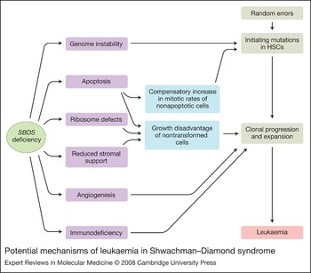
In many patients with SDS, a myelodysplasia phase can be identified before the appearance of leukaemia (Refs Reference Dror22, Reference Smith33). Since only a portion of patients with SDS develop leukaemia, it is reasonable to postulate that SDS haematopoietic cells acquire additional molecular events before malignant myeloid transformation is apparent, and these events promote the initiation or progression of malignant clones. Such a premise is congruent with the multi-hit hypothesis of cancer development. We and others have observed that bone marrow from SDS patients without morphological evidence of myelodysplasia demonstrate several characteristic features of myelodysplasia syndrome (Ref. Reference Dror35). Similarly to other IBMFSs, the initial molecular events involved in the transformation, as well as the subsequent events that allow propagation of the malignant clones, are unclear. Several potential risk factors for malignant myeloid transformation in SDS will be considered and the potential internal and external defects in the SDS bone marrow that might promote malignant myeloid transformation are discussed (Fig. 1).
Potential mechanisms of leukaemia in Shwachman–Diamond syndrome. Abbreviation: HSC, haematopoietic stem cell.

Risk factors
Of the patients with clonal marrow cytogenetic abnormalities or myelodysplasia syndrome, 68% were males; of those with leukaemia, 92% were males. Predominance of males with myelodysplasia syndrome or AML has also been described in other marrow-failure syndromes, such as severe congenital neutropaenia (Ref. Reference Rosenberg54), but not with the same proportions as observed in SDS. The cause for the male predominance is unknown, but could be related to reporting bias or to the existence of unknown protective genes on the X chromosome or cancer-promoting genes on the Y chromosome.
The influence of genotype on the risk of leukaemia in SDS is difficult to study because most of the patients have the same genotype; double heterozygosity for 183–184TA → CT/258 + 2T → C (Ref. Reference Boocock25). However, mild and severe phenotypes have been found in patients with the common mutations, and leukaemia is not confined to patients with SDS and rare SBDS genotypes (Ref. Reference Kuijpers55).
The role of granulocyte colony-stimulating factor (G-CSF) administration in promoting malignant myeloid transformation in bone marrow failure syndromes is controversial. The development of myelodysplasia syndrome and AML in SDS patients who are receiving G-CSF therapy has been reported (Refs Reference Davies56, Reference Freedman57). However, most SDS patients who develop myelodysplasia syndrome or leukaemia had not received treatment with G-CSF. Therefore, the exact role of the endogenous or therapeutically administered cytokine in leukaemic conversion in SDS is still unknown.
Predisposition at the haematopoietic stem cell level
SBDS protects cells from developing malignant myeloid transformation in a manner that is different from typical tumour suppressor genes. Development of malignant myeloid transformation in SDS does not follow the Knudson ‘two-hit’ hypothesis, and haploinsufficiency (e.g. obligate carriers in the family) has not been reported to be associated with SDS (Ref. Reference Majeed58). It is likely that a second mutation on a background of SBDS deficiency initiates the malignant clone; however, little is known about alterations of specific genes in transforming SDS cells. We identified no mutations in P53 or RAS in marrow cells from nine patients without malignant myeloid transformation and two with clonal marrow cytogenetic abnormalities (Ref. Reference Dror35).
Genomic instability
The data on chromosomal instability in SDS are inconclusive. Increased frequencies of spontaneous chromosomal aberrations in patient phytohaemagglutinin-stimulated circulating lymphocytes were demonstrated by several groups (Refs Reference Hershkovits, Dagan and Freier59, Reference Tada60), but not by others (Refs Reference Fraccaro, Scappaticci and Arico61, Reference Koiffmann62). Interestingly, transient or intermittent detection of chromosomal aberrations in SDS bone marrow cells has been reported (Refs Reference Dror35, Reference Maserati63). This might suggest that loss of SBDS leads to chromosomal instability. The recent observation that SBDS localises to the mitotic spindle (Ref. Reference Austin53) and that its deficiency results in centrosomal amplification and multipolar spindles, raises the possibility that genomic instability leads to chromosomal and molecular abnormalities, which activate oncogenes or inhibit of tumour-suppressor genes thereby bypassing the apoptotic machinery that usually eliminates such abnormal cells.
Hematopoietic cells from SDS patients have shorter mean telomere length than controls (Refs Reference Thornley64, Reference Calado65) and excessive telomere shortening results in unprotected chromosome ends, activation of DNA repair machinery (Refs Reference Hao, Strong and Greider66, Reference Counter67) and chromosome fusions (Ref. Reference Smogorzewska68). This can, in turn, result in chromosome breakage at the next cell division, further exacerbating the instability of the chromosomes. Genome instability caused by telomere shortening usually leads to cell death or crisis. However, several haematopoietic stem cells could escape from crisis through reactivation of a telomere-maintenance mechanism, such as telomerase. Genome instability before crisis might allow cells to accumulate mutations that contribute to leukaemogenesis (Ref. Reference Londono-Vallejo69). The cause of telomere attrition in SDS is unknown. It might be directly related to a loss of SBDS function in telomere maintenance or reflect a compensatory increase in cell division of nonapoptotic haematopoietic stem cells.
Ribosome biogenesis
Several IBMFSs with defects in ribosome biogenesis manifest an increased cancer risk (Refs Reference Dokal70, Reference Vlachos, Klein and Lipton71, Reference Taskinen72). Defects in ribosome biogenesis are known to be associated with malignant transformation (Refs Reference Ruggero19, Reference Ebert73). Haploinsufficiency of many small and large ribosomal proteins confers a risk of cancer in zebrafish (Ref. Reference Amsterdam74) and abnormal ribosome biogenesis as seen in SDS usually leads to translation insufficiency. However, cancers such as AML are typically characterised by upregulation of ribosome activity, an increased number and size of the nucleoli and an increase in protein synthesis. Therefore, if defects in ribosome biogenesis are related to the development of leukaemia in SDS, the leukaemia is unlikely to be directly due to translation insufficiency. In SDS, the ribosome defects are observed not only in rRNA, but also at the protein level. We have found that SDS marrow cells are characterised by downregulation of many ribosomal genes (Ref. Reference Rujkijyanont75). By contrast, cancer cell lines and some primary tumours are characterised by overexpression of these genes (Refs Reference Ferrari76, Reference Loging and Reisman77, Reference Kondoh78). These observations suggest that if predisposition to cancer in SDS is related to a defect in ribosome biogenesis, it might be due to an imbalance in either the rRNA subunits or ribosomal proteins or to a growth disadvantage of nonmalignant cells, which allows expansion of the transforming cells (see below).
Leukaemia-related gene expression
Leukaemic clones are initiated or progress because of genomic alterations and deregulation of gene expression. Using oligonucleotide microarray analysis of bone marrow mononuclear cells, we have found significant upregulation of several oncogenes, including LARG, TAL1 and MLL, and downregulation of several tumour-suppressor genes, including RUNX1, FANCD2 and DKC1, among 154 known leukaemia-related genes (Ref. Reference Rujkijyanont75). These abnormal expression patterns might result in continuous stimulation favouring evolution or progression of malignant clones.
The role of the microenvironment and neighbouring cells
Increased apoptosis of neighbouring cells
SDS bone marrow cells and SBDS-knockdown cells are characterised by accelerated apoptosis, which is characteristic of many preleukaemic IBMFSs (Refs Reference Li11, Reference Carlsson79). Two hypotheses might explain increased clonal evolution among a population of apoptotic cells. In the first model, accelerated apoptosis can lead to compensatory mechanisms through increased secretion of growth factors (Refs Reference Mempel80, Reference Ballmaier81), which leads to hyperproliferation of nonapoptotic cells. This compensatory mechanism was traditionally referred to as ‘stress haematopoiesis’. The high percentage of cycling cells might lead to statistically more frequent mutations in the patient, some of which might be in leukaemia-related genes. The second model for the development of leukaemia among an apoptotic cell population is described below.
Bone marrow stromal defects and accelerated angiogenesis
The bone marrow stroma might promote clonal evolution by either supporting leukaemic cell growth or by not supporting the growth of nonleukaemic cells. Similarly to results described in myelodysplasia syndrome (Ref. Reference Aizawa82), we have shown that the SDS stroma has a decreased ability to support haematopoiesis from normal CD34+ cells in long-term marrow stromal cultures (Ref. Reference Dror and Freedman31). This abnormality may provide growth disadvantage to the nontransforming cells over the leukaemic cells. The ability of the SDS stroma to support leukaemic cell growth has not been studied; however, we have found evidence of abnormally increased angiogenesis in bone marrow biopsies from SDS patients (Ref. Reference Leung83). Angiogenesis can be stimulated by many tumours, and promotes cancer cell growth (Refs Reference Folkman6, Reference Gasparini84). Bone marrow angiogenesis is also accelerated in haematological malignancies, including myelodysplasia syndrome (Ref. Reference Aguayo8), for which antiangiogenic factors have been shown to be an effective therapy (Refs Reference List85, Reference Pellagatti86).
Increased apoptosis of non-transformed cells results in a growth advantage
Recently, a new model for the development of leukaemia in an apoptotic cell population was developed and was also proposed to apply to IBMFSs (Refs Reference Li11, Reference Li12, Reference Bagby and Meyers15). In this model, apoptosis results in growth disadvantage of the nontransforming cells. Mutation may occur either at a ‘normal’ or increased frequency because of genomic instability. The mutations might improve the adaptation of the transforming cells, and render them resistant to factors in the microenvironment that continue to injure the surrounding nontransforming cells. The somatically mutated haematopoietic stem cells thus have a high selection coefficient. In Fanconi anaemia, the new haematopoietic stem cell clones escape cell death by destroying apoptotic responses to TNF-α, whereas the nontransforming cells are hypersensitive to these environmental cues (Ref. Reference Li12). In patients with severe congenital neutropaenias, neoplastic haematopoietic stem cells might emerge as a result of a G-CSF receptor mutation, which confers hypersensitivity to G-CSF, whereas the nontransforming haematopoietic stem cells have a lower response to the ligand (Ref. Reference Liu10). Therefore, the secondary mutations in IBMFS haematopoietic stem cells might result in aberrant activation of an antiapoptotic program, resulting in selective survival of the transformed cells and accumulation of additional mutations until leukaemia develops. In SDS, nontransforming cells are hypersensitive to Fas ligand. It is possible that the transforming SDS cells acquire resistance to apoptosis stimulus from the Fas ligand, thereby acquiring a growth advantage over the surrounding cells.
Immunodeficiency
Mild to moderate deficiencies in T cells, B cells and natural killer cells have also been described in SDS (Refs Reference Dror and Freedman49, Reference Kornfeld87). Natural killer cells are thought to have an important role in removing cancer cells. Deficiencies in these lymphocytes have been shown to be associated with an increased risk of cancer (Refs Reference Pinkerton88, Reference Guerra89). However, in contrast to what is seen in SDS, the malignancies in these disorders are of B cell or T cell origin.
Summary
Several changes that can contribute to malignant myeloid transformation have been reported in SDS. Future studies need to determine which of these abnormalities are necessary for malignant myeloid transformation and at which stage. Importantly, the changes described above can synergise to promote the initiation and progression of the malignant clones. It could be that one of these abnormalities or an undiscovered one(s) is sufficient to transform the haematopoietic stem cell and promote its progression to leukaemia. Correction of one abnormality at a time (e.g. by inhibiting angiogenesis) in animal models might help to decipher its relative contribution to malignant myeloid transformation in SDS. Research revealing how this transformation occurs in SDS is likely to uncover novel mechanisms of cancer, which may be relevant to similar processes in other inherited or acquired types of cancer.
Acknowledgements
I thank Dr David Malkin for critical review of the manuscript and also the anonymous reviewers for their comments.