As we argued recently (Depue & Lenzenweger, Reference Depue, Lenzenweger, Lenzenweger and Clarkin2005), with the exception of schizotypal and paranoid disorders, it is no longer possible to accept the notion that personality disorders (PDs) represent distinct, categorical diagnostic entities. The behavioral features of PDs are not organized into discrete diagnostic entities, and multivariate studies of behavioral criteria fail to identify factors that resemble existing diagnostic constructs (Livesley, Reference Livesley2001). Indeed, just the opposite is observed: the behavioral features of PDs merge imperceptibly in a continous fashion across diagnostic categories, resulting in (a) significant overlap of behavioral features across categories and hence diagnostic comorbidity within individuals, and (b) symptom heterogeneity within categories, and hence frequent (and most common in some studies) application of the ambiguous diagnosis of PDs not otherwise specified (Livesley, Reference Livesley2001; Saulsman & Page, Reference Saulsman and Page2004). Moreover, aside from schizotypy/schizotypic disorders, the existing latent class and taxometric analysis literature on PDs generally does not provide support for distinct entities in the PD realm, and some provisional taxonic findings for borderline and antisocial PDs are open to doubt. It is, accordingly, not surprising that diagnostic membership is not significantly associated with predictive validity as to prognosis nor psychological or pharmacological treatments (Livesley, Reference Livesley2001).
Such a state of affairs supports proponents of a dimensional approach to PDs, who note that (a) the behavioral features of PDs not only overlap diagnostic categories, but also merge imperceptibly with normality (Livesley, Reference Livesley2001; Saulsman & Page, Reference Saulsman and Page2004); and (b) the factorial structure of behavioral traits associated with PDs is similar in clinical and nonclinical samples (Reynolds & Clark, Reference Reynolds and Clark2001). Furthermore, the higher order structure of PD traits resembles four of the five major traits identified in the higher order structure of normal personality (Reynolds & Clark, Reference Reynolds and Clark2001). These findings suggest that PDs may be better understood as emerging at the extremes of personality dimensions that define the structure of behavior in the normal population (Depue & Lenzenweger, Reference Depue, Lenzenweger, Lenzenweger and Clarkin2005).
This realization has led to innumerable attempts to illustrate the association of personality traits to PD diagnostic categores. Most of these studies have relied on the five-factor model of personality, which defines a structure characterized by the higher order traits of extraversion, neuroticism, agreeableness, conscientiousness, and openness to experience. A recent meta-analysis demonstrated the limitation of the approach of correlating the first four traits with PDs (openness was uncorrelated with any category; Saulsman & Page, Reference Saulsman and Page2004). The meta-analysis showed (a) the correlation of these four traits with PD categories is moderate to weak, (b) the complex “entity” of a PD category is defined by as little as one trait but never by four traits in a significant way, and (c) single traits (especially neuroticism) characterize more than one, sometimes several, putatively distinct PDs. How helpful is it to know that histrionic PD is characterized by moderately high extraversion but by no other trait or that dependent PD is associated with moderately high neuroticism but with no other trait. Furthermore, the problem in this approach does not appear to be explained simply by use of a small number of broad traits. When the 30 facet scales of the NEO-personality inventory (PI) were correlated with PD categories, only modest gains were achieved relative to the use of the four major traits (mean difference in R 2 ~ 0.04; Reynolds & Clark, Reference Reynolds and Clark2001). What the meta-analysis did reveal is that most PDs manifest, in common, higher trait levels of neuroticism and lower trait levels of agreeableness, meaning that most individuals with PDs are subject to negative emotionality, heightened stress reactivity, and impaired interpersonal behavior. Thus, when PD categories serve as the outcome variable, personality traits provide little in the way of power to discriminate between such categories or to inform the issue of dimensional continuity from personality traits to states of disorder.
On the basis of the above discussion, we have argued that PDs as disease entities do not exist (Depue & Lenzenweger, Reference Depue, Lenzenweger, Lenzenweger and Clarkin2005). As an alternative, we proposed a new perspective, where personality disturbance represents emergent phenotypes that arise from the interaction of extreme values on critical subsets of major personality traits. That is, each individual's personality represents the interaction of all major traits. Where individuals lie at the extreme (low or high) ends of interacting trait dimensions on subsets of the major traits, emotional behavior emerges that generates social and/or occupational impairment, rendering disturbance in the individual's life resulting from personality factors. This model follows from the position that higher order traits of personality reflect the activities of neurobehavioral systems (Depue, Reference Depue and Canli2006; Depue & Collins, Reference Depue and Collins1999; Depue & Morrone-Strupinsky, Reference Depue and Morrone-Strupinsky2005). Although genetically speaking it may be possible to conceive of the neurobiological variables associated with neurobehavioral systems as subject to independent influences, it is impossible to imagine that these systems are independent at a functional level. Neurobehavioral systems, and the personality traits that reflect their influence, interact to produce complex behavior patterns (personality as a whole) in a multivariate fashion. Although this model is dimensional in nature, where personality disturbance lies at the extreme of normal, interacting personality dimensions, no assumption is made herein that phenotypic dimensions of personality are genetically continuous. The phenotypic continuity could well represent several underlying distinct genotypic distributions, as may be the case even within the normal range of variation of some personality traits (Depue, Reference Depue and Canli2006).
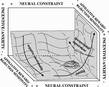
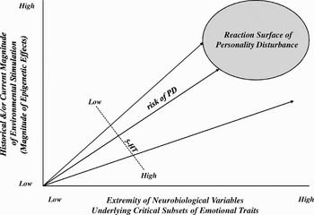
This multidimensional model of personality disturbance is illustrated in Figure 1. In the model, the axes are defined by neurobehavoral systems (which we will explicate later) rather than by personality traits, because traits are only approximate, fallible estimates of these systems. In Figure 1, behavioral approach based on positive incentive motivation (thought to underlie extraversion and positive emotionality; Depue & Collins, Reference Depue and Collins1999) and anxiety/stress reactivity (thought to underlie neuroticism and negative emotionality, Canli, Reference Canli2008; Depue & Lenzenweger, Reference Depue, Lenzenweger, Lenzenweger and Clarkin2005) are modeled as a ratio of their relative strength, because the opposing nature of their eliciting stimuli affects behavior in a reciprocal manner, such that the elicitation or expression of one system is influenced by the strength of the other. The affiliative reward dimension (representing the capacity to experience consummatory reward from social and soft-touch stimulation, and thought to underlie the trait of social closeness or agreeableness; Depue & Morrone-Stupinsky, Reference Depue and Morrone-Strupinsky2005) is placed in ratio with rejection sensitivity in the model, because they both largely influence the interpersonal domain, but can have opposite effects on social bonding. Moreover, a dimension of rejection sensitivity likely provides a novel contribution to dependent-, avoidant-, and borderline-like disturbance (all high on this dimension). The neural constraint dimension (thought to underlie the trait of constraint or conscientiousness) modulates the expressive features (especially, the threshold of stimulus elicitation) of the other systems (Carver & Miller, Reference Carver and Miller2006; Depue & Lenzenweger, Reference Depue, Lenzenweger, Lenzenweger and Clarkin2005; Spoont, Reference Spoont1992). Because of the limitation of graphing more than three dimensions, it should be noted that the model includes one additional system/trait that we suggest influences the manifestation of personality disturbance: the ratio of affective aggression to fear is necessary to account for the full range of antisocial (high aggression to low fear) behavior. Taken together, then, the model proposes that the interaction of at least six neurobehavioral systems underlying dimensional personality traits are necessary to account for the emergent phenotypes observed in personality disturbance.
A multidimensional model of personality disturbance. See text for details.

Two significant features of the model are worth emphasizing. First, the phenotypic expression of personality disturbance, represented by the gray-shaded reaction surface in the figure (accounting for ~10% of the population; Livesley, Reference Livesley2001), is continuous in nature, changing in character gradually but seamlessly across the surface in a manner that reflects the changing product of the mulitdimensional interactions. It may be that certain areas of the surface (i.e., certain system/trait interactions) are associated with increased probability (risk) of certain features of personality disturbance (perhaps having led to clinical theories of PDs based on prototypic phenotypes), and this is represented in the right-back area of the figure as an elevation in the surface that is continous rather than distinct in contour. This representation, however, is not meant to imply that a distinct disorder exists in that particular area of the surface.
Second, the extent and positioning of the gray reaction surface is weighted most heavily by increasing anxiety/stress reactivity, decreasing neural constraint, and decreasing affiliative reward to increasing rejection sensitivity. This weighting is also reflected in the lines with directional arrows overlaying the gray surface in the figure, which illustrate increasing phenotypic negative emotionality, emotional lability, and interpersonal impairment. These three factors were found to be the most common characteristics of all PDs in the meta-analysis of Saulsman and Page (Reference Saulsman and Page2004), and a recent study showed independent genetic contributions to these factors (Kendler et al., Reference Kendler, Aggen, Czajkowski, Røysamb, Tambs and Torgersen2008), thereby empirically supporting their emphasis in the model.
Implications of the model are discussed in detail elsewhere (Depue & Lenzenweger, Reference Depue, Lenzenweger, Lenzenweger and Clarkin2005), but one seems important in the consideration of the development of personality disturbance: sex differences in some forms of personality disturbance may imply differential risk (neurobiologic and/or environmental) based on gender. The increased prevalence of males with antisocial behavior may reflect the higher mean of males in the population on the ratio of traits of aggression to fear and a lower mean on constraint (Spoont, Reference Spoont1992). In addition, the increased prevalence of females with borderline- and dependent-like personality disturbance may reflect a combination of (a) their higher mean on both trait anxiety; (b) their higher mean on social closeness, thereby increasing the need for social relationships whose loss is feared; (c) their higher mean on rejection sensitivity, thereby increasing their vacillation between dependence and rejection of others; (d) their lower mean on extraversion, particularly social dominance, hence enhancing the predominance of high levels of trait anxiety; and (e) their lower mean on the ratio of aggression to fear, hence decreasing the prevalence of antisocial behavior (Depue & Collins, Reference Depue and Collins1999; Depue & Morrone-Strupinsky, Reference Depue and Morrone-Strupinsky2005; Tellegen & Waller, Reference Tellegen, Waller, Briggs and Cheek1997).
Developmental Implications of the Model
Viewing the phenotypic expression of personality disturbance within the concept of a reaction surface in Figure 1 implies that an individual's expression of disturbance is variable across time, waxing and waning with fluctuations in environmental circumstances, stressors, and interpersonal disruptions. Indeed, Lenzenweger demonstrated in a longitudinal assessment of the stability of formally defined PDs that phenotypic intensity varied markedly over time, thereby affecting the presence/absence of diagnostic status as well as level of symptomatology (Lenzenweger, Johnson, & Willett, Reference Lenzenweger, Johnson and Willett2004). This implies that one's multivariate value on the reaction surface represents a risk or potential for personality disturbance whose full expression is dependent on current life circumstances. Furthermore, that risk or potential is accordingly variable over time as well. An individual's mean level on personality traits is a function of multiple genetic and environmental variables, in keeping with recent models of the development of psychopathology (Cicchetti, Reference Cicchetti2007), and the interaction of these variables over the life span may modify the extremity of an individual's level in enduring ways. In this way, developmental experience can substantially modify an individual's level on personality traits, and may contribute to an individual's position with respect to the reaction surface.
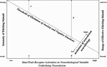
In our subsequent discussion of these experiential effects on the neurobehavioral systems underlying personality traits, it is helpful to first define a framework of how experiential factors interact with neurobiological variables to determine personality trait levels. In so doing, I will use neuroticism, which is defined as anxiety, stress reactivity, and negative emotionality, as an example. As shown in Figure 2, an individual's trait level of expressed anxiety and negative emotion is dependent on the individual's characteristic minimum threshold of stimulus elicitation of a neurobiological variable associated with anxiety/stress reactivity. This minimum threshold represents a central nervous system (CNS) weighting of the external and internal factors that contribute to initiation of the processes associated with anxiety (Depue & Collins, Reference Depue and Collins1999). Generally, the threshold is weighted most strongly by the joint function of two main variables: (a) magnitude of anxiety-inducing stimulation, and (b) level of postsynaptic receptor activation by the neurotransmitters or neuropeptides that mediate the effects of the stimulus in facilitating anxiety. The relation between these two variables is represented in Figure 2 as a trade-off function, where pairs of values (of stimulus magnitude and receptor activation) specify a diagonal representing the minimum threshold value for activation of a behavioral or emotional process; in this case, anxiety and negative emotions. Because the two input variables are interactive, independent variation in either one not only modifies the probability of behavior, but it also simultaneously modifies the value of the other variable that is required to reach a minimum threshold. Finally, individual variation in variables that significantly interact with the functioning of the anxiety-facilitating neurotransmitter would serve as a source of modulation of the threshold. We discuss this further under the section on constraint.
A minimum threshold for elicitation of anxiety and negative emotions is illustrated as a trade-off function between eliciting stimulus magnitude (left vertical axis) and postsynaptic receptor activation of a neurobiological variable underlying the personality trait of neuroticism (horizontal axis). Range of effective (eliciting) stimuli is illustrated on the right vertical axis as a function of level of receptor activation. Two hypothetical individuals with low and high trait postsynaptic receptor activation (demarcated on the horizontal axis as A and B, respectively) are shown to have narrow (A) and broad (B) ranges of effective stimuli, respectively. Threshold effects due to a modulator of neuroticism neurobiology are illustrated as well.

A trait dimension of postsynaptic receptor activation by an anxiety-facilitating neurobiological variable is represented on the horizontal axis of Figure 2, where two individuals with divergent trait levels are demarcated: A (low trait level) and B (high trait level). Trait levels would be determined by genetic factors that affect the functional activity of the neurobiological variable, as well as by developmental experiences that modify gene expression (e.g., by affecting receptor availability). These two divergent individuals may be used to illustrate the effects of trait differences in both acquisition and maintenance of anxiety, stress reactivity, and negative emotionality. As Figure 2 indicates, for any given stimulus, the degree of state (contemporaneous, stimulus-induced) neural response (receptor activation) will, on average, be larger in individual B versus A. Our proposal is that this differential responsivity is a major source of the neurobiological basis of the temperamental (i.e., nonexperiential) component of anxiety and, hence, of variation in the acquisition of stimulus control and levels of anxiety. Because the degree of state neural activity is correlated with the magnitude of subjective emotional experiences that are naturally elicited by stimuli, negative emotionality is also predicted to be more enhanced in B versus A.
This difference between individuals A and B in magnitude of stimulus-induced neural activation and accompanying subjective emotional experience may contribute to two important sources of variation in the development of anxious, stress reactive behavior. First, as noted above, independent variation in either of the two interactive variables that determine threshold (neural activation and magnitude of the stimulus) also simultaneously modifies the value of the other variable that is required to reach a minimum threshold. This suggests that the enhanced negative emotion induced by stimuli in individual B will result in an equally enhanced negative encoding in memory of that anxiety-inducing stimulus relative to individual A.
Second, if individual differences in emotional encoding apply across the full range of magnitudes of stimuli, trait differences in neural activation may have marked effects on the range of effective (i.e., anxiety-inducing) stimuli. This is illustrated in Figure 2, where the right vertical axis represents the range of effective stimuli. Increasing trait levels of neural activation (horizontal axis) are associated with an increasing efficacy of weaker stimuli and, thus, with an increasing range of effective stimuli. In Figure 2 individuals A and B are shown to have a narrow versus broad range, respectively. It is significant that the broader range for individual B suggests that on average B will experience more frequent stimulus elicitation of anxiety, stress, and negative emotions. This means that the probability at any point in time of being in an anxious, stressed state for individual B is higher than for A. Therefore, when subsequent stimuli are encountered, their subjectively evaluated emotional magnitude will show a stronger negative bias for B than A. Thus, trait differences in thresholds may proactively influence the evaluation and encoding of stimuli, and may not be restricted to reactive emotional processes.
Such a perceptual/attentional bias for negative, anxiety-eliciting cues may be mediated by the basolateral nuclei of the amygdala. Crude representations of anxiety-associated cues can reach the basolateral nuclei via monosynaptic projections from the thalamus within ~10 ms without awareness. This input then activates a dense network of backprojections from the basolateral nuclei to many regions of the brain, including those that process sensory representations, thereby facilitating the processing specifically of anxiety-producing cues. The enhanced negative encoding in memory in the basolateral amygdala of such cues by individual B throughout development would lead to frequent, rapid activation of the amygdala when such cues were reexperienced, and in turn, a perceptual/attentional biasing of anxiety cues of even weak magnitude via the amygdala's backprojections.
The overall manifestation of these various processes is likely to be more frequent negative emotional experience for individual B relative to A. A broader range of effective, anxiety-producing stimuli, each with enhanced negative emotional encoding, is expected to produce more robust neural reactivity to stimuli in individual B. Accordingly, an enhanced acquisition of a broader array of conditioned stimuli that become associated with anxiety-producing contexts can be expected for individual B. Thus, variation in neural activation by stimuli may not only influence the level of negative emotion, but also may lead to variation in the strength of associative processes that link neutral stimuli with negative emotional contexts. The outcome of these interactions may be the acquisition of a more elaborate associative network of negative emotional environmental contexts in individual B, leading to different developmental trajectories in anxiety and stress reactivity for individuals A and B.
The following discussion briefly reviews the neurobehavioral systems that underlie the major personality traits and focuses more substantially on the evidence that they are subject to experience-dependent modification that can be enduring through the effects of experience on genetic expression, mainly through processes known as epigenetics. It is through such processes that risk for personality disturbance may be modified by experience at any point in development, but perhaps especially during early critical periods of development. Because this research has been inordinately focused on anxiety and stress reactivity, the neuroticism section will be most extensive relative to other traits. This seems appropriate, however, because the trait of neuroticism is the strongest associate of most PDs.
Neurobehavioral Systems Underlying Higher Order Personality Traits and Their Modification by Experience
We focus on the five major traits in Figure 1 that are robustly identified in the psychometric literature: neuroticism, extraversion, social closeness or agreeableness, rejection sensitivity, and constraint or conscientiousness. Coherent neurobehavioral systems for each of these traits is defined below, and evidence indicating that they are significantly modified by experience is provided.
Neuroticism
Fear versus anxiety
Adaptation to aversive environmental conditions is crucial for species survival, and at least two distinct behavioral systems have evolved to promote such adaptation. One system is fear (often labeled harm avoidance in the trait literature), which is a specific behavioral system that evolved as a means of escaping unconditioned aversive stimuli that are inherently dangerous to survival, such as tactile pain, injury contexts, snakes, spiders, heights, and sudden sounds. These stimuli are specific, discrete, and explicit, and in turn elicit specific, short-latency, high-magnitude phasic responses of autonomic arousal, subjective feelings of panic, and behavioral escape. Specific, discrete neutral stimuli associated with these unconditioned events elicit conditioned fear.
There are, however, many situations in which specific aversive cues do not exist, and instead, the stimulus conditions are associated with an elevated potential risk of danger or aversive consequences. In such cases, no explicit aversive stimuli are present to inherently activate fear circuitries. Nevertheless, the stimuli can be unconditioned in nature, as in darkness, open spaces, unfamiliarity, strangers, and predator odors, or they can be conditioned contextual cues (general textures, colors, relative spatial locations, sounds) that have been associated with previous exposure to specific aversive stimuli. Conceptually, these stimuli are commonly characterized by unpredictability, uncontrollability, unavoidability, unfamiliarity, or, more simply, uncertainty, which are the same stimulus features that characterize what is referred to as “stress.”
To reduce the risk of danger in such circumstances, a second behavioral system evolved: anxiety. Anxiety is characterized by negative emotions (anxiety, depression, hostility, suspiciousness, distress) that serve the purpose of informing the individual that, although no explicit, specific aversive stimuli are present, conditions are potentially threatening (White & Depue, Reference White and Depue1999). This affective state, and the physiological arousal that accompanies it, continues or reverberates until the uncertainty is resolved. It is this prolonged negative state of anxiety that distinguishes its subjective character from the rapid, brief state of panic and fear associated with the presence of a specific fear stimulus. Associated responses, that may functionally help to resolve the uncertainty, are heightened attentional scanning of the uncertain environment and cognitive worrying and rumination over possible response–outcome scenarios. Thus, anxiety represents a general behavioral system that is activated by a broad class of stimulus (uncertainty), whereas fear represents a more specific behavioral system that evolved to respond to specific stimuli critical for survial. The trait literature supports the independence of anxiety and fear, which as personality traits are completely uncorrelated and are subject to distinct sources of genetic variation (Tellegen et al., Reference Tellegen, Lykken, Bouchard, Wilcox, Segal and Rich1988; White & Depue, Reference White and Depue1999).
Neurobiology of anxiety and stress reactivity
The neurobehavioral system of anxiety is represented in the higher order traits of personality by neuroticism, which is characterized by negative emotions, a heightened and persistent state of stress reactivity, and interpersonal alienation. Animal and human research have identified three neurobiological pathways that are associated with anxiety, stress reactivity, and neuroticism, which is consistent with the systems theory concept of multifinality (Cicchetti & Dawson, Reference Cicchetti and Dawson2002): a genetic polymorphism in the gene that codes for the serotonin transporter (5-HTT), the central corticotropin-releasing hormone (CRH) system, and the peripheral glucocorticoid system. Furthermore, all three are subject to modification by adverse events in early development.
5-HTT
5-HTT and neural circuitries
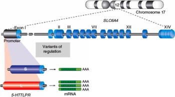
Following release 5-HT is deactivated by being taken back up into the presynaptic terminal and is then restored in vesicles. This uptake process requires active transport across the presynaptic membrane by the 5-HTT residing in that membrane. As shown in Figure 3, there is a polymorphism in the promotor region of the gene (SLC6A4) that codes for 5-HTT that results in two common alleles, referred to as long (l) and short (s) alleles because their respective promotor regions differ in the number of nucleotides (Lesch & Canli, Reference Lesch, Canli and Canli2006). Because of the facilitatory effects of the promotor region on gene production, the l allele codes for more RNA and therefore produces more transporter protein than the s allele. The s allele is characterized by reduced 5-HTT promotor activity and 5-HTT expression, resulting in a reduced rate of removing 5-HT from the synaptic cleft and increased extracellular levels of 5-HT.
Illustration of the components of the gene (SLC6A4) that codes for the serotonin transporter (5-HTT). Polymorphism of the promotor region yields two common alleles (S and L) that differ in length of nucleotide number. See text for details. [A color version of this figure can be viewed online at journals.cambridge.org/dpp]

At the behavioral level, 5-HTT knockout mice exhibit increased anxiety and reduced exploration, and primates and humans with the s allele (or the homolog of it in primates) express increased behavioral anxiety, increased scores on neuroticism scales, increased risk of depression, less effective responses to selective serotonin reuptake inhibitor (SSRI) treatment, and reduced social closeness (Hariri, Reference Hariri and Canli2006). At the neural level, the s allele has been associated with both neural function and structure. The s allele has been consistently found to be associated in both males and females with enhanced amygdala reactivity to many forms of emotional stimuli, including fear and angry faces, and negative scenes and words (Canli & Lesch, Reference Canli and Lesch2007; Hariri, Reference Hariri and Canli2006). In addition, the s allele is associated with reduced gray matter volume in the amygdala, suggesting reduced dendritic branching and axonal arborization (Hariri, Reference Hariri and Canli2006). Thus, there is a link between gene variation and basic brain mechanisms involved in processing negative emotion.
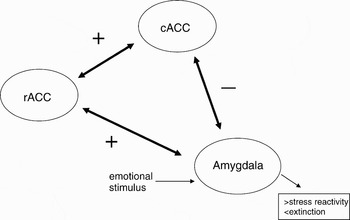
The reduced gray matter volume and increased reactivity of the amygdala in s-allele carriers (both S/S and L/S) raises the possibility that the amygdala's neural circuitry interactions may be influenced (i.e., its projections to and efferents from other brain regions). Indeed, recent research on the anterior cingulate cortex (ACC)–amygdala circuitry confirms this possibility. Specifically, the perigenual portion of the ACC, which surrounds the genua of the corpus collosum, is composed of the subgenual, rostral (rACC) and the supragenual, caudal (cACC). As shown in Figure 4, the rACC receives the most dense projections from the amygdala, conveys that information to the cACC, which in turn, sends efferents back to the amygdala. This circuitry is critical to modulating amygdala reactivity via inhibitory feedback from cACC to the amygdala during times of emotional stress.
Anterior cingulate–amygdala circuitry that provides negative feedback to modulate activity of the amygdala. See text for details.

Pezawas et al. (Reference Pezawas, Meyer-Lindenberg, Drabant, Verchinski, Munoz and Kolachana2005) found psychiatrically normal s-allele carriers to have reduced gray matter volume of both the perigenual ACC and the amygdala, with the most pronounced reduction in the rACC region, and a reduced covariance of perigenual ACC (pACC, especially rACC)–amygdala volumes. Furthermore, during the processing of fear and angry faces, there was greatly reduced positive covariance between rACC–amygdala activation, and reduced negative covariance between cACC–amygdala activation, suggesting a reduced negative feedback of the cACC on amygdala reactivity to stressful stimuli. Covariance between rACC–amygdala activation accounted for ~30% of the vairance in neuroticism scores in the entire sample, whereas no single region was significantly related to neuroticism. Thus, it is the reduced coherence of activity across brain regions associated with the s allele that most markedly affects enhanced reactivity to stress.
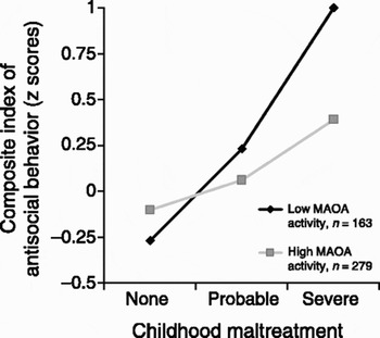
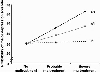
The implications of amygdala overreactivity are consistent with the phenotypic features of neuroticism. One can expect the persistent increased arousal being conveyed by amygdala efferents to other brain regions to be associated with anxiety and feelings of distress, persistent enhancement of vigilance for threat, increased rumination of negative thoughts, increased acquisition of negative emotional memories, and decreased extinction of anxiety elicited by environmental contexts. The s allele creates a reduced threshold for stimulus-induced arousal in the amygdala that is dependent on less coherent activity within its interactions with perigenual ACC circuitry. This state of persistent overreactivity apparently creates an enhanced risk for anxiety and depressive disorders in s-allele carriers, but only under conditions of stressful life events or early adverse experience such as childhood maltreatment (Caspi, Sugden, Moffitt, Taylor, & Craig, Reference Caspi, Sugden, Moffitt, Taylor and Craig2003).
Serotonin and development of neural connections
The altered coherence in gray matter volume and functional activity in the pACC–amygdala circuitry, implying reduced dendritic branching and axonal arborization, suggests the possibility that the s allele modifies connections between brain regions. Significantly, 5-HT plays a critical role in the development and plasticity of the brain (Gaspar, Cases, & Maroteaux, Reference Gaspar, Cases and Maroteaux2003). 5-HT neurons are among the earliest neurons to be generated prenatally, appearing in the first embryonic month in primates. Knockout of the 5-HT system prenatally results in no gross wiring or structural effects, but 5-HT is used as a later developmental signal, which modulates construction and plasticity of brain circuits. Major pre- and postnatal developmental effects of 5-HT include modulation of dendritic length and number of spines, and axon arborization: that is, the development of selected brain circuits.
Pre- and postnatal developmental effects of 5-HT arise from two sources. First, there is direct release of 5-HT from brain stem raphe (where 5-HT synthesizing neurons are localized) projections to cortical target regions. Second, glutamate neurons, which comprise the main excitatory neurons that interconnect brain regions, in the process of developing thalamic–limbic and corticocortical efferents capture 5-HT via transient axonal expression of the 5-HT transporter. 5-HT is then released at thalamocortical and corticocortical synapses to enhance presynaptic axon arborization and postsynaptic dendritic processes, again enhancing neural circuitries. This represents, then, one of the means by which l- and s-allele variation could make a significant difference in interconnections between brain regions.
The effects of 5-HT during development are dependent on which 5-HT receptors are activated. There are four groupings of these receptors. 5-HT1A autoreceptors, localized to soma and dendrites of raphe nuclei, and 5-HT2A receptors, one of the most widely expressed in the brain, are expressed late in development and enhance dendritic processes. 5-HT2A receptors also modulate the expression of the brain-derived neurotrophic factor (BDNF) gene in the neocortex and hippocampus. BDNF secretion from postsynaptic neurons in response to neurotransmitter activation of postsynaptic receptors sculpts glutamate innervation patterns of dendrites and axons, increases synaptic efficacy of glutamate, and modulates long-term potentiation in the hippocampus. Thus, 5-HT2A receptor activation can in this indirect way modulate dendritic and axonal processes. Conversely, 5-HT1B and 5-HT2C receptors are mainly localized to axons and axon terminals, enhancing synaptic strength via axon arborization of glutamate axonal connections through enhanced release of glutamate.
This 5-HT modulation has its strongest effects during development as opposed to later in life (Ansorge, Zhou, Lira, Hen, & Ginrich, Reference Ansorge, Zhou, Lira, Hen and Gingrich2004; Gaspar et al., Reference Gaspar, Cases and Maroteaux2003; Gross & Hen, Reference Gross and Hen2004; Leonardo & Hen, Reference Leonardo and Hen2006). In rodents, the forebrain has an experience-expectant period of dramatic synaptogenesis and dendritic growth 3–4 weeks postnatally, and hippocampal maturation of dendritic branches occurs during 2–4 weeks postnatally. In primates, this period corresponds to early postnatal months. The 5-HT1A receptor also appears in Week 2 postnatally in the hippocampus, amygdala, and ACC. If knocked out or blocked in the first 4 weeks in the early postnatal period, there is a strong enhancement of adult anxiety that first appears in Week 3, but no anxiety enhancement occurs when the receptor is blocked in adulthood (Lesch & Canli, Reference Lesch, Canli and Canli2006; Tsetsenis, Ma, Iacono, Beck, & Gross, Reference Tsetsenis, Ma, Iacono, Beck and Gross2007). 5-HT1A autoreceptors enhance dendritic maturation, and thus their blockade could affect neural circuitry; whereas 5-HT1A postsynaptic receptors induce hyperpolarization and reduce neuronal excitability in postsynaptic neurons, thus increasing the threshold for firing the postsynaptic neuron. Blockade of this latter effect could contribute to amygdala overreactivity. These findings suggest that 5-HT mediation by 5-HT1A receptors is essential to the establishment of normal anxiety-modulating circuits in the brain during early postnatal development. Knockout of monoamine oxidase A (MAOA), which provides the main degradation of 5-HT after reuptake, inordinately increases 5-HT and has many developmental effects, but only when it is knocked out in the early postnatal period (Gaspar et al., Reference Gaspar, Cases and Maroteaux2003). Consistent with these biochemical findings, adverse rearing environments in monkeys (Champoux et al., Reference Champoux, Bennett, Shannon, Higley, Lesch and Suomi2002) and in humans (Taylor et al., Reference Taylor, Way, Walch, Hilmert, Lehman and Eisenberger2006) interacts with the 5-HTT polymorphism, where the effects of the s allele on anxiety and depression is enhanced by the environment.
In sum, the hypothesis is that the 5-HTT polymorphism affects 5-HT modulated development of dendritic and axonal development and fine-tuning of connectivity patterns in the forebrain. Variation in the connectivity of the pACC–amygdala circuits manifests neurally as amygdala hyperreactivity to stress, and behaviorally as anxiety, stress reactivity, negative emotionality, and an increased risk for anxiety and depressive disorders in the context of accumulating environmental adversity. Risk factors that interact with 5-HT effects may be a polymorphic variation in the BDNF gene (val BDNF) that results in reduced BDNF postsynaptic secretion and hence reduced synaptic connectivity; low levels of MAOA activity, increasing 5-HT presynaptic levels and release; and early environmental diversity.
Central CRH functioning
Central CRH and anxiety
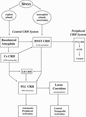
As illustrated in Figure 5, the effects of stress are mediated by a central system of CRH neurons. Stressful contexts can include both explicit, discrete stimuli that generate fear and nonexplicit stimuli associated with uncertainty that generate anxiety. It is now well established that association of discrete, explicit neutral stimuli (fear conditioned stimulus [CS]) with an aversive unconditioned stimulus (UCS) occurs via cortical uni- and polymodal sensory efferents that converge on the basolateral complex of the amygdala (Davis & Shi, Reference Davis and Shi1999; Kalin, Shelton, & Davidson, Reference Kalin, Shelton and Davidson2004; LeDoux, Reference LeDoux1998). Fear CSs elicit a host of behavioral, neuropeptide, and autonomic responses via output from the basolateral amygdala to the central amygdala (Ce), which in turn, sends functionally separable efferents to many hypothalamic and brain stem targets (LeDoux, Reference LeDoux1998).
Components of the central and peripheral corticotrophin-releasing hormone (CRH) systems. Ce, central amygdala nucleus; BNST, bed nucleus of the stria terminalis; LH, lateral hypothalamus; PGi, paragiganticocellularis; PVN, paraventricular nucleus of the hypothalamus; ACTH, adrenocorticotropin hormone from the anterior pituitary.

In contrast, the enduring potentiating effects of nondiscrete, contextually related aversive stimuli on anxiety (but not fear) is dependent on a group of structures collectively referred to as the extended amygdala, which receives massive projections from basolateral and olfactory amygdala complexes, and which represents a macrostructure that is characterized by two divisions, central and medial (Heimer, Reference Heimer2003; Kalin, Shelton, Davidson, & Kelley, Reference Kalin, Shelton, Davidson and Kelley2001). These two divisions originate from the central and medial nuclei of the amygdala, and consist of cell groups that are distributed throughout the sublenticular area, bed nucleus of the stria terminalis (BNST), around striatal and pallidal structures, and back to merge specifically with the caudomedial region of the nucleus accumbens (NAS).
The central division of the extended amygdala appears to associate general contextual features and nonexplicit, nondiscrete conditioned and unconditioned stimuli with reward or punishment (e.g., light conditions, physical features, spatial relations; Davis & Shi, Reference Davis and Shi1999; McDonald, Shammah-Iagnado, Shi, & Davis, Reference McDonald, Shammah-Lagnado, Shi and Davis1999). Similar to the outputs from the Ce, most structures of the central division of the extended amygdala can transmit this motivationally relevant information to some or all hypothalamic and brain stem structures related to emotional expression (Heimer, Reference Heimer2003). This output coordinates activation of (a) the negative affective state of anxiety to inform the individual that the current context is uncertain and potentially dangerous, (b) autonomic arousal to mobilize energy for potential action, (c) selective attention to maximize sensory input at specified locations in the visual field, and (d) cognition to derive a response strategy. Thus, two emotional learning systems may have evolved: the basolateral amygdala to associate reinforcement with explicit, specific characteristics of objects (i.e., for fear), and the BNST to associate reinforcement with nonexplicit spatial and contextual stimulus aspects (i.e., for anxiety).
As indicated in Figure 5 above, the basolateral and central amygdala and the BNST provide integration of behavioral, neuroendocrine, and autonomic responses to stressful circumstances (Leri, Flores, Rodaros, & Stewart, Reference Leri, Flores, Rodaros and Stewart2002). This integration is accomplished by the activity of both the peripheral and central CRH systems. The peripheral system involves CRH neurons located in the paraventricular nucleus (PVN) of the hypothalamus, which when activated initiates the series of events that ends in the release of cortisol from the adrenal cortex. Cortisol then circulates in the bloodstream and increases energy reserves (gluconeogenesis) and, if not excessive in quantity, can enhance neurotransmitters involved in modulating emotion and memory (Kim & Diamond, Reference Kim and Diamond2002).
In contrast, the central CRH system is composed of CRH neurons located in many different subcortical brain regions that modulate emotion, memory, and CNS arousal. Whereas the majority of CRH neurons in the CNS do not mediate the effects of stress, some of the CRH-containing regions that are important in mediating stress effects are illustrated in Figure 5. For instance, the basolateral amygdala detects discrete aversive stimuli associated with stressful circumstances and activates the extensive array of CRH neurons located in the Ce (~1,750 neurons/hemisphere; Merali, Michaud, McIntosh, Kent, & Ainsman, Reference Merali, Michaud, McIntosh, Kent and Anisman2003). These CRH neurons project to many brain regions that modulate emotion, memory, and arousal, including the BNST and the peripheral CRH neurons in the PVN. Stress variables associated with context and uncertainty activate CRH neurons in the BNST, which have similar projection targets as the Ce (Macey, Smith, Nader, & Porrino, Reference Macey, Smith, Nader and Porrino2003). Both the Ce and the BNST can activate CRH neurons in the lateral hypothalamus (LH), a region that integrates CNS arousal as a means of modulating changes in the autonomic nervous system (ANS). In turn, the LH CRH projections modulate ANS activity. As illustrated in Figure 5, all three sources of CRH projections (Ce, BNST, and LH) innervate CRH neurons in the paragiganticocellularis (PGi; Aston-Jones, Rajkowski, Kubiak, Valentino, & Shiptley, Reference Aston-Jones, Rajkowski, Kubiak, Valentino, Shiptley, Holstege, Bandler and Saper1996), which is located in the rostral ventrolateral area of the medulla in the brain stem. The PGi is a massive nucleus that provides major integration of central and autonomic arousal and, in turn, coordinates and triggers arousal responses to urgent stimuli via two main pathways eminating from its own population of CRH neurons, which make up 10% of PGi neurons (Aston-Jones et al., Reference Aston-Jones, Rajkowski, Kubiak, Valentino, Shiptley, Holstege, Bandler and Saper1996). One CRH pathway modulates the ANS via projections to the intermediolateral cell column of the spinal cord, activating sympathetic preganglionic autonomic neurons.
The other CRH pathway modulates central arousal effects via activation of the locus coeruleus (LC), where PGi CRH innervation of the LC in humans and monkeys is dense (Aston-Jones et al., Reference Aston-Jones, Rajkowski, Kubiak, Valentino, Shiptley, Holstege, Bandler and Saper1996). The LC, which is composed of ~20,000 neurons that provide the major source of norepinephrine (NE) in the brain, innervates the entire brain. LC neurons that release NE onto B-adrenergic receptors are responsible for producing a nonspecific emotional activation via broadly collateralizing axons to the CNS (Aston-Jones et al., Reference Aston-Jones, Rajkowski, Kubiak, Valentino, Shiptley, Holstege, Bandler and Saper1996), and this NE release to chronic stress is increased in inbred rat strains with high anxiety (Blizard, Reference Blizard1988) and in posttraumatic stress disorder patients (Charney, Grillon, & Brenner, Reference Charney, Grillon and Bremner1998). This nonspecific emotional activation pattern comprises a global urgent response system that responds to unpredicted events, and hence, facilitates behavioral readiness, alerting (enhanced sensory processing), and attention (enhanced selection of stimuli; Aston-Jones et al., Reference Aston-Jones, Rajkowski, Kubiak, Valentino, Shiptley, Holstege, Bandler and Saper1996), which are all characteristic of states of anxiety in highly stress-reactive rodents and monkeys (Blizard, Reference Blizard1988; Redmond, Reference Redmond and Meltzer1987). This central arousal can be enduring: PGi CRH activation of LC neurons endures and peaks 40 min after stimulation of the PGi (Aston-Jones et al., Reference Aston-Jones, Rajkowski, Kubiak, Valentino, Shiptley, Holstege, Bandler and Saper1996). This persistent activation of spontaneous activity in the LC renders the effects of short-lived, nonsalient stimuli of little effect on the LC, such that salient stimuli have an inordinate effect on LC activity, and hence in turn, on central activation. Thus, taken together, the central CRH neuron system is capable of activating a vast array of behaviorally relevant neural and hormonal processes during stressful conditions, including activation of the peripheral CRH system.
Early research demonstrated that CRH in the BNST rather than in the Ce mediates anxiety as opposed to fear, whereas the Ce expresses fear but not anxiety. Thus, injection of CRH into the BNST of rats potentiates startle (an index of anxiety) in a dose-dependent manner for up to 2 hr duration (Davis & Shi, Reference Davis and Shi1999). Subsequent work has confirmed a specific role for CRH in the BNST in the mediation of anxiogenic effects. Intense inescapable stressors induce a persistent anxious state in rats characterized by increased sensory reactivity and enhanced associative learning with aversive UCSs. This is accompanied by elevated levels of CRH in the BNST. Marked anxiety effects lasting longer than 24 hr are produced after administration of three doses of CRH over 1.5 hr, despite no lasting effect on peripheral release of corticosterone (Servatius et al., Reference Servatius, Beck, Moldow, Salameh, Tumminello and Short2005). The source of naturally occurring CRH in the BNST is from the Ce's population of CRH neurons: CRH release from the Ce after a psychogenic stressor causes long-term anxiety effects in rats, which correlates with cerebrospinal fluid CRH concentrations (Merali et al., Reference Merali, Michaud, McIntosh, Kent and Anisman2003).
The anxiogenic effects of CRH are specific to the lateral region of the BNST. Intralateral BNST (but not intra-Ce) administration of CRH in rats elicits dose-dependent anxiogenic effects via CRH-R1 receptors, including reduced exploration of an unfamiliar cage and an elevated T-maze (which are both aversive UCSs); reduced sleep, food intake, and sexual activity; and increased sympathetic and behavioral signs of anxiety (shaking, crouching, cautious approach; Nie et al., Reference Nie, Schweitzer, Roberts, Madamba, Moore and Siggins2004). Furthermore, intralateral BNST (but not intra-Ce) administration of CRH in rats provides a dose-dependent aversion to the CRH-paired environment via CRH-R1 receptors, indicating that the lateral BNST is a critical site for associative conditioning of context and aversive signals mediated by CRH (Sahuque et al., Reference Sahuque, Kullberg, Mcgeehan, Kinder, Hicks and Blanton2006). The CRH-R1 receptors in the forebrain mediate these anxiogenic effects. Transgenic mice with elevated CRH-R1 (but not with elevated R2) receptors in the central forebrain (but with no elevation peripherally in the hypothalamus or pituitary) show extreme indications of anxiety; whereas mice with knockout of the CRH-R1 (but not R2) receptor gene in the central forebrain (but not peripherally) show little anxiety in the elevated T-maze or in the light–dark test (both being aversive UCSs; Deussing & Wurst, Reference Deussing and Wurst2005). Moreover, conditional activation of CRH-R1 gene expression centrally (but not peripherally) produces anxiogenic effects, whereas conditional inhibition of CRH-R1 gene expression centrally (but not peripherally) produces anxiolytic effects. Finally, a CRH-R1 antagonist administered orally to male monkeys impaired development and expression of contextual anxiety conditioning, and blocked social stress-induced (two unfamiliar male monkeys placed in attached plexiglass cages) increases in anxiety behaviors, cortisol, cerebrospinal fluid CRH concentrations, and LC neuronal firing (Habib et al., Reference Habib, Weld, Rice, Pushkas, Champoux and Listwak2000).
These findings, taken together, suggest that anxiety is essentially a stress response system that relies on a central network of CRH neuron populations acting via CRH-R1 receptors that provide integrated responses (hormonal, behavioral, autonomic, and central arousal) to the stressor. CRH especially in the lateral BNST mediates what is a defining characteristic of anxiety as opposed to fear: prolonged anxiogenic effects, which last as long as uncertainty is unresolved, and aversive contextual conditioning. These CRH-induced psychological effects appear to strongly match the content of trait neuroticism measures, where the strongest primary or facet scale in the higher order factor of neuroticism in Tellegen's personality questionnaire is termed stress reactivity because of the persistent negative emotional responses reported to stress-related events in items loading on the scale (Tellegen & Waller, Reference Tellegen, Waller, Briggs and Cheek1997).
Finally, although trait levels of neuroticism are very stable over as many as 20 years, it is less stable up to adolescence. This suggests that neural factors that contribute to neuroticism may be more influenced by experience earlier in life. In support of this notion, a recent study found that early postnatal separations from mother rats daily for 3 hr caused in pups later in life enhanced reactivity to brief stressors in anxiety behaviors and corticosterone secretion, but also in CRH mRNA expression in the Ce (Gillespie & Nemeroff, Reference Gillespie and Nemeroff2007). The latter implies that early adverse experience may result in a persistent Ce-induced enhanced reactivity via its CRH efferents throughout stress-related neural pathways shown in Figure 5, including in the lateral BNST.
Peripheral glucocorticoid system
Peripheral pathways
As shown on the far right side of Figure 5, the BNST via CRH efferents activates CRH neurons in the PVN under stressful conditions. CRH is then released from the hypothalamic CRH neurons and is transported via a portal system to the anterior pituitary, where adrenocorticotropin hormone (ACTH) is released into the blood stream, activates the adrenal cortex, which then secretes cortisol (mainly corticosterone in rodents). Cortisol travels through the blood stream and has a diverse set of effects on energy metabolism and nerual functioning. In humans and rodents, there are two types of stress-induced cortisol receptors. Type I or mineralocorticoid receptors (MRs) have a high affinity for cortisol and thereby mediate the acute stress-induced effects of cortisol on bodily responses. Type II or glucocorticoid receptors (GRs) have a low affinity for cortisol, and thereby become bound to cortisol only at high levels of cortisol concentration. Such high levels of cortisol in the hippocampus, acting via GRs, activate inhibitory output from the hippocampus to the hypothalamic CRH secretory neurons, thereby reducing the activity of the CRH–cortisol stress response system. This pathway represents the major negative feedback system for controlling stress-induced CRH–cortisol responses.
Peripheral glucocorticoid system and early adversity
Noting that the relations between early adversity and environmental poverty with adulthood health (including stress disorders later in life) appear to be mediated by parental influences, researchers have begun to explore whether these parental influences operate via effects on the development of neural systems that underlie the expression of behavioral and endocrine responses to stress (Gross & Hen, Reference Gross and Hen2004; Meaney, Reference Meaney2001; Sapolsky, Reference Sapolsky2004). Postnatal handling studies in rats provided early support for this notion. Postnatal handling of 3–15 min/day resulted in reduced stress reactivity and fearfulness, and increased exploration; decreased hippocampal aging, and more intact cognitive functioning in old age; increased GRs in the hippocampus; and increased gamma aminobutyric acid (GABA) A receptors (the activation of which reduces anxiety behavior in rats) in the amygdala, LC, and hippocampus. In contrast, maternal separation of rat pups had the opposite effects of postnatal handling. For instance, pups separated from their mothers for 180 min/day for 2–14 days (which simulates a scarce environment in which the mother leaves the nest for days at a time to forage for food) show significant increases in CRH mRNA expression in the Ce and PVN; decreased GR mRNA and reduced 5-HT1A responses in the hippocampus; and reduced GR binding in the hippocampus, hypothalamus, and frontal cortex, all of which blunts the CRH negative feedback system. Of importance, these pups also manifested a lifetime hyperresponsivity to unfamiliar stressors.
A series of studies of individual differences in rat maternal behavior toward pups has also supported these findings (Champagne et al., Reference Champagne, Bagot, Hasselt, Ramakers, Meaney and de Kloet2008; Leonardo & Hen, Reference Leonardo and Hen2006; Meaney, Reference Meaney2001; Weaver et al., Reference Weaver, Cervoni, Champagne, D'Alessio, Sharma and Seckl2004). Rat mothers naturally differ in their levels of licking/grooming (LG) and arched-back nursing (ABN; which is less stressful for pups than being compressed by the mother's body during nursing), creating two groups of mothers: high versus low LG-ABN. These different maternal patterns are stable over litters, so they appear to be traitlike under normal conditions. Behaviorally, pups of high LG-ABN mothers show reduced startle responses, increased open-field exploration, and reduced latencies to eat food in a novel environment. Neurobiologically, these pups show reduced plasma ACTH and corticosterone responses to acute stress, increased hippocampal GR mRNA expression, and hence, increased glucocorticoid negative feedback sensitivity, decreased CRH mRNA levels in the hypothalamus, increased affinity for GABA at its A receptor in the amygdala and LC, and an increased 5-HT tone in the hippocampus. Moreover, as pups and adults, offspring of high LG-ABN mothers also show less cell death and increased glutamate receptor levels (resulting in enhanced synaptogenesis) in the hippocampus and enhanced memory on hippocampal-dependent tasks.
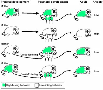
Thus, the behavior of the mother toward offspring can apparently “program” behavioral and endocrine responses to stress that endure the entire lifespan. Moreover, as illustrated in Figure 6, individual differences in stress reactivity mediated by maternal care are transmitted across generations (Gross & Hen, Reference Gross and Hen2004). Indeed, although adult mothers must overcome initial neophobia to pups, female offspring of high LG mothers are initially less averse to pups. When offspring of low LG mothers are raised from birth by high LG mothers (i.e., cross-fostering), these offspring manifest low stress reactivity and anxiety as pups and adults. Of importance, however, Meaney (Reference Meaney2001) reported that raising offspring of high LG mothers with low LG mothers produced stress reactive offspring, but this has not been replicated: others have found that these offspring show adult stress phenotypes that are more similar to their high LG biological mother (Figure 6; Gross & Hen, Reference Gross and Hen2004). We return to this issue later. What is clear is that variation in maternal care can affect development of neural systems that mediate stress reactivity, which subsequently influences offsprings' maternal behavior.
Rats raised by mothers that display low licking/grooming behavior exhibit more anxiety-related behavior than rats raised by high licking/grooming mothers. Cross-fostering studies show that the offspring of low licking/grooming mothers raised by high licking/grooming mothers are less prone to anxiety-related behavior as adults. This indicates that the effect is mediated by postnatal maternal environment. However, offspring of high licking/grooming mothers raised by low licking/grooming mothers do not have an increased tendency to develop anxiety-related behavior in adulthood, indicating that specific factors inherited by the high licking/grooming offspring protect them from the effects of being mothered by low licking/grooming females. From “Developmental Origins of Anxiety,” by E. Gross and R. Hen, Reference Gross and Hen2004, Nature Reviews Neuroscience, 5. Copyright 2004 by Nature Publishing Group. Reprinted with permission. [A color version of this figure can be viewed online at journals.cambridge.org/dpp]

It is important to note that there is a convergence between the roles of 5-HT and CRH in these studies that may be significant with respect to the above discussion of the 5-HTT and CRH in anxiety and stress reactivity. Recall that high LG mothers and their offspring showed increased 5-HT tone in the hippocampus, and that maternal separation reduced 5-HT1A responses in hippocampus. Stress-induced CRH release onto CRH-R1 receptors in the dorsal raphe nucleus (a major brain stem site of 5-HT neurons) elevates 5-HT concentrations in the hippocampus by 350%. 5-HT activity in the hippocampus activates GR gene expression, providing increased negative feedback to the PVN and Ce CRH neurons. In turn, GR activation enhances postsynaptic effects of 5-HT1A activation, which increases inhibition of excitation in the postsynaptic neuron. Furthermore, 5-HT activation of postsynaptic glutamate neurons in the hippocampus activates BDNF secretion into the synapse, increasing synaptic processes and decreased aging effects in the hippocampus. Therefore, variation in the functional properties of the 5-HT and/or the CRH-R1 systems could influence the efficacy of the GR-induced negative feedback system for controlling stress-induced cortisol secretion.
Early experience and epigenetic processes
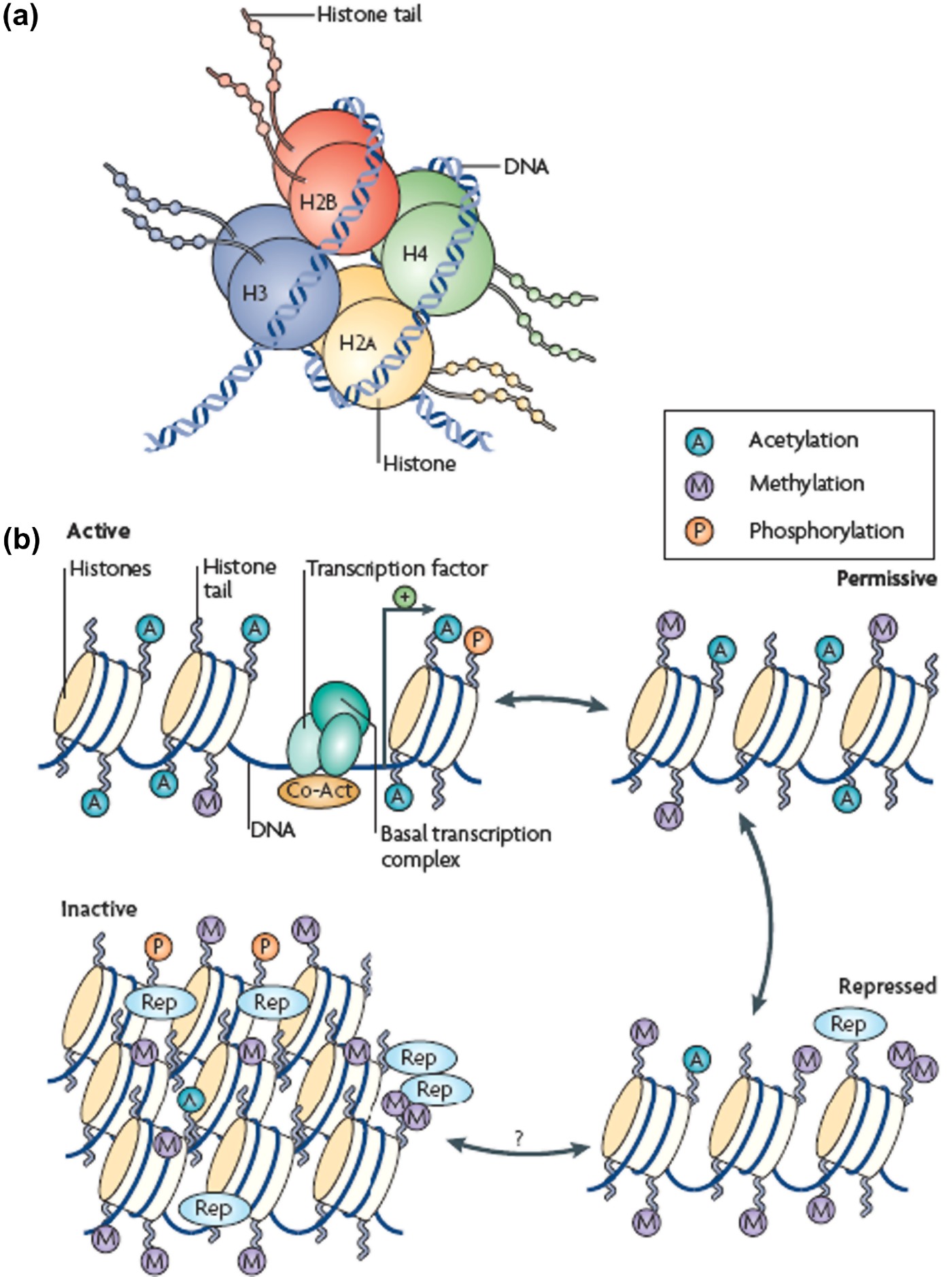
The critical question concerning the early handling, separation, and maternal care findings is how these influences on stress reactivity and anxiety become long lasting. Research thus far has supported the role of two mechanisms: epigenetic effects and DNA methylation, both of which involve an alteration of gene expression by experience. In terms of epigenetic effects, as shown in Figure 7, DNA is wrapped around proteins called histones, with the DNA and histones collectively comprising chromatin (Tsankova, Renthal, Kumar, & Nestler, Reference Tsankova, Renthal, Kumar and Nestler2007). The functional unit of chromatin is the nucleosome, which is composed of 147 base pairs of DNA wrapped around a core histone group, where there are two copies of each of the four histones (Figure 7a). The compactness (openness vs. closedness) of histones in a particular region of chromatin determines how readily transcription factors can access promoters in the DNA (Figure 7b). A wide variety of regulators can alter chromatin structure (i.e., its openness), and hence the access to genes. Neurotransmitter (e.g., 5-HT) or hormone (e.g., cortisol) action at receptors can elicit activity in postsynaptic regulators of chromatin structure, and hence access to genes. For instance, activation of MRs and GRs alters the expression of 70–100 genes, 50% of which are upregulated. As we shall see, experience, via neurotransmitter and hormone effects, can permanently alter chromatin regulators and openness to gene transcription, and hence behavior.
The general scheme of chromatin remodeling. (a) A picture of a nucleosome showing a DNA strand wrapped around a histone octamer composed of two copies each of the histones. (b) Chromatin can be conceptualized as existing in two primary structural states: as active or open (top left) in which histone acetylation (A) is associated with opening the nucleosome to allow binding of the basal transcriptional complex and other activators of transcription; or as condensed or closed where all gene activity is permanently silenced (bottom left). In reality, chromatin exists in a continuum of several functional states (active; permissive (top right); repressed (bottom right; and inactive). Adapted from “Epigenetic Regulation in Psychiatric Disorders,” by S. Tsankova, D. Renthal, A. Kumar, and E. Nestler, Reference Tsankova, Renthal, Kumar and Nestler2007, Nature Reviews Neuroscience, 8. Copyright 2007 by Nature Publishing Group. Adapted with permission. [A color version of this figure can be viewed online at journals.cambridge.org/dpp]
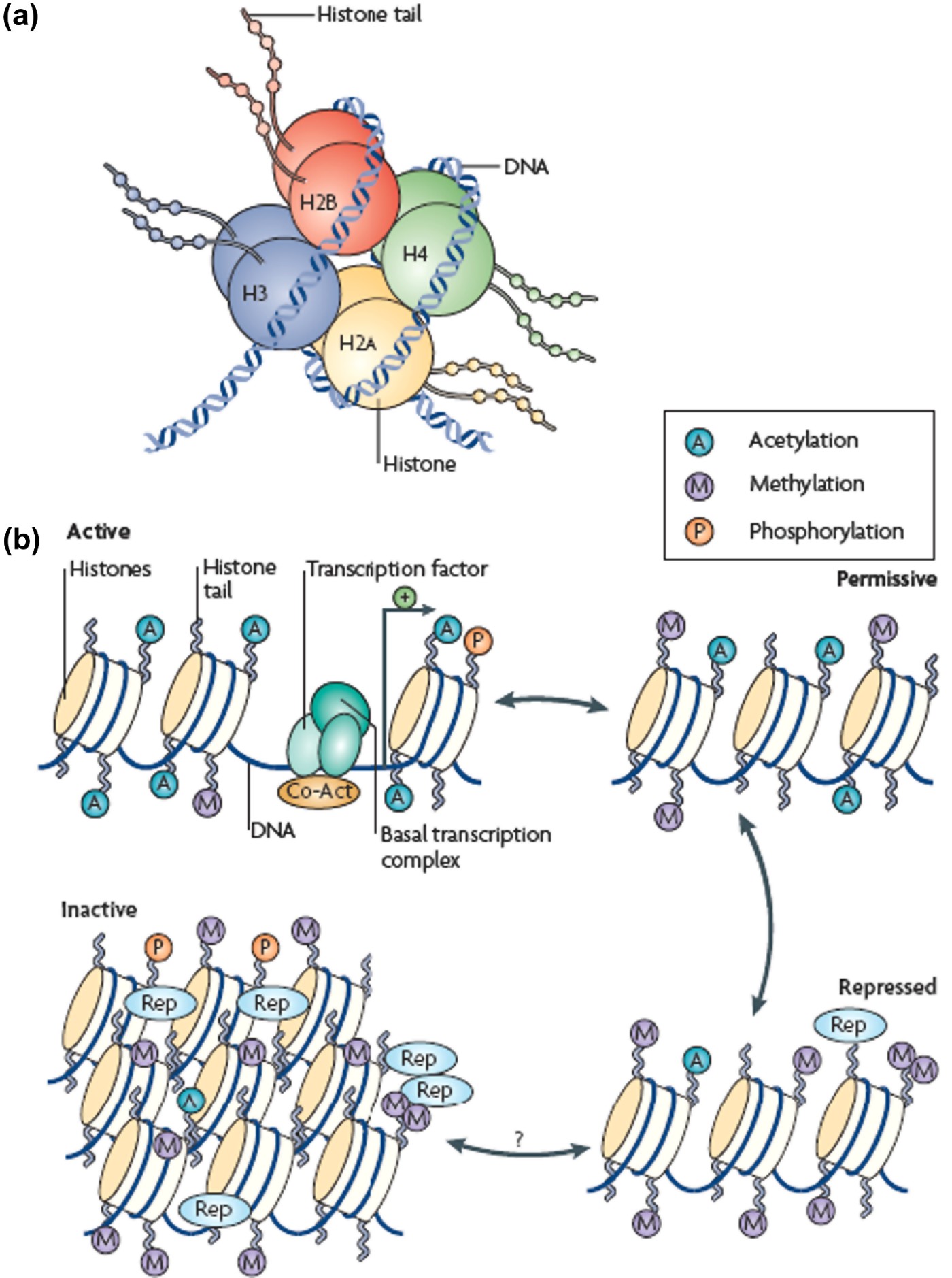
Alternatively, in terms of DNA methylation, genes can be stably silenced for substantial periods of time (a lifetime in the case of the second X chromosome in females) when a methyl group is attached to nucleotides in a gene's promotor or exon regions, where the methyl group then tightly binds a repressor protein (Figure 7b, inactive and repressed sections). Demethylation of a gene leads to enhanced expression of its protein product.
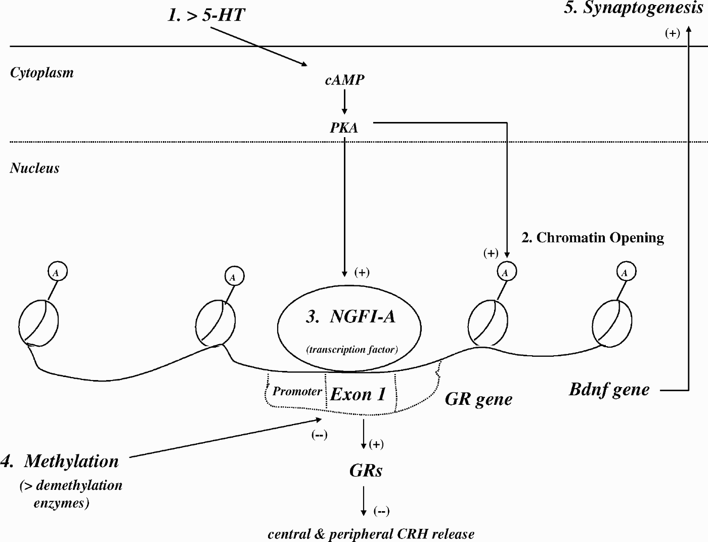
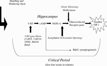
Changes in both chromatin remodeling and DNA methylation state are implicated strongly in the effects of handling and maternal style. Acetylation of histones decreases their binding to DNA (increases chromatin openness), thereby increasing access of transcription factors to promoters and exons. Histones surrounding the GR gene were more acetylated in high LG mothers and their offspring. As illustrated in Figure 8, a hypothesized chain of events (Champagne et al., Reference Champagne, Bagot, Hasselt, Ramakers, Meaney and de Kloet2008; Sapolsky, Reference Sapolsky2004; Tsankova et al., Reference Tsankova, Renthal, Kumar and Nestler2007) is that high LG rearing results in increased 5-HT neurotransmission in the hippocampus of pups (Point 1, Figure 8). The resulting increased 5-HT activation of postsynaptic neurons in the hippocampus leads to strong activation of cAMP, protein kinase A, and its cascade of events within the postsynaptic neuron. In turn, an increased acetylation of histones and opening of chromatin occurs (Point 2, Figure 8). Also, increased expression of the transcription factor nerve growth factor-inducible protein A (NGFI-A) occurs, which results in increased NFGI-A binding to its binding site located on the first exon of the GR gene (Point 3, Figure 8). Such binding enhances the expression of GRs in the hippocampus, providing enhanced negative feedback effects on central and peripheral CRH release. 5-HT via its postsynaptic effects activates the BDNF gene, which increases synaptogenesis in the hippocampus when released into the synaptic cleft (Point 5, Figure 8).
Five effects of high licking/grooming maternal care in the hippocampus on gene expression. See text for details.
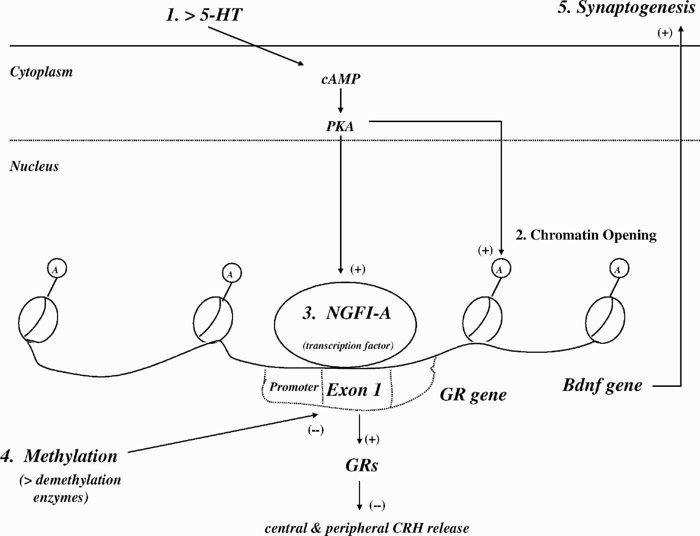
In terms of DNA methylation, it has been found that the methylation state of the NGFI-A binding site on the first exon of the GR gene is plastic around the time of birth. The site is then methylated the first day after birth, decreasing transcription of the hippocampal GR gene. Over the next week, exposure to high LG mothering (by biological or cross-fostering females) caused the binding site to be demethylated, increasing transcription of the GR gene, and hence, hippocampal GRs (Point 4, Figure 8). Recent evidence supports a causative role for the above histone actylation and methylation effects. A pharmacological inhibitor of histone deacetylation opens up chromatin and facilitates access of demethylating enzymes, shifting the balance in chromatin toward acetylation. Administration of this inhibitor to adult rats raised in a low LG environment transformed them into a high LG adult in terms of stress reactivity. This behavioral effect was accompanied by an increase in NGFI-A access to the now demethylated promotor and exon 1 of the GR gene and by reduced levels of circulating glucocorticoids.
It is significant that most of the effects of handling and maternal style occur only within a critical developmental period (Champagne et al., Reference Champagne, Bagot, Hasselt, Ramakers, Meaney and de Kloet2008; Gross & Hen, Reference Gross and Hen2004; Leonardo & Hen, Reference Leonardo and Hen2006; Meaney, Reference Meaney2001). Handling, maternal style, and methylation status all must be present in the first week of life to have an effect in rodents. This is consistent with the findings discussed above, where the first 3 weeks in rodents are critical in the developmental effects of 5-HT, the effects of knocking out 5-HT1A receptors and MAOA, and in CRH reactivity. Similarly, SSRIs (which result in enhanced 5-HT availability) administered during Weeks 1–3 (but not during Weeks 6–8) normalize the glucocorticoid response in parentally stressed mice, indicating that this critical developmental period may have a relatively narrow window. Thus, as summarized in Figure 9, experience during early postnatal weeks in rodents, and perhaps early months in primates, appear to interact with genetic polymorphisms in 5-HT, BDNF, and MAOA functioning, creating a critical period for establishing effective synaptic development in the forebrain structures that mediate anxiety in response to experience-dependent signals.
A summary of the effects of maternal experience and genetic polymorphisms on gene expression of glucocorticoid receptors during an early postnatal period. See text for details.

Extraversion
Dopamine (DA) contributions to extraversion
Through empirical studies and integrative reviews, we have demonstrated that extraversion reflects the activity of a mammalian behavioral approach system based on positive incentive motivation (Depue, Reference Depue and Canli2006; Depue & Collins, Reference Depue and Collins1999; Depue & Morrone-Strupinsky, Reference Depue and Morrone-Strupinsky2005). This system is activated by, and serves to bring an animal in contact with, unconditioned and conditioned rewarding incentive stimuli. Incentives are inherently evaluated as positive in valence, and activate incentive motivation, increased energy through sympathetic nervous system activity, and forward locomotion as a means of bringing individuals into close proximity to rewards. The incentive state is clearly evident in the phenotypic features of extraversion, and is associated with subjective feelings of desire, wanting, excitement, elation, enthusiasm, potency, and self-efficacy that are distinct from, but typically cooccur with, feelings of pleasure and liking.
Animal research demonstrates that the positive incentive motivation and experience of reward that underlies a behavioral system of approach is dependent on the functional properties of the midbrain ventral tegmental area (VTA) DA projection system. DA agonists or antagonists in the VTA or NAS, which is a major terminal site of VTA DA projections, in rats and monkeys facilitate or markedly impair, respectively, a broad array of incentive motivated behaviors. Furthermore, dose-dependent DA receptor activation in the VTA–NAS pathway facilitates the acute rewarding effects of stimulants, and the NAS is a particularly strong site for intracranial self-administration of DA agonists. DA agonists injected in the NAS also modulate behavioral responses to conditioned incentive stimuli in a dose-dependent fashion. In single-unit recording studies, VTA DA neurons are activated preferentially by appetitive incentive stimuli, and DA cells, most numerously in the VTA, respond vigorously to and in proportion to the magnitude of both conditioned and unconditioned incentive stimuli and in anticipation of reward (Schultz, Reference Schultz2007). Similarly, in human neuroimaging studies, increased activation in the VTA and NAS correlates with the amount of reward anticipated, and with ratings of excitement preceding the reward (Canli, Reference Canli and Canli2006; Knutson & Bhanji, Reference Knutson, Bhanji and Canli2006). Morevover, during acute administration the intensity of a subject's subjective euphoria increased in a dose-dependent manner in proportion to cocaine binding to the DA uptake transporter (and hence to DA levels) in the striatum (Drevets et al., Reference Drevets, Gautier, Price, Kupfer, Kinahan and Grace2001; Volkow et al., Reference Volkow, Wang, Fischman, Foltin, Fowler and Abumrad1997). Hence, taken together, the animal and human evidence demonstrates that the VTA DA–NAS pathway is a primary neural circuit for incentive motivation and its accompanying subjective state of reward.
Consistent with these findings are results with a polymorphism of the DA D4 receptor gene on chromosome 11 (Munafo et al., Reference Munafo, Clark, Moore, Payne, Walton and Flint2003). The D4 receptor is part of the D2 family of DA receptors, which are generally inhibitory in nature. The D4 polymorphism is characterized by variation in the number of repeats of a 48 base pair unit in exon 3. The most common repeats in the population are 4 and 7 repeats, where the 7-repeat allele is characterized by a twofold reduction in inhibitory effects on postsynaptic second-messenger cAMP relative to the 4-repeat allele. Several small studies have found the 7-repeat allele to be associated with increased extraversion levels relative to the 4-repeat allele, and this was also found using behavioral features of positive emotion in a study of 8- to 12-month-old infants (Lakatos et al., Reference Lakatos, Nemoda, Birkas, Ronai, Kovacs and Ney2003). When this polymorphism is taken in interaction with a polymorphism of the DA transporter (one allele being associated with reduced DA uptake and hence increased time of DA in the synapse to activate postsynaptic receptors), impulsivity was significantly increased in terms of number of comission errors in a go–no-go task.
In the discussion above of a threshold model of behavioral responding, it was argued that higher levels of neurobiological traits underlying personality would enhance the association of neutral cues with emotional state. We found that high extraverts more readily associate specific and general contextual cues to DA-agonist induced subjective reward than low extraverts, who showed no associative conditioning at the dose level of methylphenidate used in the studies (Depue, Reference Depue and Canli2006). These findings are consistent with an enhanced reward encoding of positive cues, and are consistent with the fact that extraversion is correlated with increased amygdala activation to happy versus neutral and negative faces. Moreover, the latter finding implies early detection of the enhanced weighted positive stimuli in the amygdala, which through backprojections to many areas of the brain, including sensory processing regions, can bias perceptual processing and attention to positive cues in the environment. Several studies support just such a bias in extraversion (Canli, Reference Canli and Canli2006).
Early adversity and DA functioning
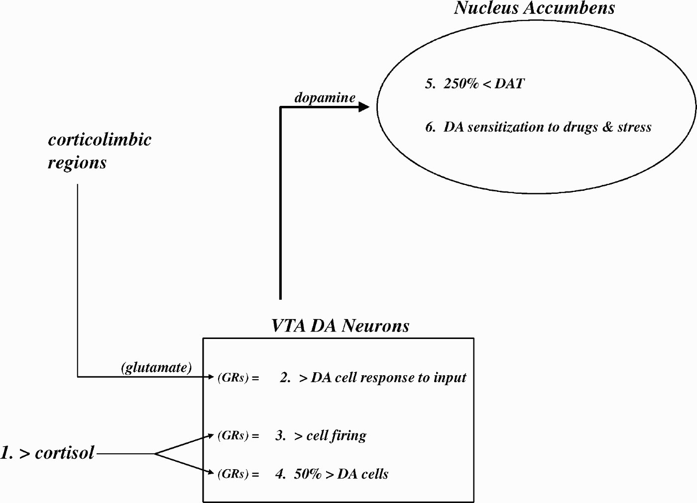
Early adverse experience in animals has a marked effect on DA functioning, as summarized in Figure 10. Single intense stressors or repeated, prolonged stressors, including maternal separations in neonatal rats and monkeys, lead to a sensitization of NAS DA release to repetition of the same stressor or to novel stressors and DA agonists later in life (Point 6, Figure 10; Depue & Collins, Reference Depue and Collins1999; Meaney, Brake, & Cratton, Reference Meaney, Brake and Cratton2002). There is also an increase in the acquisition of self-administration, and a reduced threshold in terms of effective dosage of psychostimulants, alcohol, and opiates, and an increased intensity of effort (craving?) to obtain these drugs (Meaney et al., Reference Meaney, Brake and Cratton2002). Furthermore, rat pups exposed to maternal separations have a 250% reduction in DA transporters in the NAS as adults, which increases time of DA availability at postsynaptic receptors, whereas rats with a history of handling show reduced DA release in the NAS to stress and DA-agonist drugs (Point 5, Figure 10; Meaney et al., Reference Meaney, Brake and Cratton2002).
Six effects of early aversive experience on glucocorticoid and dopamine (DA) systems in the ventral tegmental area (VTA) and nucleus accumbens (NAS). See text for details.
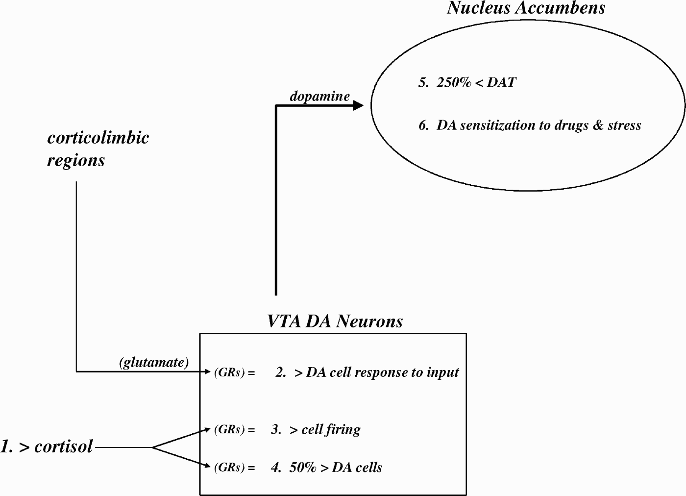
These effects appear to be enhanced by an interaction of glucocorticoid activation of GRs with DA. Amphetamine and stress both increase corticosterone secretion and NAS DA release. Administration of dexamethasone (synthetic cortisol) prenatally and during the first 7 postnatal days in rats results in a 50% increase in VTA DA neurons (Point 4, Figure 10), and increased corticosterone and NAS DA release to stress and DA-agonist drugs in adulthood (McArthur, Dalley, Buckingham, & Gillies, Reference McArthur, Dalley, Buckingham and Gillies2005). Even adult humans who report poor maternal care as children have stress-induced increased cortisol levels that correlate (r = .78) with increased NAS DA release (Pruessner, Champgane, Meaney, & Dagher, Reference Pruessner, Champagne, Meaney and Dagher2004). This effect is enhanced by stressor-induced prolonged oversecretion of cortisol (Point 1, Figure 10) that activates GRs located on VTA DA neurons (Oswald et al., Reference Oswald, Wong, McCaul, Zhou, Kuwabara and Choi2005). Such activation has the effect of increasing DA cell responses to input from corticolimbic regions (Point 2, Figure 10), possibly from an enhancement of glutamate release mediated by GRs on glutamate terminals that synapse on VTA DA neurons. In addition, cortisol activation of GRs on VTA DA neurons also enhances the frequency of DA cell firing (Point 3, Figure 10).
Thus, prolonged stressors, such as poor, conflictual, violent, or inconsistent familial environments or physical and/or sexual abuse early in life, can sensitize the VTA–NAS DA system in an enduring manner. This may have significance for personality disturbance. High levels of extraversion are often associated with histrionic–impulsive behavioral features that can lead to both occupational and interpersonal impairment. In addition, if extreme levels of extraversion reflect a sensitized DA system because of early adverse conditions, the risk for increased acquisition of addiction to even low doses of DA-agonist drugs (psychostimulants, alcohol, and opiates) later in life may be significantly increased. The increased associative conditioning of context to reward in high extraverts that we found may contribute to rapid conditioning of drug-associated contexts that enhance drug craving and maintenance of addiction. All of these effects would aggravate personality disturbance that is created in combination with other subsets of critical traits, such as neuroticism.
Social closeness/agreeableness
In contrast to the highly motivating incentive state associated with extraversion, the core content of social closeness and agreeableness scales reflects the operation of neural processes that create a warm, affectionate, gratifying subjective emotional state elicited by affiliative stimuli, such as soft touch (kissing, caressing, stroking, play); sexual touch; hair grooming; psychological and physical warmth; a caring, smiling, friendly face; soft, caring vocalizations; and shared intimacy (symbolic touching). Our hypothesis is that the subjective emotional experience of warmth and affection reflects the capacity to experience consummatory reward that is elicited by a broad array of affiliative stimuli (Depue & Morrone-Strupinsky, Reference Depue and Morrone-Strupinsky2005). This reward capacity is viewed as providing the key element utilized in associative conditioning processes that permit the development and maintenance of longer term affective bonds that are characteristic of social organization in human and other primate societies.
A broad range of evidence suggests a role for the endogenous opiate neuropeptide in reward that is exerted by opiate neurons in the hypothalamic arcuate nucleus that project to many brain regions. Most relevant to affiliative reward is the μ-opiate receptor (μOR) family, which is the main site of exogenously administered opiate drugs (e.g., morphine) and of endogenous endorphins (particularly, β-endorphin; Depue & Morrone-Strupinsky, Reference Depue and Morrone-Strupinsky2005). Rewarding properties of μOR agonists are directly indicated by the fact that animals will work for the prototypical μ-agonists morphine and heroin and that they are dose-dependently self-administered in animals and humans. β-Endorphin release on μOR receptors is increased in rats, monkeys, and humans by lactation and nursing, sexual activity, vaginocervical stimulation, maternal social interaction, brief social isolation, and grooming and other nonsexual tactile stimulation (Depue & Morrone-Strupinsky, Reference Depue and Morrone-Strupinsky2005).
In monkeys and rodents, social bonding is not possible without soft tactile stimulation from the mother (Fleming, Korsmit, & Deller, Reference Fleming, Korsmit and Deller1994), and this may involve activation of the μ-opiate system. Mammals, and especially primates, have evolved particular receptors on the skin and a sensory pathway for soft touch (Liu et al., Reference Liu, Vrontou, Rice, Zylka, Dong and Anderson2007; Olausson et al., Reference Olausson, Lamarre, Backlund, Morin, Wallin and Starck2002) that projects to the anterior insular cortex, a structure that mediates awareness of the subjective experience of emotions (Craig, Reference Craig2009). Via projections to the rACC, the anterior insula can activate β-endorphin neurons in the arcuate nucleus that activate reward and physiological quiescence via projections to the VTA–NAS and brain stem regions.
The rewarding effect of opiates may be especially mediated by μORs located in the NAS and VTA, both of which support self-administration of μOR agonists that is attenuated by intracranially administered μOR antagonists. Destruction of DA terminals in the NAS also showed that opiate self-administration is independent of DA function, at least at the level of the NAS (Laviolette, Gallegos, Hentriksen, & van der Kooy, Reference Laviolette, Gallegos, Henriksen and van der Kooy2004). Rewarding effects of opiates are also directly indicated by the fact that a range of μOR agonists, when injected intracerebroventricularly or directly into the NAS, serve as unconditioned rewarding stimuli in a dose-dependent manner in producing a conditioned place preference, a behavioral measure of reward. VTA-localized μORs particularly in the rostral zone of the VTA mediate (a) rewarding effects such as self-administration behavior and conditioned place preference, (b) increased sexual activity and maternal behaviors, and (c) the persistently increased play behavior, social grooming, and social approach of rats subjected to morphine in utero. Transgenic mice lacking the μOR gene show no morphine-induced place preferences nor physical dependence from morphine consumption. Significantly, opiate but not oxytocin antagonists block the development of partner preference that is induced specifically by repeated exposure and repeated sexual activity in rodents (Carter, Lederhendler, & Kirkpatrick, Reference Carter, Lederhendler and Kirkpatrick1997).
Individual differences in social closeness are subject to strong genetic influence (Tellegen et al., Reference Tellegen, Lykken, Bouchard, Wilcox, Segal and Rich1988). If personality disturbance occurs in the region located at the extreme tails of individual difference distributions in the trait of social closeness, it is important to show that the neuromodulators associated with the trait are sources of individual differences in these behavioral systems. Individual differences in humans and rodents have been demonstrated in levels of μOR expression and binding that are associated with a preference for μOR-agonists such as morphine (Uhl, Sora, & Wang, Reference Uhl, Sora and Wang1999; Zubieta et al., Reference Zubieta, Smith, Bueller, Xu, Kilbourn and Jewett2001). In humans, individual differences in CNS μOR densities show a range of up to 75% between lower and upper thirds of the distribution (Uhl et al., Reference Uhl, Sora and Wang1999), differences that appear to be related to variation in the rewarding effects of alcohol in humans and rodents.
One source of this individual variation is different single nucleotide polymorphisms in the μOR gene, OPRM1 (Bond et al., Reference Bond, LaForge, Tian, Melia, Zhang and Borg1998; Gelernter, Kranzler, & Cubells, Reference Gelernter, Kranzler and Cubells1999). The most prevalent of these is A118G, with an allelic frequency of ~10%. This single nucleotide polymorphism bound β-endorphin three times more tightly than the most common allelic form of the receptor. Furthermore, β-endorphin is three times more effective in agonist-induced activation of G-protein coupled potassium channels at the A118G variant receptor compared to the most common allelic form. It is important that this genetic variation in μOR properties is related to the response to rewarding drugs, such as morphine, alcohol, and cocaine, and to opiate self-administration behavior in animals (Zhang et al., Reference Zhang, Shao, Shao, Yan, Wang and Liu2007). Taken together, these studies suggest that genetic variation in μOR properties in humans and rodents is (a) substantial, (b) an essential element in the variation in the rewarding value of opiate agonists, and (c) critical in accounting for variation in the Pavlovian learning that underlies the association between contextual cues and reward, as occurs in partner and place preferences (Van den Oever et al., Reference Van den Oever, Goriounova, Li, Van der Schors, Binnekade and Schoffelmeer2008).
Two recent studies have found that μORs are critical in modulating the strength of attachment behaviors in animals. Transgenic mice lacking the μOR gene showed markedly reduced (a) opiate self-administration reward, (b) acquisition of place and mate/pup preferences, (c) maternal behaviors (pup tactile stimulation, pup retrieval, nursing), (d) play and social grooming, and (e) NAS DA release to ethanol and amphetamine (Moles, Kieffer, & D'Amato, Reference Moles, Kieffer and D'Amato2004). In addition, juvenile monkeys having the homolog of the μOR gene polymorphism that is characterized by enhanced β-endorphin functioning showed greatly enhanced distress and a lack of attachment behavior as a function of a series of maternal separations (Barr et al., Reference Barr, Schwandt, Lindell, Higley, Maestripieri and Goldman2008). Finally, we have shown that a μOR antagonist in subjects with high trait levels of social closeness reversed their heightened ratings of affection to film scenes and their greater tolerance to heat (mediated by μORs) such that they now expressed levels not different from low trait social closeness subjects (Depue, Reference Depue and Canli2006).
In terms of effects of experience on social closeness, it is the only higher order trait that shows significant within-family experience effects (Tellegen et al., Reference Tellegen, Lykken, Bouchard, Wilcox, Segal and Rich1988), indicating that both familial and nonfamilial relationships have a marked effect on trait levels. Of course, there is an immense animal and human literature that demonstrates that level of attachment is affected by interpersonal experience starting in the postnatal period (Fleming, O'Day, & Kraemer, Reference Fleming, O'Day and Kraemer1999). Unfortunately, no studies of experience-dependent modulations of the μOR system have been reported, so it is unclear whether experience operates on social bonding via this system. There is emerging evidence, however, that social interactions during the neonatal period organize the subsequent expression of affiliative behavior by altering sensitivity to neuropeptides (Cushing & Kramer, Reference Cushing and Kramer2005). What is clear is that low trait levels of social closeness/agreeableness accompany neuroticism as being the strongest personality associates of a range of personality disturbances. Whether these low trait levels are attributable to adverse early experience or an interaction of genetic and experiential factors needs empirical attention.
Social rejection sensitivity (SRS)
Items covering the nature of SRS have not been included in personality questionnaires. I view this as a serious oversight, because close interpersonal relationships are modulated by at least two very strong emotional systems: social closeness and bonding, which draws people together, and SRS, which either pulls people closer to avoid separation anxiety or pushes people away. SRS is also a central feature in personality disturbance, appearing in classical PD categories as borderline, dependent, and avoidant.
SRS is elicited by social separation because of group exclusion and whenever social relationships are threatened, damaged, or lost. It is also elicited by more symbolic forms of withdrawal of love or acceptance, including negative feedback about the self; verbal devaluation; disapproving, frowning faces; averted gazes; and expressions of the social emotions of contempt, indignation, and disgust by others. In response, people experience hurt, pain, anxiety and agitation, sadness, and the social emotions of guilt, shame, and embarrassment. Such a neurobehavioral system allows others to control social transgressions, and the rejected person avoids social ostracization that would follow if no anxiety or guilt were experienced, which in monkeys leads to death within weeks. From an evolutionary standpoint, SRS may have developed to maintain closeness of long-developing toddlers to nourishment- and safety-providing caregivers (Eisenberger & Lieberman, Reference Eisenberger and Lieberman2004). Thus, SRS may be based on social pain, which is the distressing experience arising from the perception or potential psychological distance from close others or a social group (Eisenberger & Lieberman, Reference Eisenberger and Lieberman2004).
The description of the distress associated with social rejection is dominated by the terms pain and hurt, terms that are not ordinarily associated with other negative emotions. Physical pain has two components: physical sensory experience, and affective experience of unpleasantness and psychological pain and hurt. The latter component is associated with activation in the dorsal region of the ACC (dACC). This region is also the location of neurons that provide an aversive, punishment signal to the basolateral amygdala, where that aversive signal is paired with neutral cues in emotional associative learning (Johansen & Fields, Reference Johansen and Fields2004). Thus, the dACC may provide the foundation for affectively aversive signals during the experience of harm, either physical harm or social harm. Indeed, recent studies have found that during the experience of social rejection, humans show intense activation of the dACC, which presumably activates the amygdala to generate the bodily responses that create the unpleasant emotional experience (Eisenberger & Lieberman, Reference Eisenberger and Lieberman2004; Somerville, Heatherton, & Kelley, Reference Somerville, Heatherton and Kelley2006).
Although I am not aware of any research on the effects of experience on SRS levels or dACC activation levels, it is clear that familial and nonfamilial experiences of social rejection affect social trust and security of attachments. The entire area of SRS is understudied in psychology and neuroscience.
Constraint/conscientiousness
The nature of constraint
Constraint represents a broad trait of impulsivity that modulates the expression of many domains of behavior, including emotional, motor, cognitive, and sensory reactivity (Carver & Miller, Reference Carver and Miller2006; Depue & Collins, Reference Depue and Collins1999; Depue & Lenzenweger, Reference Depue, Lenzenweger, Lenzenweger and Clarkin2005; Depue & Spoont, Reference Depue and Spoont1986; Spoont, Reference Spoont1992; Zald & Depue, Reference Zald and Depue2001). Whereas the four traits described above all reflect the activity of neurobehavioral systems, constraint lacks ties to any specific motivational-emotional system (Carver & Miller, Reference Carver and Miller2006; Depue & Collins, Reference Depue and Collins1999; Spoont, Reference Spoont1992). Thus, I have proposed that constraint is the personality trait that reflects the greatest CNS weight on the construct of a minimum response threshold (Depue & Collins, Reference Depue and Collins1999; Depue & Lenzenweger, Reference Depue, Lenzenweger, Lenzenweger and Clarkin2005; Depue & Spoont, Reference Depue and Spoont1986). This can be visualized in Figure 2 above, where the trait of constraint would be the major modulator of the threshold for behavioral manifestation. In this model, emotional higher order personality traits reflect the influence of neurobiological variables that strongly contribute to the threshold for responding, such as DA in the facilitation of incentive motivated behavior, μ-opiates in the experience of affiliative reward, and CRH in the potentiation of anxiety. Constraint modulates the probability of elicitation of all of those variables.
Significantly, as shown in Figure 11, constraint in its modulatory interaction with an emotional personality trait creates a diagonal of stability of emotional behavior. This diagonal extends from lability where the emotional trait's level is high and constraint is low (i.e., increasingly easy stimulus elicitation of behavior because of a low threshold created by the interactions in Figure 2) to rigidity when the emotional trait's level is low and constraint is high (i.e., increasingly difficult stimulus elicitation of behavior because of a high threshold created by the interaction).
The interaction of an emotional trait with the higher order trait of constraint, forming a diagonal of behavioral stability extending from labile to rigid. See text for details.
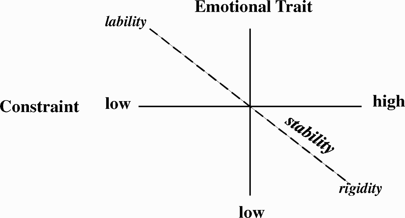
Constraint and serotonin functioning
Functional levels of neurotransmitters that provide a strong, relatively generalized tonic inhibitory influence on behavioral responding would be good candidates as significant modulators of a response elicitation threshold, and hence, would likely account for a large proportion of the variance in the trait of constraint. Numerous researchers previously (Carver & Miller, Reference Carver and Miller2006; Coccaro & Siever, Reference Coccaro and Siever1991; Depue, Reference Depue1995; Depue & Spoont, Reference Depue and Spoont1986; Lesch & Canli, Reference Lesch, Canli and Canli2006; Panksepp, Reference Panksepp1998; Spoont, Reference Spoont1992; Zald & Depue, Reference Zald and Depue2001; Zuckerman, Reference Zuckerman, Halverson, Kohnstamm and Marten1994) suggested that 5-HT, acting at multiple receptor sites in most brain regions, is such a modulator. 5-HT modulates a diverse set of functions, including emotion, motivation, motor, affiliation, cognition, food intake, sleep, sexual activity, and sensory reactivity (Carver & Miller, Reference Carver and Miller2006; Depue & Spoont, Reference Depue and Spoont1986; Lesch & Canli, Reference Lesch, Canli and Canli2006; Spoont, Reference Spoont1992). Furthermore, reduced 5-HT functioning in animals (Depue & Spoont, Reference Depue and Spoont1986; Spoont, Reference Spoont1992) and humans (Coccaro & Siever, Reference Coccaro and Siever1991) is also accompanied by irritability and hypersensitivity to stimulation in most sensory modalities (Spoont, Reference Spoont1992). Thus, 5-HT plays a substantial modulatory role in general neurobiological functioning that affects many forms of motivated behavior. In this sense, constraint might be viewed as reflecting the influence of the CNS variable of 5-HT, which we refer to as neural constraint in Figure 1. In support of this notion, we found that 5-HT agonist-induced increases in serum prolactin secretion were correlated significantly and specifically only with the Constraint/Impulsivity (control) Scale (−0.44, p < .01) from Tellegen's personality questionnaire of 13 primary scales (Depue, Reference Depue1995).
Constraint, instability, and personality disturbance
Along with neuroticism and low social closeness, constraint is well correlated with personality disturbance, particularly of the type related to unstable behavior (as in the classical unstable group of histrionic, antisocial, and boderline PDs). A vast body of animal and human evidence consistently associates reduced functioning of 5-HT neurotransmission with behavioral instability. In addition, recent research on two polymorphisms of the gene for tryptophan hydroxylase (TPH2, which is localized in brain rather than the periphery), the rate limiting enzyme in the synthesis of 5-HT, indicates marked behavioral instability (particularly in affective aggression and impulsive suicide) and increased amygdala reactivity to positive and negative emotional slides in one of the alleles that reduces brain 5-HT by 50% (Hennig, Reuter, Netter, & Buck, Reference Hennig, Reuter, Netter and Burk2005; Manuck et al., Reference Manuck, Flory, Ferrell, Dent, Mann and Muldoon1999; Rujescu et al., Reference Rujescu, Giegling, Bondy, Gietl, Zill and Moller2002). Furthermore, reduced levels of 5-HT are associated with many clinical conditions, including impulsive, violent suicide across several types of disorder, obsessive–compulsive disorder, disorders of impulse control, aggression and irritability, depression, anxiety and enhanced stress reactivity, arson, unconstrained sexual behavior, and substance abuse (Carver & Miller, Reference Carver and Miller2006; Coccaro & Siever, Reference Coccaro and Siever1991; Depue & Spoont, Reference Depue and Spoont1986; Winstanley, Theobald, Dalley, & Robbins, Reference Winstanley, Theobald, Dalley and Robbins2005).
The nature of this instability depends on the neurobehavioral system being elicited. An example of the interaction of neurobiological variables associated with constraint (5-HT) and extraversion (DA) helps to illustrate this point (for a discussion of the behavioral consequences of an interaction of each of the higher order traits with constraint, see Depue & Lenzenweger, Reference Depue, Lenzenweger, Lenzenweger and Clarkin2005). 5-HT is an inhibitory modulator of a host of DA-facilitated behaviors, including the reinforcing properties of psychostimulants, novelty-induced locomotor activity, the acquisition of self-administration of cocaine, and DA utilization in the NAS (Depue & Collins, Reference Depue and Collins1999; Depue & Spoont, Reference Depue and Spoont1986; Spoont, Reference Spoont1992). This modulatory influence arises in large part from the dense dorsal raphe 5-HT efferents to the VTA and NAS, connections that are known to modulate DA activity (Spoont, Reference Spoont1992). A 5-HT-related reduction in the threshold of DA facilitation of behavior results in an exaggerated response to incentive stimuli that is most apparent in reward–punishment conflict situations. In such situations, exaggerated responding to incentives results in (a) a greater weighting of immediate versus delayed future rewards, (b) increased reactivity to the reward of safety or relief associated with active avoidance (e.g., suicidal behavior), (c) impulsive behavior, that is, a propensity to respond to reward when withholding or delaying a response may produce a more favorable long-term outcome, and (d) various attempts to experience the increased magnitude and frequency of incentive reward, for example, self-administration of DA-active substances (Coccaro & Siever, Reference Coccaro and Siever1991; Depue & Iacono, Reference Depue and Iacono1989; Lesch & Canli, Reference Lesch, Canli and Canli2006; Winstanley et al., Reference Winstanley, Theobald, Dalley and Robbins2005). All of these effects impair the ability to sustain long-term goal-directed behavior programs (such as obtaining a college degree) by mental representations of expected rewards that can be repeatedly accessed, held on-line in the prefrontal cortex, and thereby symbolically motivate behavior (Depue & Collins, Reference Depue and Collins1999).
This discussion suggests an important role for 5-HT in the model of personality disturbance shown in Figure 1. Reduced levels of 5-HT create a highly reactive individual in terms of hyperactivity of the ANS to stress, hyperirritability to sensory overload and life frustrations, enhanced affective aggression to even minor provocations, unplanned suicidal attempts when stressors become unbearable, instability of attachment to others (vacillating between approach and avoidance), and labile positive and anxious moods. This behavioral instability may reflect the effect of 5-HT's reduced neural constraint over many CNS variables (Spoont, Reference Spoont1992), which might explain how alterations of 5-HT functioning is found in so many different clinical disorders. Such weak constraint over CNS variables might render the effects of stress as markedly increased on biology and hence behavior. In other words, 5-HT constraint may serve as a general modulator of expression on the reaction surface, increasing expression of personality disturbance during times of life stress and interpersonal chaos. At the same time, stress may increase the probability that 5-HT constraint will disinhibit the elicitation of the neurobehavioral systems of other emotional traits: animal research has shown that stressors of many types, but especially perceived or actual social aggression and social isolation (both of which are common in the life of individuals with personality disturbance), further decrease 5-HT functioning. Thus, 5-HT constraint may serve as a strong modulator of the extremity of an individual's position on a trait and, in turn, the course of intensity of personality disturbance over the life span.
Concluding Remarks
The above discussion has suggested a number of variables that contribute to the development of a position within the reaction surface (gray area, Figure 1) of personality disturbance. In my view, the sine qua non of being within that reaction surface, being susceptible to personality disturbance, is extremity on the neurobiological variables underlying a critical subset of emotional traits. The reason for this is that being at the extreme has marked effects on the threshold for eliciting those traits under stimulus conditions. Depending on the phenotypic features of personality disturbance (which is dependent not only on extremity but also on the subset of interacting major traits, as outlined in Figure 1), the impairing threshold value may be low, as in the case of neuroticism, SRS, affective aggression, fear, and extraversion (in terms of histrionic, impulsive behavior), or high, as in the case of affiliative bonding and extraversion (in terms of depressive behavior). As noted in the discussion of Figure 2, extreme threshold values lead to variation in the range of stimuli that are subject to negative or positive associative conditioning and to subsequent biasing of perceptual and attentional processes to stimuli that elicit a certain trait. Both of these latter effects likely help to maintain and increase the extremity of trait values over time.
A second important contributor to being within the reaction surface is the functioning of 5-HT, which as shown in Figure 2 modulates the value of the threshold required for trait elicitation. As shown in Figure 11, especially reduced functioning of 5-HT creates instability when interacting with extreme levels of an emotional trait. It is important to note that 5-HT-induced instability is manifested at two different levels: (a) at the phenotypic level as in Figure 11, but also (b) at the neurobiological level. Recall the discussion of 5-HT × DA interactions: reduced 5-HT functioning in the NAS results in a very low threshold for eliciting DA facilitation of behavior by incentive stimuli. This illustrates that the impact of environmental stimulation on neurobiological variables underlying emotional traits is modulated by 5-HT functioning.
Both the extremity of neurobiological variables underlying emotional traits and 5-HT functioning are modified by genetic polymorphisms, with substantial effects noted so far for the genes of the DAD4 receptor, DA transporter, the μOR, MAOA, BDNF, 5-HTT, 5-HT1A receptor, and TPH2. Thus, genetic contributions to position within the reaction surface are likely to be significant.
There is a growing literature that indicates that stressful environmental experiences have a robust effect on the functioning of neurobiological variables underlying personality traits. The stressors may derive from evironmental conditions that are external (e.g., poverty, dangerous neighborhoods), within the family (e.g., conflict, violence, sexual abuse, caregiver separation, or neglect), or within the caregiver (e.g., maternal style). The exciting aspect of this area of research is the beginning of an understanding of how these environmental influences affect neurobiological functioning and hence behavioral variation in enduring ways. This was discussed in terms of sensitization in extraversion, and a vareity of other epigenetic effects in neuroticism. Thus, this raises the possibility that environmental history may strongly affect an individual's extremity on critical personality traits, and hence position within the reaction surface of personality disturbance.
Both genetic and environmental stress variables appear to be most profound and enduring when they exert their effects during early developmental periods. This may be because brain development has critical developmental windows in postnatal, childhood, and adolescent periods. Thus, the s allele, other genetic variations in 5-HT functioning, and MAOA activity have substantial effects on the development of neural circuitry in the postnatal period when the forebrain is undergoing exuberant experience-expectant dendritic and axonal growth. These early effects endure life long, but are not significant when introduced in adulthood. There are other periods of experience-expectant brain growth in childhood, and robust growth in corticocortical connections between prefrontal and posterior regions in adolescence (Blakemore, Reference Blakemore2008). It is perhaps for this reason that the well-documented stability of most higher order personality traits is valid mainly after adolescence.
The role of individual differences in the interaction of genes and environment
In consideration of these genetic and environmental effects, the role of individual differences in trait neurobiological functioning is often neglected. However, as indicated in the discussion of Figure 2, the effects of experience on neural functioning are dependent on preexisting trait levels of genetically influenced neural functioning. This dependency has been demonstrated recently several times (Caspi et al., Reference Caspi, Sugden, Moffitt, Taylor and Craig2003; Caspi, McClay, Moffitt, Mill, & Martin, Reference Caspi, McClay, Moffitt, Mill and Marin2002), but the influence of individual differences is often neglected in discussions of the effects of experience on neural systems (e.g., Meaney, Reference Meaney2001). A dramatic example of this dependency is illustrated in Figure 12, which is based on a body of research with rats who differ in DA functioning in the NAS, the site of DA's facilitation of incentive reward (Depue & Collins, Reference Depue and Collins1999). In this example, the stimulus is not environmental but rather a direct activation of DA receptors by amphetamine (a DA agonist), and its effect on the acquisition of self-administration of amphetamine is plotted in two groups of rats differing markely in trait DA functioning. Whereas the high DA group readily acquires self-administration behavior, the low DA group is unaffected by the stimulus (amphetamine). Apparently the amphetamine has too weak an effect on the rats with low DA reactivity to reach the threshold of experiencing a subjective sense of reward. Thus, these two groups of rats will encode their surrounding context in opposite ways (as rewarding vs. neutral), such that the subsequent efficacy of the context will only be enhanced for the high DA group. These findings also mean that stress-induced epigenetic effects on the DA system (e.g., sensitization) will be much greater in the high versus low DA group. One can imagine dramatic differences in the developmental trajectories of these two groups in terms of (a) the elaborateness of their associative networks of positive emotional environmental contexts, and (b) the capacity of these contexts to elicit epigenetic effects and positive incentive motivated behavior.
Rats differing in dopamine (DA) functioning show a different course of acquisition of self-administration of amphetamine over days. See text for details.
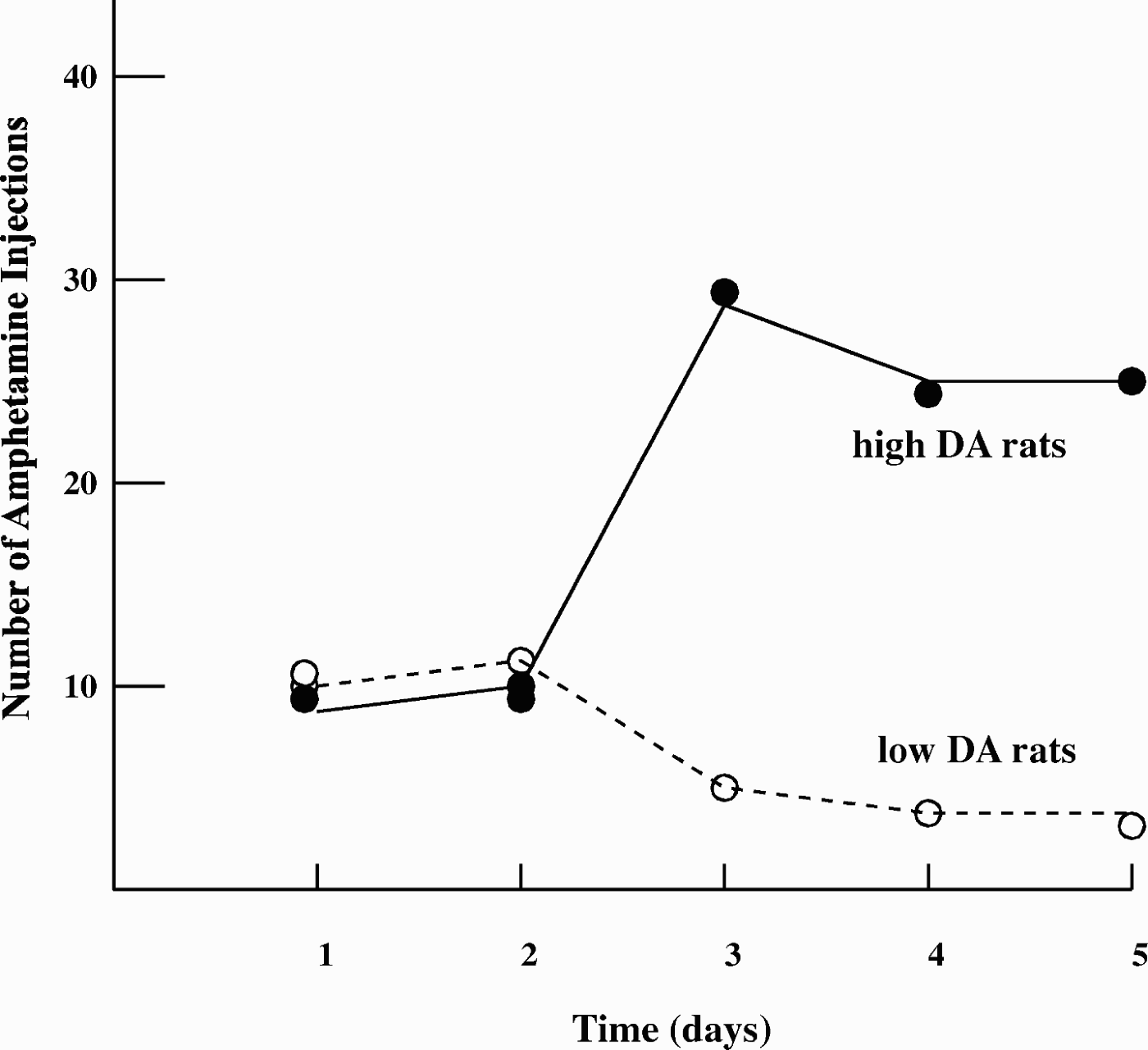
The same point can be made from the findings illustrated in the last two rows of Figure 6. The cross-fostered offspring of low LG mothers raised by high LG mothers are less prone to anxiety-related behavior as adults. This indicates that the effect is mediated by postnatal maternal environment. However, offspring of high LG mothers raised by low LG mothers do not have an increased tendency to develop anxiety-related behavior in adulthood, indicating that specific factors inherited by the high LG offspring modify or protect them from the effects of being mothered by low LG females.
Similarly, from the results in Figure 13, it was concluded that the genotypic variation in MAOA activity was only expressed as significant antisocial behavior with experience of severe childhood maltreatment: the genotype's effects on behavior are dependent on level of environmental adversity. An alternative perspective suggests that, because there is a dimension of childhood maltreatment from none to severe and the level of antisocial behavor is correlated with that dimension, the extent of environmental influence on antisocial behavior is dependent on genetically influenced neurobiological trait levels (i.e., those created by variation in MAOA activity levels), once environmental adversity rises above “none.” Both MAOA groups show increasing behavioral responses to increasing stress; it is just that stress activates a greater response in the neurobiology created by low MAOA conditions and hence in behavior. A similar case can be made in Figure 14: the effect of maltreatment on depression is dependent on the interaction of stress with different neurobiological trait levels created by genetic variation in 5-HTT (s vs. l alleles).
Effects of increasing levels of childhood maltreatment on antisocial behavior as a function of a genetic polymorphism in monoamine oxidase A (MAOA). From “Role of Genotype in the Cycle of Violence in Maltreated Children,” by A. Caspi, J. McClay, T. Moffitt, J. Mill, and J. Marin, Reference Caspi, McClay, Moffitt, Mill and Marin2002, Science, 297. Copyright 2002 by the American Association for the Advancement of Science. Reprinted with permission.

Effects of increasing levels of childhood maltreatment on depression as a function of a genetic polymorphism in the serotonin transporter (5-HTT; short vs. long alleles). From “Influence of Life Stress on Depression: Moderation by a Polymorphism in the 5-HTT Gene,” by A. Caspi, K. Sugden, T. Moffitt, A. Taylor, and I. Craig, Reference Caspi, Sugden, Moffitt, Taylor and Craig2003, Science, 301. Copyright 2003 by the American Association for the Advancement of Science. Reprinted with permission.

Finally, these various considerations can be modeled more formally as in Figure 15, where the risk of lying within the reaction surface of personality disturbance is a function of three variables. First, extremity of the trait values of neurobiological variables underlying a critical subset of emotional traits (such as a combination of high neuroticism and SRS, and low affiliative reward) is on the horizontal axis. Again, this serves as the foundation of risk, the sine qua non for the development of personality disturbance. This foundation is set by genetic factors (but, of course, later reflects a history of genetic–environment interaction), which may have their greatest impact on neural development during critical early life periods up to adolescence. Second, an individual's magnitude of historical and/or current environmental stress is a substantial contributor to development of personality disturbance. However, as shown in the figure, the environmental effect varies in its magnitude as a function of neurobiological trait levels on the horizontal axis. This implies that the magnitude of epigenetic effects are dependent on neurobiological trait levels, being greatest at extreme neurobiological levels. At low neurobiological trait levels, environmental impact via epigenetic effects are hypothesized, as illustrated in Figures 6 (bottom row), 12, 13, and 14, to be limited. Third, the level of 5-HT functioning is expected to modify risk for lying within the reaction surface of personality disturbance, particularly by its effect on neurobiological reactivity to stimulus provocation, that is, the magnitude of epigenetic effects induced by environmental provocation. Reduced 5-HT functioning is hypothesized to (a) enhance the reactivity of neurobiological variables to stress, thereby (b) increasing enduring epigenetic effects and thus extremity of trait neurobiological functioning of variables on the horizontal axis, and (c) risk of lying within the reaction surface for personality disturbance, where reduced intensities of environmental stress are required to induce epigenetic effects in the neurobiological variables on the horizontal axis, an effect represented by elevation of the upper diagonal in the figure. Thus, in this model of personality disturbance, both genetic and environmental factors play key roles, the relative influence of which probably varies as a function of type of genes, cumulative environments, developmental period, and behavioral patterns.
Nature of the interaction between environmental and neurobiological variables in the development of personality disturbance. See text for details.