




No CrossRef data available.
Article contents
Frontal lobes and attention: Processes and networks, fractionation and integration
Published online by Cambridge University Press: 22 March 2006
Abstract
The frontal lobes (FL), are they a general adaptive global capacity processor, or a series of fractionated processes? Our lesion studies focusing on attention have demonstrated impairments in distinct processes due to pathology in different frontal regions, implying fractionation of the “supervisory system.” However, when task demands are manipulated, it becomes evident that the frontal lobes are not just a series of independent processes. Increased complexity of task demands elicits greater involvement of frontal regions along a fixed network related to a general activation process. For some task demands, one or more anatomically distinct frontal processes may be recruited. In other conditions, there is a bottom-up nonfrontal/frontal network, with impairment noted maximally for the lesser task demands in the nonfrontal automatic processing regions, and then as task demands change, increased involvement of different frontal (more “strategic”) regions, until it appears all frontal regions are involved. With other measures, the network is top-down, with impairment in the measure first noted in the frontal region and then, with changing task demands, involving a posterior region. Adaptability is not just a property of FL, it is the fluid recruitment of different processes anywhere in the brain as required by the current task. (JINS, 2006, 12, 261–271.)
Keywords
Information
- Type
- SYMPOSIUM
- Information
- Journal of the International Neuropsychological Society , Volume 12 , Issue 2 , March 2006 , pp. 261 - 271
- Copyright
- © 2006 The International Neuropsychological Society
INTRODUCTION
“From the first examination of the patient, the disorder of attention is noticeable” (Hécaen & Albert, 1978, p. 368). Modern theorists support the role of the frontal lobes in attention (e.g., Heilman & Watson, 1977; Mesulam, 1985; Norman & Shallice, 1986; Posner & Petersen, 1990; Knight, 1991). Different models have been proposed. One position is that the frontal lobes act globally to support attentional functions. An apparent opposing position is that there are specific roles of distinct frontal regions for the varied attentional functions (e.g., arousal, selective attention, inhibition) studied by cognitive psychologists.
Our contribution to this theoretical debate is evidence from human lesion research (imaging research will not be reviewed, but see e.g., Paus et al., 1997; Sturm & Willmes, 2001). Neuropsychological data from our lab is presented that have some relevance to both theoretical positions. An interpretation is proposed in the last section.
The Potential Conflict
Duncan and colleagues (1996) questioned how one could reconcile a modular view of frontal functions (e.g., switching, inhibition) controlled by separate frontal systems with the undifferentiated psychometric concept of general intelligence or Spearman's “g” (Spearman, 1927). Based on a series of four experiments on goal neglect, three in normal subjects emphasizing low “g” and one in a mixed sample of “frontal” patients, they concluded that “simple goal activation is a central element of frontal impairment [with] a close link between frontal lobe function and Spearman's ‘g’” (p. 293). They offered two possible mechanisms for this linkage. First, that many but not all frontal regions may have general cognitive roles and “g” is associated with those regions, or second, that global function is the inevitable result of the coordinated activity of several different distinct frontal systems, each with the intrinsic neural flexibility to be recruited for different tasks.
A modified version of this concept was presented by Duncan and Miller (2002). Although some level of regional specialization within the frontal lobes was accepted, their emphasis again was on the role of the frontal lobes in “adaptive coding,” a term used for the general and pervasive capacity of the frontal lobe structures to adapt to any goal-directed activity. For example, in single-cell studies in animals, a single neuron can represent different information, depending on the specific task conditions (e.g., Freedman et al., 2001). This adaptive ability of the frontal lobes is also suggested by functional imaging research, since there are very similar activation patterns for different cognitive demands (for a review, see Duncan & Owen, 2000). Duncan and Miller imply that this convergence of prefrontal activation with different tasks is due to adaptability of a global attention system that responds to information as required for task completion.
This approach implies that it is difficult, or perhaps fruitless, to search for different roles for different regions, since there is substantial flexibility of neural properties. The prefrontal region is perhaps best viewed as a “general computational resource, freely adapting to solve many quite different cognitive problems” (Duncan & Miller, 2002, p. 289).
However, the prefrontal adaptability model does not address how the prefrontal region selects or discards information or even what selects and discards means in neural terms. A decade ago, in an attempt to address the specific question of “how,” we adapted the model of Norman and Shallice, suggesting that there are different attentional processes served by distinct frontal brain regions (Stuss et al., 1995), each having specific roles to play that are revealed in the unfolding performance of a task. Lesion research was used as one way to shed light on this dilemma, since this approach provides the opportunity to determine whether a particular brain region is necessary for the performance of a function, as opposed to merely being active when the function occurs. If a specific brain region is consistently necessary for a particular process, this in all likelihood suggests functional fractionation. If frontal fractionation can be demonstrated, then task difficulty can be manipulated to evaluate frontal adaptability.
How To Discover Frontal Functional Regions: A Primer
One potential reason why there might be an apparent conflict between the global and fractionation models of frontal lobe functioning may lie in the inherent problems in doing studies of fractionation of processes in lesion research. There is a particular practical problem for assembly of patient groups representing lesions in all regions of the frontal lobes, because different etiologies—trauma, stroke, hemorrhages—have such different regional predilections; thus, location effects become hard to separate from etiology effects. In our study of patients with chronic lesions, we have found virtually no effect of etiology (Stuss et al., 1994; Alexander et al., 2005; see also Elsass & Hartelius, 1985; Burgess & Shallice, 1996). The test of regional specialization within the frontal lobes compared to a more general adaptability model requires assessment of focal lesion frontal lobe patients representing many different frontal regions, and comprehensive comparison of the site of frontal lesions on behavior will require mixing etiologies.
There has also been conceptual confusion between functions that are specifically associated with the frontal lobes and “executive” functions. These are not synonymous terms. The term “executive functions” is a psychological construct, with no necessary relation to anatomical structure (although the frontal lobes may be the best instantiation of executive functions). Impairment on tests of executive function may occur after diffuse brain damage without focal frontal injury (e.g., many patients with traumatic brain injury), inefficient integrative functioning (such as may occur in confusional states), and after damage to many different non-frontal brain regions (the latter is likely secondary to impairment in the myriad additional functions required to perform the multifaceted tests often used to test executive functions). Without anatomical localization, specific relationships cannot be examined.
Another problem in studying fractionation of processes is defining what exactly a frontal function might be. There are different categories of functions within the frontal lobes, such as behavioral/emotional self-regulation, and metacognition (Stuss & Levine, 2002; Stuss, in press); not all frontal lobe functions are “executive,” if one defines this term to mean functions such as task-setting, planning, monitoring, and shifting. This review is limited to the attentional processes related to the frontal lobes, which fall most closely within the category of “executive” functions.
The close association between the terms “frontal functions” and “executive abilities” has led to the use of tests that emphasize novelty and complexity, an approach espoused (at least historically) by many authors (Stuss & Benson, 1984, 1986; Shallice, 1991; Rabbitt et al., 2001). However, lesion studies using this approach have often been ambiguous. Patients with frontal lesions are often impaired, but so are patients with nonfrontal lesions (e.g., Wisconsin Card Sorting Test; Anderson et al., 1991). In recent years, there has been a transition to simpler measures or tests in an attempt to see if this might provide evidence for fractionation of processes within the frontal lobes (e.g., Decary & Richer, 1995; Godefroy et al., 1999).
The other side of the behavior–lesion formula is anatomy. In early studies, simple frontal versus posterior comparisons evolved to evaluate left versus right versus bilateral frontal involvement (e.g., Della Malva et al., 1993). More recently we have developed methods for increasingly fine-grained lesion localization. When lesion sites become the dependent variable and performance on a specific test is the independent variable, there are several possible methods to define critical lesions: overlaps (Shammi & Stuss, 1999), split-half performance (Stuss et al., 1994), correlations of defined anatomical regions with performance (Stuss et al., 2001b), the Classification and Regression Tree (Stuss et al., 1998), and, most recently, “hotspotting”—for any test measure, the score of individuals with damage to a defined architectonic region (Petrides & Pandya, 1994) is compared to all those without damage to that area (Alexander et al., 2005; Stuss et al., 2005). The importance of this more fine-grained anatomical approach has been demonstrated in both case and group studies (e.g., Damasio & Damasio, 1989; Bigler et al., 1994; Godefroy & Rousseaux, 1996; Bechara et al., 1998; Godefroy et al., 1998; Richer & Boulet, 1999; Tranel et al., 2002; Aron et al., 2003; Hornak et al., 2004; Fellows & Farah, 2005; Simons et al., 2005).
Fractionation of Attentional Processes within the Frontal Lobes
The evidence presented for fractionation of frontal attentional processes has accumulated over time. Although data from our laboratory are highlighted for purposes of illustration, lesion research evidence for fractionation can be found in many sources (Milner, 1963, 1964; Godefroy et al., 1999; Burgess et al., 2000; Mirsky & Duncan, 2001; Burgess et al., 2005). Clinical tests provided some preliminary evidence (see Stuss et al., 2002a, for an overview). Lesions of the superior medial region impaired maintenance of an activated response mode over time in the blocked interference condition of the Stroop test (Stuss et al., 2001b) and reduced the initial “push” or activation to generate words in the first 15 seconds in verbal fluency tests (Stuss et al., 1998). Lesions of either the right or the left lateral regions affected working memory and shifting responses on the Wisconsin Card Sorting Test (Stuss et al., 2000) and Part B of the Trail Making Test (Stuss et al., 2001a).
Our 1995 theoretical paper (Stuss et al., 1995) led to the development of ROBBIA—the ROtman-Baycrest Battery to Investigate Attention, so named by Terry Picton after the Italian sculptor Lucca della Robbia who emphasized attention to salient information in his work. We had several objectives in this project: to replicate the findings from our previous research; investigate further the potential fractionation of frontal attentional processes; and specify with even greater precision the potential localization of such different attentional processes. The same basic reaction time (RT) paradigm was used (Simple RT), with stepwise elaborations (Choice RT, Prepare RT) to isolate distinctions between tasks (Stuss et al., 2005). In all tasks, speed-accuracy equivalency was reinforced. In a simple RT task, one symbol (“A”) was presented repeatedly, with the instructions to press a response key with the dominant hand as quickly as possible after seeing the stimulus. In a Choice RT task, four letters (A, B, C, D) were presented, each with a 25% probability. The subject pressed button 1 for the target (again, “A”) and button 2 for the other three nontargets. The Prepare RT test was identical to the Choice RT, apart from a warning stimulus being presented either 1 or 3 seconds before the onset of the letter. These two warning conditions were compared to the no warning Choice RT. Interstimulus intervals (ISI) within each condition were variable from 3–7 seconds, allowing modulation of expectancy to anticipate the change, with a hypothesized decrease in RT with increasing ISI. We also examined the ability of the patients to concentrate, defined by their ability to make frequent, rapid responses requiring effort at a particularly high level of response readiness (Alexander et al., 2005). Five LEDs, with a button response under each, were randomly illuminated, with the requirement to respond as quickly as possible by pressing the button under the illuminated LED. The Concentrate test requires setting attention to response options, because the continuous rapid responses are predefined and limited.
This approach provided the opportunity to assess context changes in a basic RT response. Against the background of specific deficits related to different frontal brain regions, the normal performance of many patients with large, chronic frontal lobe lesions was surprising. This normal performance also provided corroborating evidence for the specificity of deficits. When impairment was observed, there was a consistent relationship between the apparent underlying impaired process and regional lesion localization.
First, on all of the tasks, greatest slowing in RT was observed in patients with lesions of the superior medial (SM) regions, and the slowing was greatest in the most resource demanding (but not necessarily most complex) tests (see Figure 1). We have labelled the function associated with this region as activation or energization, the process that allows a subject to concentrate on a particular task. Energization can be considered as the allocation of arousal's energy to the neural systems needed to rapidly initiate the responses for a specific task, a concept similar to the thresholding function proposed by Paus (2001). It is not fatigue or drowsiness, which have more general effects. Moreover, the SM group was not significantly different from other frontal groups in their reported level of sleepiness or motivation (Stuss et al., 2005).

Patients with superior medial damage are slower in reaction time (RT) tests compared to control groups, and also often compared to other frontal groups without damage to the superior medial area. The results from four different RT tests are illustrated: (1) Simple RT (SRT)—the rapid response to a simple repeated stimulus; (2) Choice RT (CRT)—four letters are presented (A,B,C,D) with a 25% probability of appearing, the letter “A” designated as the target letter. The RTs are the correct responses to the target, presented 25% of the time; (3) Prepare RT (PRT)—comparison of the CRT without a warning signal (0) and after a warning stimulus presented either 1 or 3 seconds prior; (4) In the Concentrate task, 5 buttons are positioned under 5 LEDs, and the participant must respond as quickly as possible by pressing the button under the one LED that is illuminated, which occurs in random order. Note that on this simpler but more demanding task, the more obvious the impairment in rapid responding in the SM group.
Supportive evidence for this energization process was found in the Prepare RT results (see Figure 1). The one-second preparation interval improved RT in all groups, but the group with lesions in SM uniquely did not benefit when the warning interval was three seconds, suggesting that an activated state of alertness could not be maintained. This result confirmed our previous findings of slow RTs after lesions of the SM region on other RT tests (Stuss et al., 2002b), and on the Stroop test (Stuss et al., 2001b; see also Cohen et al., 1990), results compatible with other research on changes in activity of the cingulate cortex as a function of sleep stages (Hofle et al., 1997), vigilance (Paus et al., 1997), and alertness (Luu et al., 2000a, 2000b).
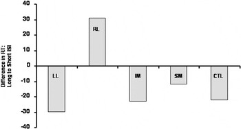
A second process was unveiled in the study of the effect of interstimulus interval (ISI) on RT (see Figure 2). The normal foreperiod effect is defined as decreasing RT with increasing ISIs (Niemi & Näätänen, 1981). The only patient group that failed to demonstrate this foreperiod effect was the group with right lateral frontal damage. This group had normal performance with a fixed warning interval (discussed later), but if they fail to track whether a stimulus has occurred over a few seconds, they will not increase their readiness to respond. Similar explanations have been offered for the functional role of this region in vigilance experiments (Wilkins et al., 1987; Pardo et al., 1991) and in functional imaging studies of monitoring (Henson et al., 1999; Shallice, 2002). According to the proposed model (Stuss et al., 1995), this deficit would be due to decreased monitoring.

The difference in reaction time (RT) between the long (6 or 7 sec) and short (3 or 4 sec) interstimulus intervals (ISIs) are presented for each of the frontal lesion groups compared to the Control (CTL) group. All patient groups except the Right Lateral (RL) have faster RTs with longer ISI. The RL group has a slower RT with a longer ISI, suggesting a monitoring deficit.
A third attentional process related to a specific region within the frontal lobes was revealed in the Concentrate (continuous rapid performance) test. As noted earlier only damage to the SM region significantly slowed RT. Errors were generally rare. Only patients with lesions in left lateral frontal regions, particularly areas 44, 45, and 47/12, made significantly more errors, and the increase was only observed in the first 20% of the trials. This deficit was interpreted as defective setting of specific stimulus-response contingencies.
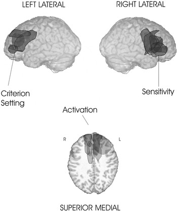
Very similar findings were demonstrated with a feature integration task that was similar to the simple reaction time/choice reaction time (SRT/CRT) with the exception that there was a third level of difficulty based on the complexity of feature integration. In this more complex task, patients had to identify and respond to a target that carried three distinct features (color, shape, and orientation of lines within the shape). Nontargets had none, one, or two features similar to the target. Three distinct attentional processes were demonstrated and each was associated with lesions in a specific frontal region (Stuss et al., 2002b; see Figure 3 for an illustration). Patients with damage to the superior medial area had the slowest reaction time, replicating the other data that show that this region is important for the energization of the required response. If the pathology was in the left lateral region, a task-setting (impaired bias or criterion setting) problem was observed, with a tendency to respond “yes” to all stimuli, reflected in greater false positives. This is similar to the task-setting deficit reported in Concentrate RT. Damage in the right lateral area, on the other hand, resulted in a significant impairment in sensitivity; patients with pathology here made errors of all kinds (omissions, false negatives, false positives), indicating a deficit in discriminating targets from nontargets. This was interpreted as a problem in monitoring the difference between targets and nontargets to diminish erroneous choices. Both the impaired foreperiod effect (ISI) and difficulty in distinguishing targets from nontargets were interpreted as impaired monitoring, with both deficits observed after right frontal damage. It is uncertain yet if these are dissociable types of monitoring, with even finer anatomical distinctions.

Analysis of performance in a complex feature integration task requiring the identification of, and rapid response to, a three-feature target presented randomly within nontargets with either zero, one, or two features revealed the involvement of three separate frontal regions associated with different processes. Damage in the right lateral area resulted in errors of all types, an impaired monitoring of the distinction between targets and nontargets. Damage to the left lateral frontal lobe caused a bias problem with more false positive responses, suggesting a tendency to respond “yes” to both targets and nontargets (Stuss et al., 2002b). Pathology in the superior medial region resulted in a significant slowing of reaction time, interpreted as deficient energization of the required response set. This figure was first published in the APA Monitor 33(11), 17, December, 2002.
These data strongly suggest that that there are different attentional functional regions within the frontal lobes; there is fractionation of frontal processes. We believe that one feature that differentiates these frontal lobe attentional processes is that they are domain general. Domain-general processes are those that are not linked to a single cognitive domain such as language or perception, but are deployed across any and all cognitive domains when a task demands it. For example, contingent response setting deficits have been observed in different types of RT tasks (described earlier), but also in memory tasks where increased false positives have been observed in similar frontal brain regions (Stuss et al., 1994; Alexander et al., 2003). Activation or “energization” deficits were seen in a wide variety of tasks, in three separate experimental groups, but only after lesions in superior medial regions. Monitoring deficits, at least on cognitive tasks, appear to result from lesions in the right lateral frontal region.
A Different View of Adaptability
Two sources of data from our own research provide a potential insight into the current “riddle of the frontal lobes” as to whether the frontal lobes function in a general adaptability mode, or whether the frontal lobes consist of a series of distinct processes. The objective here is not to integrate all the information derived from identification of different processes but to investigate if the findings here shed some light on why the frontal lobes might be interpreted as a general processor. To preface the general conclusion—the lesion research suggests that there is fractionation of processes that are flexibly assembled into different networks as required by task context and complexity. That is, there are different kinds of adaptability.
Intra-frontal Lobe Networks
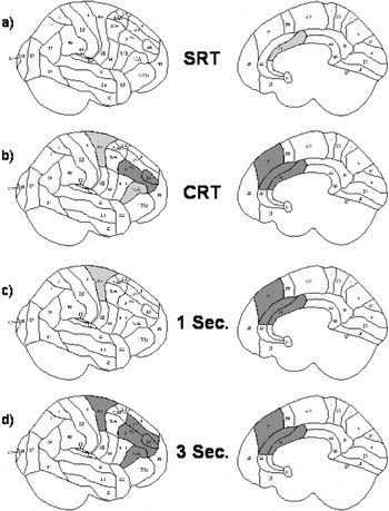
Two different frontal lobe networks, and two different types of adaptability, are illustrated in intrafrontal lobe networks. The evidence derives from the exact same ROBBIA tasks that were used to demonstrate fractionation of frontal attentional processes: the Simple RT, Choice RT, and Prepare RT tasks described earlier. In Figure 4, the lesion specificity is illustrated for these three tasks, including the 1 and 3 second warning.There are two main observations in this four-panel figure. First, shown in the right half of the figure depicting the medial view, as the demands of the task increased [from Simple RT to Choice RT (panels b, c, and d are all Choice RT)], impairments were seen with lesions in larger areas of the SM region, particularly on the right, from anterior cingulate areas 24/32 to involve medial area 9 with increasing task complexity (see right-side panels). Others have also stressed the importance of medial frontal lesions in different RT tasks (Luria, 1973; Drewe, 1975; Leimkuhler & Mesulam, 1985; Godefroy et al., 1994), without specifying a relationship of SM lesion size to task complexity. The adaptability here is the apparent importance of larger areas of the superior medial frontal cortex to tasks of increasing complexity.

The right lateral (left side) and medial (right side) views of the brain are illustrated. The highlights demonstrate the regions of the brain in which there is a significant slowing in RT compared to other regions of the brain, the lighter highlighting indicating a p < .10 significant difference, the darker highlighting p < .05 (our “hotspotting” localization method, see text). SRT = Simple RT; CRT = Choice RT, with no warning tone preceding; 1 sec = the CRT with a 1 sec warning stimulus presented prior to the stimulus presentation; 3 sec = the CRT with a warning stimulus presented 3 sec prior to the stimulus. The rights side of the figure illustrates best the impairment in activation or energization in SM pathology. The impairment is greater with CRT than SRT, but is relatively consistent across all three versions of the CRT task, with or without the warning stimulus. On the right side of the panel, the effect of the choice is illustrated. There is impairment in the right lateral region with choice, but this is minimized if a warning stimulus is presented. There is the recruitment of a second process added with the demands of a Choice RT.
A second example of the effect of task context, and a different type of adaptability, was seen with lesions of the right lateral region (left side of panel in Figure 4, right lateral view). With Simple RT (panel a), or with a Choice RT task (panel c) given a warning signal one second prior to the stimulus presentation, there was no evidence of impairment after damage to this region; however, if a choice was required without a warning (panel b), or the warning signal for the Choice RT came too early (three seconds before the Choice—panel d), dysfunction is noted in this right lateral area. This is not a general adaptability, but a more specific adaptability with the fluid recruitment of additional processes related to a different frontal brain region, dependent on task demand.
Frontal–Nonfrontal Networks
A “select what, respond where” task also informs the issue of adaptability (Stuss et al., 1999). The objective in presenting these data is not to compare the different types of attentional processes (nor their definitions—these data preceded the newer definitions), but to establish that relatively small changes in task context may require the recruitment of different processes not just within the frontal lobes, but within frontal-posterior networks. The basic demand of the task was to identify a target at a central location (select what), find the target in one of four predefined locations in the form of a cross around the central location, and then move a joy stick in the direction of that position (respond where). At times, a distracting stimulus was also present in one of the other locations. The stimuli were presented in pairs occurring in a fixed time sequence, each of the pairs having the same basic demands. Comparison of various items in the trials yielded three different attentional measures, defined as in the original article: interference (the effect of the presence of a distracting stimulus); negative priming (the effect on a second item in a trial, if the target was in the same location as the distractor was on the first item); and inhibition of return (the effect on a second item in a trial, if the target was in the same location as the distractor was on the first item, but in this instance when no target had been present—a “no go” trial). Precise definitions are available in the original article.
Manipulation of the target–distractor relationships added another layer of complexity. At the lowest level of difficulty, the target and distractor were constant (O–X, respectively). At the next level, the target was defined centrally by one of four letters that changed on every trial; the distractor was always one of the other capital letters. All letters were upper case: (UU: Upper–Upper). At the highest level of difficulty the target was defined by one of the letters presented in the central location, but this time in lower case, and the actual target and distractors were in capital letters (LU: Lower–Upper). Patients with pathology in the left frontal, right frontal, bilateral frontal, left posterior, and right posterior regions were tested.
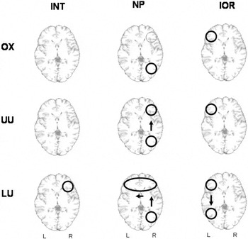
Figure 5 summarizes the results as related to the area of significant impairment. The effect of interference was observed only in the patient group with damage in the right frontal area, differing from the control group, and only at the highest level of difficulty on the measure of interference. This is a selective focal deficit, and appears to follow the “rule” that only demanding tasks are sensitive to frontal lesions.

This figure illustrates the effect of increasing task demands on three different attentional measures (see text for explanation): Interference (INT), Negative Priming (NP), and Inhibition of Return (IOR). The level of task demand increases from OX to LU. For INT, there is a relatively localized impairment in the right lateral area; for NP, the deficit is noted first in the right hemisphere for the demands of OX, but greater in the right posterior area for this “select what, respond where” task. As the task demands increase, there is increasing involvement of the frontal lobes, first of the right lateral, and then of the entire frontal regions. For IOR, the impairment in the left lateral region is always present, and remains relatively localized. However, at the highest level of task demands, when differentiation of lower and upper case letters is required, patients with damage to the left posterior region are now impaired. Three different types of processes and networks are depicted: relatively localized (INT and left frontal IOR); a bottom-up network, with increasing involvement of the frontal lobes (NP); and a top-down network (IOR), with the addition of a domain specific area as required by the task.
The effect of lesion site on negative priming was more complex. For the undemanding OX task, maximum impairment was observed after right posterior damage, with a lesser deficit after right frontal. As the task became more complex, more frontal regions were involved, until for the LU task, impairment was noted with lesions in all frontal regions. The results suggested a right hemisphere/frontal lobe network, possibly moving bottom-up from right posterior to right frontal, and at the greatest complexity all the frontal regions being involved. At first blush, if only the complex condition had been administered, the frontal lobe results with the negative priming measurement could be interpreted as compatible with the adaptability model of Duncan and Miller, in which different frontal regions work together in a common manner. Another interpretation, suggested by the use of three conditions of increasing difficulty, is that larger and more topographically diverse lesions are associated with impairment on related tasks of increasing complexity.
The Inhibition of Return measure followed a different pattern. The group with left frontal lesions was most impaired with markedly different patterns of facilitation and inhibition than the control group. The group with left posterior lesions had the same pattern of abnormal inhibition as the left frontal group on the more demanding task. No other group was significantly different from the control group under any condition. These results again suggested a network, but one that appeared to be more top-down, from left frontal to left posterior.
There is indeed evidence for adaptability within the frontal lobes. However, both the evidence for functional specificity, and the data on the complex interactions of frontal regions with changes in task demands both within the frontal lobes and in interaction with posterior brain regions, suggests that considering the frontal lobes as a general computational resource does not adequately acknowledge the functional complexity of this large region of the brain. More specifically, adaptability can be viewed as the fluid recruitment of different processes under different conditions. What is perhaps most relevant is the considerable complexity of this adaptability.
DISCUSSION
There are distinct functional regions within the frontal lobes; and there is fractionation of frontal lobe functions (more are likely to be identified). Based on adult lesion research, these processes appear to be lawful and regular. These frontal lobe functions are domain general (perhaps a better term than “executive” or “supervisory”?), that is, they are applicable to many domain-specific modules.
Are different processes organized in different manners in frontal regions—some quite focal and others more distributed? Does one process spread out to encompass different frontal brain regions, in a manner similar to a wave spreading out on still water from one dropped stone? Is it a harmonic resonation, with the different frontal brain regions drawn on for additional capacity if the task is complex? Or is adaptability merely the effect of adding one or more processes as required, either from within the different attentional processes related to the frontal lobes or from nonfrontal areas, not because of general task complexity, but because different processes, frontal (domain general) or nonfrontal (modular domain specific), are required to complete the task at hand?
The answer does not appear to be just one of the alternatives. Some frontal processes appear to be rather “fixed” and narrow, in the sense that impairment in the same process can be related to the same focal frontal region, regardless of the content domains of the task (e.g., reaction time vs. memory); such as the right lateral region monitoring impairment and susceptibility to distracting stimuli; and the left lateral region monitoring task setting. The most replicable function in this regard is the energization related to the superior medial region. This process appears to be a quite focused network, with increased task demands in a basic task involving more of the network (e.g., RT as demonstrated in the effects of Choice RT compared to Simple RT on the superior medial region, and the Concentrate task in which the number of processes involved are limited), but not other frontal networks unless a different process is required. The observation that a single neuron within the frontal lobes can be shown to carry different information, depending on task conditions, is accepted (e.g., Freedman et al., 2001), but this is difficult to demonstrate in human lesion research. Does the same type of general adaptability work on the level of assemblies of cells? Some of our past data seem to support this concept. For example, in a word-list learning task, we had demonstrated that one organization measure, subjective organization, was impaired after damage virtually anywhere in the frontal lobes (Alexander et al., 2003; Stuss et al., 1994—but note the one exception in the Alexander paper). However, the spatial negative priming example summarized in this article suggests a more complex story. There is a rather limited network between the right frontal and nonfrontal regions for simple task demands (for the OX condition) with apparently all frontal regions becoming involved for the LU condition. Had we administered only the LU condition, we might have indeed concluded (and apparently replicated the word-list learning results) that the frontal lobes are a functional general adaptability unit. However, when all three conditions are considered, adaptability would appear to be just a term for fluid recruitment of different processes under different task demands. In the task demands of the “select what, respond where” paradigm with significant spatial demands, this adaptability appeared to be driven by a bottom-up network interaction of posterior and anterior attentional systems, starting with maximum impairment in the right parietal region, and then encompassing eventually all frontal regions at the highest level of complexity. This complex interaction would not have been revealed if only the OX condition had been used or only the LU condition. With other task demands, such as the motor response inhibition of return, the anterior posterior network drives top-down (see also Tomita et al., 1999), with impaired performance limited to the left lateral frontal region with the simple task, but with increased task demands, then revealing a deficit associated with the left posterior area. In this latter network there does not appear to be frontal adaptability as the task demands increase; rather, the specificity within the frontal lobes is maintained, and the adaptability relates to anterior/posterior network interactions and the added involvement of a process associated with the left posterior region. Adaptability then is not just a property of the frontal lobes; it is a property of the brain, and in cognitive functioning it is most reflected in networks.
Implications
These findings have implications for future experimentation (and, to draw an obvious corollary, for clinical neuropsychological assessment). Defining the experimental measurements more precisely is a given, but sometimes not fully employed in neuropsychological research. In conjunction with precise lesion documentation, dissociations among processes and their particular relation to a specific brain region can be unveiled, including what brain regions are necessary for a particular function, and not just whether the region is activated.
Although Duncan and Miller (2002) had suggested that there is no further reason to study the roles of different regions, if fractionation of frontal processes is correct, and if different frontal processes do have different properties, then maybe we do indeed need first to do more “divide and conquer” research, and then study how these regions work together under different task conditions. Context, including task complexity, as well as the use of different types of tasks (e.g., visual vs. verbal; spatial/nonspatial attention, RT/memory) must be manipulated. This approach provides a window into several questions: What role does a brain region play; does this role change with changing task demands; do different brain regions become involved in a network; how do anterior/posterior attentional systems work under these different conditions? Lesion research, and functional network imaging research (Grady et al., 2001; McIntosh et al., 2003), together might more rapidly advance these concepts.
Summary
In this article, I have argued for a less common view of the complex nature of the frontal lobes. The controversy between the fractionation and general adaptability roles of the frontal lobes is a false debate. Neither model is adequate to explain the true sophistication of organization. There are fractionated processes (anatomically and functionally separable domain general processes not related to any particular knowledge domain) within the frontal regions. Some appear to maintain a fair degree of regional specificity, others appear to be more “adaptable.” Within the frontal lobes there are networks of frontal processes that work together as required by a specific task demand. These frontal processes also interact with posterior brain regions, either in a top-down, or bottom-up, fashion. It is important to investigate how networks are both locally segregated and functionally integrated. Perhaps the more adaptable the network, the higher its segregation and integration. This complexity may be the true importance of the frontal lobes.
ACKNOWLEDGMENTS
Thanks to the coauthors on the various articles referenced, all of our staff who have worked on these studies over the years, and the patients and participants who contributed their time. Fergus Craik and Mick Alexander are thanked for insightful feedback on early drafts of the paper. Susan Gillingham assisted in the figures and manuscript preparation. This research was funded by the Canadian Institutes of Health Research, # MT–12853 and # MRC–GR–14974, with additional support from the Heart and Stroke Foundation Centre for Stroke Recovery. D.T. Stuss is the Reva James Leeds Chair in Neuroscience and Research Leadership.
References
REFERENCES
Aron, A.R.,
Fletcher, P.C.,
Bullmore, E.T.,
Sahakian, B.J., &
Robbins, T.W.
(2003).
Stop-signal inhibition disrupted by damage to right inferior frontal
gyrus in humans.
Nature Neuroscience,
6,
115–116.Google Scholar
Alexander, M.P.,
Stuss, D.T., &
Fansabedian, N.
(2003).
California verbal learning test: Performance by patients with focal
frontal and non-frontal lesions.
Brain,
126,
1493–1503.Google Scholar
Alexander, M.P.,
Stuss, D.T.,
Shallice, T.,
Picton, T.W., &
Gillingham, S.
(2005).
Impaired concentration due to frontal lobe damage from two distinct
lesion sites.
Neurology,
65,
572–579.CrossRefGoogle Scholar
Anderson, S.W.,
Damasio, H.,
Jones, R.D., &
Tranel, D.
(1991).
Wisconsin Card Sorting Test performance as a measure of frontal lobe
damage.
Journal of Clinical and Experimental Neuropsychology,
13,
909–922.Google Scholar
Bechara, A.,
Damasio, H.,
Tranel, D., &
Anderson, S.W.
(1998).
Dissociation of working memory from decision making within the human
prefrontal cortex.
Journal of Neuroscience,
18,
428–437.Google Scholar
Bigler, E.D.,
Burr, R.,
Gales, S.,
Norman, M.,
Kurth, S.,
Blatter, D., &
Abildskov, T.
(1994).
Day of injury CT scan as an index to pre-injury brain
morphology.
Brain Injury,
8,
231–238.CrossRefGoogle Scholar
Burgess, P.W. &
Shallice, T.
(1996).
Response suppression, initiation and strategy use following frontal
lobe lesions.
Neuropsychologia,
34,
263–272.CrossRefGoogle Scholar
Burgess, P.W.,
Simons, J.S.,
Dumontheil, I., &
Gilbert, S.J.
(2005).
The gateway hypothesis of rostral prefrontal cortex (area 10)
function. In
J. Duncan,
L. Phillips, &
P. McLeod (Eds.),
Measuring the mind: Speed, control, and age (pp.
217–248).
Oxford, UK:
Oxford University Press.
Burgess, P.W.,
Veitch, E.,
de Lacy Costello, A., &
Shallice, T.
(2000).
The cognitive and neuroanatomical correlates of multitasking.
Neuropsychologia,
38,
848–863.CrossRefGoogle Scholar
Cohen, J.D.,
Dunbar, K., &
McClelland, J.L.
(1990).
On the control of automatic processes: A parallel distributed
processing account of the Stroop effect.
Psychological Review,
97,
332–361.CrossRefGoogle Scholar
Damasio, H. &
Damasio, A.R.
(1989).
Lesion analysis in neuropsychology.
New York:
Oxford University Press.
Decary, A. &
Richer, F.
(1995).
Response selection deficits in frontal excisions.
Neuropsychologia,
33,
1243–1253.CrossRefGoogle Scholar
Della Malva, C.L.,
Stuss, D.T.,
D'Alton, J., &
Willmer, J.
(1993).
Capture errors and sequencing after frontal brain lesions.
Neuropsychologia,
31,
363–372.CrossRefGoogle Scholar
Drewe, E.A.
(1975).
Go-no go learning after frontal lobe lesions in humans.
Cortex,
11,
8–16.CrossRefGoogle Scholar
Duncan, J.,
Emslie, H.,
Williams, P.,
Johnson, R., &
Freer, C.
(1996).
Intelligence and the frontal lobe: The organization of goal-directed
behavior.
Cognitive Psychology,
30,
257–303.CrossRefGoogle Scholar
Duncan, J. &
Miller, E.K.
(2002).
Cognitive focus through adaptive neural coding in the primate
prefrontal cortex. In
D.T. Stuss &
R.T. Knight (Eds.),
Principles of frontal lobe function (pp.
278–291).
New York:
Oxford University Press.
Duncan, J. &
Owen, A.M.
(2000).
Common regions of the human frontal lobe recruited by diverse
cognitive demands.
Trends in Neurosciences,
23,
475–483.CrossRefGoogle Scholar
Elsass, P. &
Hartelius, H.
(1985).
Reaction time and brain disease: Relations to location, etiology and
progression of cerebral dysfunction.
Acta Neurologica Scandinavica,
71,
11–19.Google Scholar
Fellows, L.K. &
Farah, M.J.
(2005).
Different underlying impairments in decision making following
ventromedial and dorsolateral frontal lobe damage in humans.
Cerebral Cortex,
15,
58–63.Google Scholar
Freedman, D.J.,
Riesenhuber, M.,
Poggio, T., &
Miller, E.K.
(2001).
Categorical representation of visual stimuli in the primate
prefrontal cortex.
Science,
291,
312–316.Google Scholar
Godefroy, O.,
Cabaret, M.,
Petit-Chenal, V.,
Pruvo, J.P., &
Rousseaux, M.
(1999).
Control functions of the frontal lobes. Modularity of the
central-supervisory system?,
Cortex,
35,
1–20.Google Scholar
Godefroy, O.,
Cabaret, M., &
Rousseaux, M.
(1994).
Vigilance and effects of fatigability, practice and motivation on
simple reaction time tests in patients with lesion of the frontal
lobe.
Neuropsychologia,
32,
983–990.CrossRefGoogle Scholar
Godefroy, O.,
Duhamel, A.,
Leclerc, X.,
Saint Michel, T.,
Henon, H., &
Leys, D.
(1998).
Brain-behaviour relationships. Some models and related statistical
procedures for the study of brain-damaged patients.
Brain,
121,
1545–1556.Google Scholar
Godefroy, O. &
Rousseaux, M.
(1996).
Binary choice in patients with prefrontal or posterior brain damage.
A relative judgement theory analysis.
Neuropsychologia,
34,
1029–1038.Google Scholar
Grady, C.L.,
McIntosh, A.R.,
Beig, S., &
Craik, F.I.M.
(2001).
An examination of the effects of stimulus type, encoding task, and
functional connectivity on the role of right prefrontal cortex in
recognition memory.
Neuroimage,
14,
556–571.Google Scholar
Hécaen, H. &
Albert, M.L.
(1978).
Human neuropsychology.
New York:
Wiley.
Henson, R.N.A.,
Shallice, T., &
Dolan, R.J.
(1999).
Right prefrontal cortex and episodic memory retrieval: A functional
MRI test of the monitoring hypothesis.
Brain,
122,
1367–1381.Google Scholar
Heilman, K.M. &
Watson, R.T.
(1977).
The neglect syndrome—A unilateral defect of the orienting
response. In
S. Harnad,
R.W. Doty,
J. Jaynes,
L. Goldstein, &
G. Krauthamer (Eds.),
Lateralization in the nervous system (pp.
285–302).
Academic Press.
Hofle, N.,
Paus, T.,
Reutens, D.,
Fiset, P.,
Gotman, J.,
Evans, A.C., &
Jones, B.E.
(1997).
Regional cerebral blood flow changes as a function of delta and
spindle activity during slow wave sleep in humans.
Journal of Neuroscience,
17,
4800–4808.Google Scholar
Hornak, J.,
O'Dherty, J.,
Bramham, J.,
Rolls, E.T.,
Morris, R.G.,
Bullock, P.R., &
Polkey, C.E.
(2004).
Reward-related reversal learning after surgical excisions in
orbito-frontal or dorsolateral prefrontal cortex in humans.
Journal of Cognitive Neuroscience,
16,
463–478.Google Scholar
Knight, R.T.
(1991).
Evoked potential studies of attention capacity in human frontal
lobe lesions. In
H. Levin,
H. Eisenberg, &
F. Benton (Eds.),
Frontal lobe function and dysfunction (pp.
139–153).
Oxford:
Oxford University Press.
Leimkuhler, M.E. &
Mesulam, M.-M.
(1985).
Reversible go-no go deficits in a case of frontal lobe tumor.
Annals of Neurology,
18,
617–619.CrossRefGoogle Scholar
Luria, A.R.
(1973).
The working brain: An introduction to neuropsychology.
New York:
Basic Books.
Luu, P.,
Collins, P., &
Tucker, D.M.
(2000a).
Mood, personality, and self-monitoring: Negative affect and
emotionality in relation to frontal lobe mechanisms of error
monitoring.
Journal of Experimental Psychology: General,
129,
43–60.Google Scholar
Luu, P.,
Flaisch, T., &
Tucker, D.M.
(2000b).
Medial frontal cortex in action monitoring.
Journal of Neuroscience,
20,
464–469.Google Scholar
McIntosh, A.R.,
Rajah, M.N., &
Lobaugh, N.J.
(2003).
Functional connectivity of the medial temporal lobe relates to
learning and awareness.
Journal of Neuroscience,
23,
6520–6528.Google Scholar
Mesulam, M.-M.
(1985).
Principles of behavioral neurology.
Philadelphia:
Davis.
Milner, B.
(1963).
Effects of different brain lesions on card sorting: The role of the
frontal lobes.
Archives of Neurology,
9,
100–110.Google Scholar
Milner, B.
(1964).
Some effects of frontal lobectomy in man. In
J.M. Warren &
K. Akert (Eds.),
The frontal granular cortex and behaviour (pp.
313–334).
New York:
McGraw-Hill.
Mirsky, A.F. &
Duncan, C.C.
(2001).
A nosology of disorders of attention. Adult Attention Deficit
Disorder.
Annals of The New York Academy of Sciences,
931,
17–32.Google Scholar
Niemi, P. &
Näätänen, R.
(1981).
Foreperiod and simple reaction time.
Psychological Bulletin,
89,
133–162.Google Scholar
Norman, D.A. &
Shallice, T.
(1986).
Attention to action: Willed and automatic control of
behaviour. In
R.J. Davidson,
G.E. Shwartz, &
D. Shapiro (Eds.),
Consciousness and self-regulation: Advances in research and
theory
(Vol. 4, pp.
1–18).
New York:
Plenum.
Pardo, J.V.,
Fox, P.T., &
Raichle, M.E.
(1991).
Localization of a human system for sustained attention by positron
emission tomography.
Nature,
349,
61–64.Google Scholar
Paus, T.
(2001).
Primate anterior cingulate cortex: Where motor control, drive and
cognition interface.
Nature Reviews Neuroscience,
2,
417–424.CrossRefGoogle Scholar
Paus, T.,
Zatorre, R.J.,
Hofle, N.,
Caramanos, Z.,
Petrides, M., &
Evans, A.C.
(1997).
Time-related changes in neural systems underlying attention and
arousal during the performance of an auditory vigilance task.
Journal of Cognitive Neuroscience,
9,
392–408.CrossRefGoogle Scholar
Petrides, M. &
Pandya, D.M.
(1994).
Comparative architectonic analysis of the human and macaque frontal
cortexy. In
F. Boller &
J. Grafman (Eds.),
Handbook of neuropsychology
(Vol. 9, pp.
17–57).
Amsterdam:
Elsevier.
Posner, M.I. &
Petersen, S.E.
(1990).
The attention system of the human brain.
Annual Review of Neuroscience,
13,
25–42.CrossRefGoogle Scholar
Rabbitt, P.,
Lowe, C., &
Shilling, V.
(2001).
Frontal tests and models for cognitive ageing.
European Journal of Cognitive Psychology,
13,
5–28.CrossRefGoogle Scholar
Richer, F. &
Boulet, C.
(1999).
Frontal lesions and fluctuations in response preparation.
Brain and Cognition,
40,
234–238.Google Scholar
Shallice, T.
(1991).
From neuropsychology to mental structure.
Behavioral and Brain Sciences,
14,
429–437.CrossRefGoogle Scholar
Shallice, T.
(2002).
Fractionation of the supervisory system. In
D.T. Stuss &
R.T. Knight (Eds.),
Principles of frontal lobe function (pp.
261–277).
New York:
Oxford University Press.
Shammi, P. &
Stuss, D.T.
(1999).
Humour appreciation: A role of the right frontal lobe.
Brain,
122,
657–666.CrossRefGoogle Scholar
Simons, J.S.,
Gilbert, S.J.,
Owen, A.M.,
Fletcher, P.C., &
Burgess, P.W.
(2005).
Distinct roles for lateral and medial anterior prefrontal cortex in
contextual recollection.
Journal of Neurophysiology,
94,
813–820.CrossRefGoogle Scholar
Spearman, C.
(1927).
The abilities of man.
New York:
Macmillan.
Sturm, W. &
Willmes, K.
(2001).
On the functional neuroanatomy of intrinsic and phasic
alertness.
NeuroImage,
14,
S76–S84.CrossRefGoogle Scholar
Stuss, D.T.,
(in press).
New approaches to prefrontal lobe testing. In
B. Miller &
J. Cummings (Eds.),
The human frontal lobes
(2nd ed.).
New York:
Guilford Publications, Inc.
Stuss, D.T.,
Alexander, M.P.,
Floden, D.,
Binns, M.A.,
Levine, B.,
McIntosh, A.R.,
Rajah, N., &
Hevenor, S.J.
(2002a).
Fractionation and localization of distinct frontal lobe processes:
Evidence from focal lesions in humans. In
D.T. Stuss &
R.T. Knight (Eds.),
Principles of frontal lobe function (pp.
392–407).
New York:
Oxford University Press.
Stuss, D.T.,
Alexander, M.P.,
Hamer, L.,
Palumbo, C.,
Dempster, R.,
Binns, M.,
Levine, B., &
Izukawa, D.
(1998).
The effects of focal anterior and posterior brain lesions on verbal
fluency.
Journal of the International Neuropsychological Society,
4,
265–278.Google Scholar
Stuss, D.T.,
Alexander, M.P.,
Palumbo, C.L.,
Buckle, L.,
Sayer, L., &
Pogue, J.
(1994).
Organizational strategies of patients with unilateral or bilateral
frontal lobe injury in word list learning tasks.
Neuropsychology,
8,
355–373.CrossRefGoogle Scholar
Stuss, D.T.,
Alexander, M.P.,
Shallice, T.,
Picton, T.W.,
Binns, M.A.,
MacDonald, R.,
Borowiec, A., &
Katz, D.
(2005).
Multiple frontal systems controlling response speed.
Neuropsychologia,
43,
396–417.Google Scholar
Stuss, D.T. &
Benson, D.F.
(1984).
Neuropsychological studies of the frontal lobes.
Psychological Bulletin,
95,
3–28.CrossRefGoogle Scholar
Stuss, D.T. &
Benson, D.F.
(1986).
The frontal lobes.
New York:
Raven Press.
Stuss, D.T.,
Binns, M.A.,
Murphy, K.J., &
Alexander, M.P.
(2002b).
Dissociations within the anterior attentional system: Effects of
task complexity and irrelevant information on reaction time speed and
accuracy.
Neuropsychology,
16,
500–513.Google Scholar
Stuss, D.T.,
Bisschop, S.M.,
Alexander, M.P.,
Levine, B.,
Katz, D., &
Izukawa, D.
(2001a).
The Trail Making Test: A study in focal lesion patients.
Psychological Assessment,
13,
230–239.Google Scholar
Stuss, D.T.,
Floden, D.,
Alexander, M.P.,
Levine, B., &
Katz, D.
(2001b).
Stroop performance in focal lesion patients: Dissociation of
processes and frontal lobe lesion location.
Neuropsychologia,
39,
771–786.Google Scholar
Stuss, D.T. &
Levine, B.
(2002).
Adult clinical neuropsychology: Lessons from studies of the frontal
lobes.
Annual Review of Psychology,
53,
401–433.CrossRefGoogle Scholar
Stuss, D.T.,
Levine, B.,
Alexander, M.P.,
Hong, J.,
Palumbo, C.,
Hamer, L.,
Murphy, K.J., &
Izukawa, D.
(2000).
Wisconsin Card Sorting Test performance in patients with focal
frontal and posterior brain damage: Effects of lesion location and test
structure on separable cognitive processes.
Neuropsychologia,
38,
388–402.CrossRefGoogle Scholar
Stuss, D.T.,
Shallice, T.,
Alexander, M.P., &
Picton, T.W.
(1995).
A multidisciplinary approach to anterior attentional
functions.
Annals of the New York Academy of Sciences,
769,
191–211.Google Scholar
Stuss, D.T.,
Toth, J.P.,
Franchi, D.,
Alexander, M.P.,
Tipper, S., &
Craik, F.I.M.
(1999).
Dissociation of attentional processes in patients with focal frontal
and posterior lesions.
Neuropsychologia,
37,
1005–1027.CrossRefGoogle Scholar
Tomita, H.,
Ohbayashi, M.,
Nakahara, K.,
Hasegawa, I., &
Miyashita, Y.
(1999).
Top-down signal from prefrontal cortex in executive control of
memory retrieval.
Nature,
401,
699–703.Google Scholar
Tranel, D.,
Bechara, A., &
Denburg, N.L.
(2002).
Asymmetric functional roles of right and left ventromedial
prefrontal cortices in social conduct, decision making and emotional
processing.
Cortex,
38,
589–612.CrossRefGoogle Scholar
Wilkins, A.J.,
Shallice, T., &
McCarthy, R.
(1987).
Frontal lesions and sustained attention.
Neuropsychologia,
25,
359–365.CrossRefGoogle Scholar

Patients with superior medial damage are slower in reaction time (RT) tests compared to control groups, and also often compared to other frontal groups without damage to the superior medial area. The results from four different RT tests are illustrated: (1) Simple RT (SRT)—the rapid response to a simple repeated stimulus; (2) Choice RT (CRT)—four letters are presented (A,B,C,D) with a 25% probability of appearing, the letter “A” designated as the target letter. The RTs are the correct responses to the target, presented 25% of the time; (3) Prepare RT (PRT)—comparison of the CRT without a warning signal (0) and after a warning stimulus presented either 1 or 3 seconds prior; (4) In the Concentrate task, 5 buttons are positioned under 5 LEDs, and the participant must respond as quickly as possible by pressing the button under the one LED that is illuminated, which occurs in random order. Note that on this simpler but more demanding task, the more obvious the impairment in rapid responding in the SM group.
The difference in reaction time (RT) between the long (6 or 7 sec) and short (3 or 4 sec) interstimulus intervals (ISIs) are presented for each of the frontal lesion groups compared to the Control (CTL) group. All patient groups except the Right Lateral (RL) have faster RTs with longer ISI. The RL group has a slower RT with a longer ISI, suggesting a monitoring deficit.
Analysis of performance in a complex feature integration task requiring the identification of, and rapid response to, a three-feature target presented randomly within nontargets with either zero, one, or two features revealed the involvement of three separate frontal regions associated with different processes. Damage in the right lateral area resulted in errors of all types, an impaired monitoring of the distinction between targets and nontargets. Damage to the left lateral frontal lobe caused a bias problem with more false positive responses, suggesting a tendency to respond “yes” to both targets and nontargets (Stuss et al., 2002b). Pathology in the superior medial region resulted in a significant slowing of reaction time, interpreted as deficient energization of the required response set. This figure was first published in the APA Monitor 33(11), 17, December, 2002.
The right lateral (left side) and medial (right side) views of the brain are illustrated. The highlights demonstrate the regions of the brain in which there is a significant slowing in RT compared to other regions of the brain, the lighter highlighting indicating a p < .10 significant difference, the darker highlighting p < .05 (our “hotspotting” localization method, see text). SRT = Simple RT; CRT = Choice RT, with no warning tone preceding; 1 sec = the CRT with a 1 sec warning stimulus presented prior to the stimulus presentation; 3 sec = the CRT with a warning stimulus presented 3 sec prior to the stimulus. The rights side of the figure illustrates best the impairment in activation or energization in SM pathology. The impairment is greater with CRT than SRT, but is relatively consistent across all three versions of the CRT task, with or without the warning stimulus. On the right side of the panel, the effect of the choice is illustrated. There is impairment in the right lateral region with choice, but this is minimized if a warning stimulus is presented. There is the recruitment of a second process added with the demands of a Choice RT.
This figure illustrates the effect of increasing task demands on three different attentional measures (see text for explanation): Interference (INT), Negative Priming (NP), and Inhibition of Return (IOR). The level of task demand increases from OX to LU. For INT, there is a relatively localized impairment in the right lateral area; for NP, the deficit is noted first in the right hemisphere for the demands of OX, but greater in the right posterior area for this “select what, respond where” task. As the task demands increase, there is increasing involvement of the frontal lobes, first of the right lateral, and then of the entire frontal regions. For IOR, the impairment in the left lateral region is always present, and remains relatively localized. However, at the highest level of task demands, when differentiation of lower and upper case letters is required, patients with damage to the left posterior region are now impaired. Three different types of processes and networks are depicted: relatively localized (INT and left frontal IOR); a bottom-up network, with increasing involvement of the frontal lobes (NP); and a top-down network (IOR), with the addition of a domain specific area as required by the task.
 You have
Access
- 121
- Cited by