Introduction
After fertilization, both pronuclei undergo nuclear envelope breakdown (NEBD) and the microtubule-organizing centres (MTOCs) (Schatten et al., Reference Schatten, Simerly and Schatten1985) form a barrel-shaped mitotic spindle. This spindle is a highly-dynamic cell structure controlled by complex regulatory protein kinases. Subsequently, both maternal and paternal chromosomes align to the metaphase plate and proceed to the first anaphase and cytokinesis, termed embryo cleavage (Schatten et al., Reference Schatten, Schatten, Mazia, Balczon and Simerly1986). One of the most remarkable features of this transition between meiosis and mitosis in rodents is the gradual shift from acentriolar MTOC-driven spindle formation to the formation of the spindle supported by true centrosome composed of two apposed centrioles surrounded by pericentriolar material (PCM). Consequently, the mitotic spindle of 1-cell mouse embryo is formed in the absence of centrioles because sperm centrioles are degraded during spermiogenesis (Manandhar et al., Reference Manandhar, Schatten and Sutovsky2005). In rodents, in which the spermatozoon lacks a centriole, these early developmental stages depend entirely on maternal centrosomal components (Sutovsky & Schatten, Reference Sutovsky and Schatten2000). Spindle morphology and the dynamics of cell division change only gradually towards a centrosome-driven division during mouse preimplantation embryo development. The first three embryonic divisions in the mouse employ a centriole-free mitotic spindle and the acentriolar MTOCs are only later replaced by true centrosomes as the centrioles are produced de novo (Courtois et al., Reference Courtois, Schuh, Ellenberg and Hiiragi2012). The exact mechanism of this transition remains to be investigated. Correct segregation of chromosomes into daughter cells is controlled by spindle-assembly checkpoint (SAC) machinery (Musacchio & Salmon, Reference Musacchio and Salmon2007). The SAC remains active in the presence of improper kinetochore–microtubule attachment, causing absence of microtubule tension and chromosome misalignment. In the absence of tension, the entry into anaphase and the completion of mitosis is delayed. When the correct microtubule–kinetochore attachment is formed and chromosomes are exposed to the tension, the SAC is satisfied and switched-off, and the cell enters anaphase. It was shown earlier that natural prolongation of the first embryonic metaphase is independent of the SAC (Sikora-Polaczek et al., Reference Sikora-Polaczek, Hupalowka, Polanski, Kubiak and Ciemerych2006), but still SAC is essential for the correct chromosome segregation during cleavage-stage mouse embryo (Wei et al. Reference Wei, Multi, Yang, Ma, Zhang, Wang, Li, Wei, Ge, Zhang, Ouyang, Hou, Shatten and Yuang2011).
Aurora kinases are a highly-conserved family of oncogenic serine/threonine protein kinases that have critical regulatory roles throughout the mitotic cell-cycle phase. In vertebrates, Aurora A (AURKA) is a key mitotic protein kinase that is mainly related to centrosome maturation (De Luca et al., Reference De Luca, Lavia and Guarguaglini2006), bipolar spindle assembly (Kufer et al., Reference Kufer, Silje, Korner, Gruss, Meraldi and Nigg2002; Barr & Gergely, Reference Barr and Gergely2007), chromosome alignment and cytokinesis (Marumoto et al., Reference Marumoto, Honda, Hara, Nitta, Hirota, Kohmura and Saya2003). In the late G2 phase of the cell cycle, AURKA is recruited to the centrosome. This process is promoted with Polo-like kinase (PLK1). Initially, AUKRA is self-activated and, in complex with its cofactor Bora, is responsible for the initial activation of PLK1 before mitotic entry (Macurek et al., Reference Macurek, Linquist, Lim, Lampson, Klompmaker, Freire, Clouin, Taylor, Yaffe and Medena2008; Seki et al., Reference Seki, Coppinger, Jang, Yates and Fang2008) and recruitment of γ-tubulin (Hannak et al., Reference Hannak, Kirkham, Hymann and Oegema2011) and the CDK1–cyclin B complex (Hirota et al., Reference Hirota, Kunitoku, Sasayama, Marumoto, Zhang, Nitta, Hatakeyama and Saya2003) to centrosomes. Immediately before mitotic entry, activated PLK1 phosphorylates Wee1 and thus activation of CDK1–cyclin B is induced. In addition, AURKA-mediated phosphorylation of Cdc25B at centrosomes is an important step contributing to the earliest events inducing mitosis, upstream of Cdk1–cyclin B activation (Cazales et al., Reference Cazales, Schmitt, Montembault, Dozier, Prigent and Ducommun2005), which is crucial for promoting mitotic entry and NEBD. During mitosis, active CDK1 primes Bora for interaction with PLK1, which induces degradation of Bora following interaction of TPX2 with AURKA. The TPX2–AURKA complex plays an important role in bipolar spindle assembly (Lens et al., Reference Lens, Voest and Medena2010). Recently it was shown that a small amount of AURKA activity is sufficient to phosphorylate and activate PLK1 in mitosis. In addition, a fraction of Bora is retained in mitosis in complex with AURKA, which is sufficient to sustain PLK1 activity (Bruinsma et al., Reference Bruinsma, Macurek, Freire, Lindquist and Medena2014). An integrated computational model of the cell cycle revealed that AURKA and aPLK1 are redundant in activation of CDK1–cyclin B during mitotic entry, but PLK1 is essential for activation of CDK1–cyclin B during G2/M-checkpoint recovery. The concurrent targeting of AURKA and PLK1 achieves a synergistic effect (Zhou et al., Reference Zhou, Singh, Mir, Parker, Lindner, Dreicer, Ecsedy, Zhang, Teh, Almasan and Hansel2013). Activation of AURKA first occurs at centrosomes at late G2 and is required for centrosome separation independently of CDK1 activity. Upon entry into mitosis, AURKA then becomes fully activated downstream of CDK1 activation. Inactivation of AURKA or PLK1 individually has no effect on CDK1 activation and entry into mitosis. But, simultaneous inactivation of both AURKA and PLK1 delays CDK1 activation and entry into mitosis (van Horn et al., Reference Van Horn, Chu, Fan, Yin, Du, Beckmann, Mader, Zhu, Toth, Blanchard and Ye2010). Selective inhibition of AURKA leads to abnormal mitotic spindle and chromosome segregation defects in both cancerous and non-transformed mammalian cells (Manfredi et al., Reference Manfredi, Ecsedy, Meetze, Blanai, Burenkova, Chen, Galvin, Hoar, Huck, LeRoy, Ray and Sells2007; van Horn et al., Reference Van Horn, Chu, Fan, Yin, Du, Beckmann, Mader, Zhu, Toth, Blanchard and Ye2010). AURKA is over-expressed in many tumours and is being investigated as a target of anti-mitotic components for cancer therapy. Therefore, MLN8327 (Alisertib) as a second generation AURKA selective inhibitor with ATP-site competence was created and has been used for Phase-I/-III clinical trials. This inhibitor displays higher specify (300-fold in in vitro assays) for AURKA over Aurora-B (Manfredi et al., Reference Manfredi, Ecsedy, Chakravarty, Silverman, Zhang, Hoar, Stroud, Chen and Shide2011). MLN8237-mediated inhibition results in disruption of mitotic spindle apparatus assembly, disruption of chromosome segregation, and breakdown of cell proliferation. Selective inhibition of AURKA activity with the small-molecule kinase inhibitor also gave rise to a similar mitotic phenotype as siRNA (Manfredi et al., Reference Manfredi, Ecsedy, Meetze, Blanai, Burenkova, Chen, Galvin, Hoar, Huck, LeRoy, Ray and Sells2007). MLN8237 treatment affected multiple processes that are differentially sensitive to the loss of AURKA function (Asteriti et al., Reference Asteriti, di Cesare, de Mattia, Hilsenstain, Neumann, Cundari, Lavia and Guarguaglini2014). In addition, the inhibition of AURKA resulted in impaired microtubule nucleation, disorganized spindles and multipolar or failed cell division, and finally variable levels of aneuploidy in daughter cells. AURKA knock-out mice died early during embryonic development before the 16-cell stage. These AURKA null embryos had defective mitosis, supporting important functions of AURKA during mitotic transitions of the early cleaving embryo. In addition, AURKA heterozygous mouse embryonic fibroblasts had higher rates of aneuploidy (Lu et al., Reference Lu, Wood, Ye, Minter-Dykhouse, Sauders, Yu and Chen2008). In this context it was shown in mice that germ line AURKA deficiency causes embryonic death at the blastocyst stage with pronounced cell proliferation failure, mitotic arrest, and monopolar spindle formation. AURKA deletion in mid-gestation embryos increased the presence of apoptotic cells (Cowley et al., Reference Cowley, Rivera-Pérez, Schliekelman, He, Oliver, Lu, O'Quinn, Salmon, Magnuson and Van Dyke2009) followed by inhibition of embryo growth, causing lethality during early post-implantation stages (Yoon et al., Reference Yoon, Cowley, Gallant, Jones, van Dyke and Rivera-Pérez2012). AURKA is a critical regulator of cell-cycle progression and microtubule organization also in the mouse oocyte during meiotic maturation, fertilization and cleavage embryo during resumption of mitosis (Yao et al., Reference Yao, Zhong, Zhang, Chen, Schatten and Sun2004). Consistently, AURKA regulates MTOCs (centrosome equivalent) number and spindle length and also γ-tubulin recruitment to MTOCs, but it is not a trigger kinase for G2/M transition during oocyte maturation (Saskova et al., Reference Saskova, Solc, Baran, Kubelka, Schultz and Motlik2008; Solc et al., Reference Solc, Baran, Mayer, Bohmova, Havlova-Panenkova, Saskova, Schultz and Motlik2012). Whereas the mechanism of spindle assembly and chromosome segregation in somatic cells have been studied intensively, there is no detailed analysis of AURKA inhibition effect on the spindle formation during the first embryonic mitosis. Using confocal time-lapse microscopy we have shown that AURKA supports spindle formation and is essential for correct chromosome segregation during the first mitosis.
Materials and methods
Embryo collection and culture
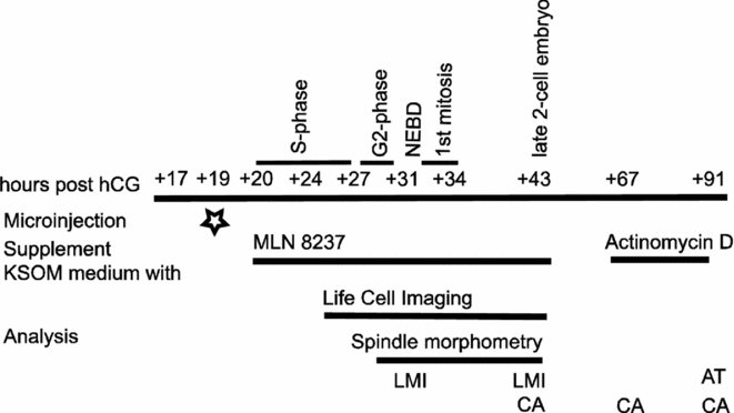
The experimental design is shown in Fig. 1. Six- to 8-week-old female mice of the C57BL/6J strain were superovulated with 5 IU of pregnant mares' serum gonadotropin (eCG) followed by 5 IU human chorionic gonadotropin (hCG) 46–47 h later and then mated in vivo with males of the BALB/c strains. Fertilized 1-cell embryos were obtained from the ampulla oviduct of plug-positive females 17 h post hCG, and the cumulus cells were dispersed with 1 mg/ml hyaluronidase. These embryos were collected into defined M2 medium (Sigma-Aldrich, St. Louis, MO, USA), followed immediately by culturing in vitro in defined potassium simplex optimized medium (KSOM) (Millipore Co., Billerica, MA, USA) at 37°C in humidified air containing 5% CO2. After 2 h of culture in pure KSOM (19 h post hCG administration), collected embryos were microinjected. At time interval 20 h post hCG, collected embryos were divided into two separate groups: (i) control embryos; and (ii) AURKA-inhibited embryos. The control embryos were cultured in pure KSOM. The AURKA-inhibited embryos were cultured in KSOM supplemented with MLN8237 (SelleckAxon Medchem, Groningen, Germany). Two concentrations of MLN8237 (500 or 1000 nM) were used. Cleavage ability of the embryos was evaluated under an inverted microscope (Nikon-Eclipse-90i; Nikon Instruments Europe B.V.) at defined time intervals 43, 67 and 91 h post hCG administration. In the time-lapse experiments, a 500 nM concentration of MLN8237 was applied.
Experimental design. All 1-cell embryos collected at time +17 h post hCG were microinjected at time +19 h post hCG. Culture medium was supplemented with AURKA A inhibitor MLN8237 at the start of the S-phase (+20 h post hCG). The control and MLN8237-treated embryos were time-lapse recorded up to +34 h post hCG. Other embryos were cultured up to +91 h post hCG. LMI (light microscopic immunocytochemistry), CA (cleavage ability), life cell imaging (time-lapse recording), AT (apoptosis tests) and spindle morphometry were applied for analysis.

mRNA in vitro transcription and microinjection
Fertilized 1-cell embryos were microinjected with mRNAs for MAP4–EGFP (Schuh & Ellenberg, Reference Schuh and Ellenberg2007) and H2B–mCherry produced by in vitro transcription as described previously (Solc et al., Reference Solc, Saskova, Baran, Kubelka, Schultz and Motlik2008) to visualize spindle formation and chromosome dynamics in live cells. The embryos were injected with 4 pl of 125 ng/μl MAP4–EGFP and 75 ng/μl H2B–mCherry in M2 medium at 19 h post hCG.
Time-lapse confocal microscopy and image analysis
For live imaging of chromosome dynamics during the first mitosis, we used CAG::H2B–GFP transgenic mice (Hadjantonakis & Papaioannou, Reference Hadjantonakis and Papaioannou2004). For live imaging of spindle formation during the first mitosis, we used the microinjected 1-cell embryos. Time-lapse image acquisitions were performed using Leica TCS SP5 confocal system equipped with an acousto-optical beam splitter (AOBS) with the following settings: lens PlanNeoFluor ×40 1.25 oil objective, 1024–1024 px resolution, 16-bit depth, 12 stacks with 7.4 μm thickness, time frame 5 min. During scanning, 488 nm and 561 nm lasers were used at 3% power. GFP and EGFP signals were detected at 500–560 nm, the mCHERRY signal was detected at 580–630 nm. The time-lapse imaging was performed at 5-min intervals to capture chromosome rearrangements at time points between the pronuclear stage and first mitosis. NEBD, normally coupled with chromosome condensation, was accounted for up to the onset of mitosis. Thus well time-defined NEBD in both control and inhibited embryos was assigned as time 0. Moreover, the onset of anaphase is very well detectable process on live-cell imaging record. Four-dimensional (3D + time) image datasets of individual embryos were processed by the Correct 3D Drift plug-in in FiJi software (Schindelin et al., Reference Schindelin, Aranda-Carreras, Frise, Kaying, Longair, Pietzsch, Preibisch, Ruenden, Saalfeld, Schmid, Tinevez, White, Hartenstein, Eliceri, Tomancak and Cardona2012). The datasets were de-noised using median filters, and images were presented as maximum Z-projection for H2B–mCherry, MAP4–EGFP, and H2B–GFP or as single confocal section for bright field. The time of NEBD was defined as the time when pronucleus chromatin began to be condensed. The spindle volume was measured from 3D-reconstructed images when the MAP4 signal was intensity thresholded (Kitajima et al., Reference Kitajima, Ohsugi and Ellenberg2011; Iannuccelli et al. Reference Iannuccelli, Mompart, Gellin, Lahbib-Mansais, Yerle and Boudier2010).
Immunocytochemistry
The in vitro cultured embryos were harvested at 29, 31 or 43 h post hCG administration. After brief washing in phosphate-buffered saline (PBS), the embryos were fixed in 3.7% paraformaldehyde diluted in PBS for 50 min at room temperature (RT). After washing in PBS (3 × 20 min), the embryos were permeabilized with 0.5% Triton X-100 in PBS for 45 min at RT and pre-incubated with inactive 2% normal goat serum (Sigma) in PBS containing 0.2% bovine serum albumin (PBS/BSA) for 2 h at RT. For indirect immunofluorescence, the embryos were incubated overnight at 4°C with the primary antibodies diluted in PBS/BSA. Acetylated α-tubulin was detected with mouse monoclonal antibody (Sigma, T7451) at 1:1000 dilution and γ-tubulin with rabbit polyclonal antibody (Biolegend, 620901) at 1:200 dilution.
The active form of AURKA was detected with rabbit anti-phosphorylated T288 AURKA antibody (Novus Biological, NB100–2371) at dilution 1:1000. For immunolabelling of pericentrin (centrosome marker), mouse antibody (BD Biosciences, 611851) was used at dilution 1:50. After washing, the embryos were stained with affinity-purified secondary antibodies coupled with fluorescent dye [fluorescein isothiocyanate (FITC) or Texas Red] for 1 h at RT. Specificity of immunostaining was established by omitting the primary antibodies or using another species-specific fluorescein conjugate; DNA was counter-stained with 4′,6-diamidino-2-phenylindole (DAPI). Finally, the embryos were mounted in Mowiol mounting medium (Merck, 475904) and examined under a Nikon-Eclipse 90i epifluorescence microscope. Images were recorded using NIS Elements imaging software.
Apoptosis assay
The in vitro cultured embryos were harvested during the interval 91–115 h post hCG administration and fixed in 3.7% paraformaldehyde as described above. After permeabilization, the embryos were incubated in terminal deoxynucleotidyl transferase dUTP nick end labelling (TUNEL) assay reagents (TUNEL, In Situ Cell Death Detection Kit; Roche, Penzberg, Germany) for 1 h at 37°C. Non-specific immunoreactions were blocked with 10% normal goat serum (NGS) for 2 h at RT followed by overnight incubation with a primary caspase-3 antibody (polyclonal rabbit cleaved caspase-3 – Asp175; Cell Signaling Tech., USA) at 1:100 dilution in 10% NGS at 4°C. After washing, the embryos were stained with affinity-purified secondary antibodies coupled with a fluorescent dye (Texas Red) for 1 h at RT. Afterwards the embryos were washed twice in PBS/BSA and cell nuclei were counter-stained with Hoechst 33342 (20 mg/ml; Sigma-Aldrich, Germany) and mounted with Vectashield (Vector Laboratories, Burlingame, CA, USA) on glass slides, and observed using an epifluorescence microscope (BX 51 Olympus, Japan).
Several control 4-cell embryos cultured in pure KSOM medium were treated with actinomycin D (0.05 mg/ml diluted in 100% ethanol; Sigma–Aldrich). Actinomycin D is a well known and standardized apoptosis inductor that interferes with RNA polymerase and inhibits RNA synthesis. It can also interfere with DNA topoisomerases I and II, and can directly damage DNA. In our analysis, the number of cell nuclei was determined for each embryo and phenotypes of nuclear morphology and also incidence of DNA fragments in the nucleoplasm were classified as follows: (i) normal (without morphological changes in nuclei, without TUNEL or cleaved caspase-3 labelling in the nucleoplasm); (ii) necrotic (fragmented or condensed chromatin, degenerated nuclei, without TUNEL or cleaved caspase-3 labelling); and (iii) apoptotic (showing at least one of specific features of apoptosis). These are typical fragments or condensed apoptotic bodies, positive TUNEL or cleaved caspase-3 labelling. For statistical analysis the numbers of normal, damaged and apoptotic cells were calculated in relation to the total number of evaluated cells. Statistical difference between groups was assessed using one-way ANOVA followed by the Tukey post hoc test.
Results
Phosphorylated AURKA associates with MTOCs during resumption of first mitosis
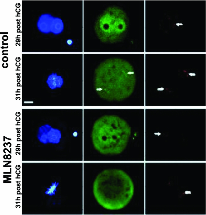
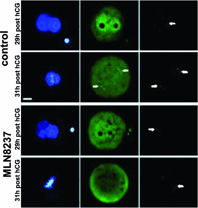
Immediately before NEBD of first mitosis, phosphorylated AURKA was detected in both male and female pronuclei (Fig. 2, control – 29 h post hCG; green). After NEBD, pAURKA Thr288 was detected in MTOCs (Fig. 2, control – 31 h post hCG, arrows). The MTOCs, detected by immunolabelling of pericentrin, were scattered throughout the cytoplasm (before NEBD) or arranged in spindle poles (Fig. 2, double arrows). No immunostaining of phosphorylated AURKA (pAURKA Thr288) was detected in MLN8237-treated 1-cell embryos immediately before NEBD or during spindle formation. Neither the male and female pronuclei nor the MTOCs displayed detectable fluorescence of pAURKA Thr288 (Fig. 2, MLN8237 – green).
Immunofluorescence of pAURKA Thr288 (green, arrows) and pericentrin (red, double arrows) immediately before NEBD (29 h post hCG) or during metaphase (31 h post hCG) in control and MLN8237-treated embryo. DNA staining with DAPI (blue). Scale bar = 10 μm.
Pharmacological inhibition of AURKA activity reduced the cleavage ability of early embryo
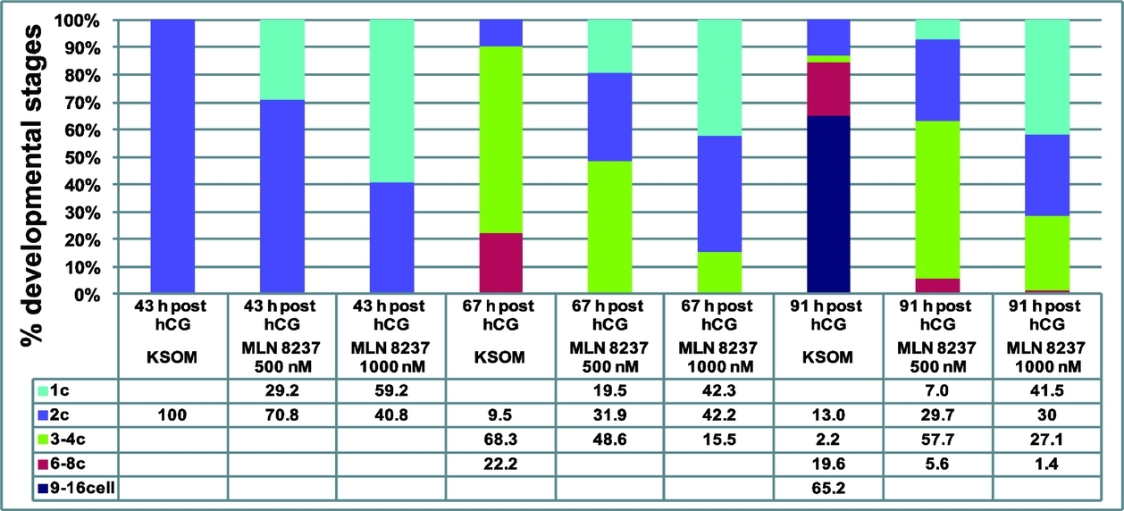
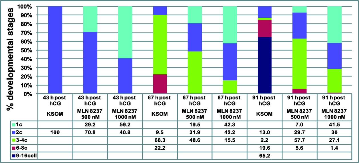
The specific AURKA inhibitor MLN8237 was used to assess the involvement of AURKA in regulation of embryo cleavage ability and mitotic spindle assembly during early embryo stages. The progression of zygotic development following culture with MLN8237 is shown in Fig. 3. In the control group, all 1-cell embryos (100%) completed first cleavage at time 43 h post hCG administration. Such embryos displayed the 2-cell stage. At the same time point, two-thirds (70.8%) of embryos treated with 500 nM MLN8237 had completed first mitosis and approximately one-third (29.2%) were still at the 1-cell stage. At higher concentrations (1000 nM) of MLN8237, 40.8% were cleaved into the 2-cell stage and 59.2% were still at the 1-cell stage. At time interval 67 h post hCG, nearly 70% (68.3%) of control embryos displayed the 3- or 4-cell stage, and 22.2% of control embryos displayed the 6- or 8-cell stage. At the same time (67 h post hCG), the MLN8237-treated embryos were significantly delayed in cleavage ability. As shown in Fig. 3, less than half (48.6%) of the embryos reached the 3- or 4-cell stage when they were inhibited with 500 nM MLN8237, and 31.9% were still at the 2-cell stage and 19.5% at the 1-cell stage. In the same period, only 15.5% of embryos cleaved into the 3- or 4-cell stage when cultured in 1000 nM MLN8237. At the higher concentration of the inhibitor, 42.2% embryos were still in 2-cell stage and 42.3% in the 1-cell stage. At time interval 91 h post hCG, nearly 70% (65.2%) of control embryos reached the early morula stage (from the 9- to 16-cell stages) and nearly 20% (19.6%) the 6- or 8-cell stage. By the same time interval, no inhibited embryo had reached more than the 8-cell stage. When the culture medium was supplemented with 500 nM MLM8237, 5.6% embryos had cleaved into the 6- or 8-cell stage, but more than half (57.7%) of the embryos were at the 3- or 4-cell stage; 7% of inhibited embryos were still at the 1-cell stage (not cleaved into 2-cell stage) and 29.7% embryos were at 2-cell stage, only. Thus, in total, 36.7% inhibited embryos were arrested or delayed in development. There were 29.7% of embryos at the 2-cell stage and 7% of embryos at the 1-cell stage. At the 1000 nM concentration of the inhibitor, only 1.4% embryos were cleaved into the 6- or 8-cell stage and less than one-third (27.1%) into the 3- or 4-cell stage. In this group, 71.5% of embryos were arrested in the earlier stages of embryogenesis. There were 30% of embryos at the 2-cell stage and 41.5% of embryos at the 1-cell stage. Taken together, these data suggest that inactivation of AURKA during the early stages of embryogenesis slows the development and gradually reduces the cleavage ability of the mouse embryo.
Cleavage of control 1-cell mouse embryos and AURKA-inhibited 1-cell embryos cultured in different concentrations (500 nM or 1000 nM) of MLN8237. Figure shows the percentage of embryo developmental stages at individual time periods after hCG administration.
MLN8237-reduced cleavage ability of early embryos results in cell death
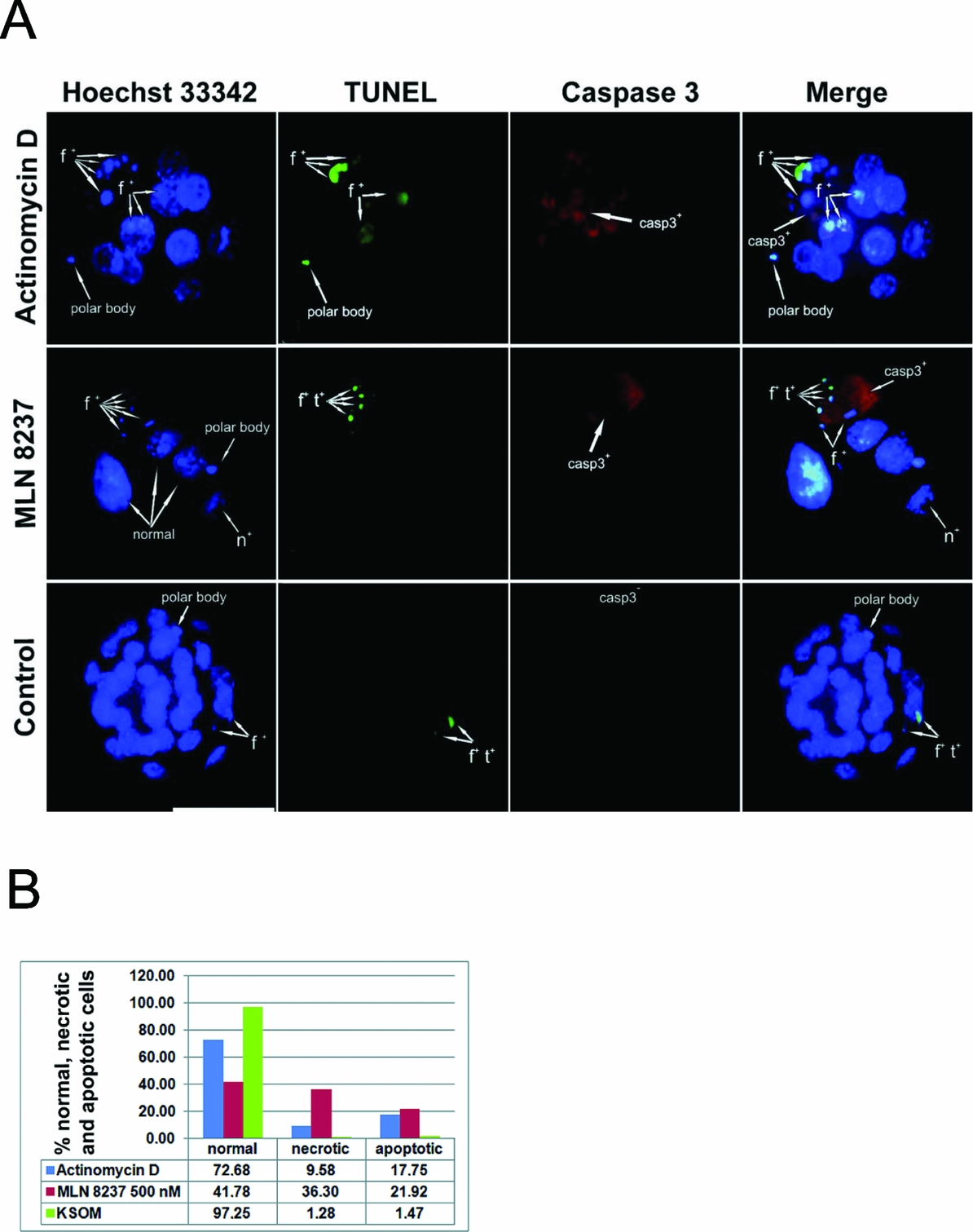
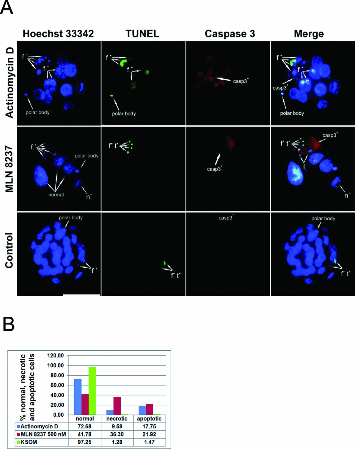
Fluorescence staining patterns of cultured embryos are shown in Fig. 4A . Nuclear morphology visualized by chromatin staining with Hoechst 33342 revealed chromatin condensation (n+) or fragmentation (f+). The chromatin fragments were also detected by TUNEL assay (t+). In addition, the presence of active forms of caspase-3 (the enzyme marker of apoptosis) assessed with immunocytochemistry were visible in cytoplasm (casp3+). Fluorescence analysis (Fig. 4B ) showed that incidence of apoptosis (21.92% MLN8237 vs. 1.47% KSOM; p < 0.01) and necrosis (36.30% MLN8237 vs. 1.28% KSOM; p < 0.001) was significantly increased in MNL8237-treated embryos (in 500 nM concentration) compared with controls. Consequently the rate of normal nuclei was reduced in MLN8237-treated mice embryos in comparison with embryos cultured in control medium (41.78% MLN8237 vs. 97.25% KSOM; p < 0.001). Actinomycin D treatment increased apoptosis (17.75%; P < 0.01) and decreased proportion of normal nuclei (72.68%; P < 0.01) but only slightly increased proportion of necrotic nuclei (9.58%; without statistical significance). The rate of apoptosis in MLN8237-treated embryos was almost at the same level as in the embryos treated with the apoptotic inductor – actinomycin D. We can conclude that growth blockage of cleaving embryos cultured in the presence of MLN8237 was caused by cell death, which is quite high (58.22%) at 91–105 h post hCG.
Fluorescence micrographs of mouse actinomycin D-treated embryo, MLN8237-treated embryo and control embryo (A). Nuclear phenotypes are visualized by chromatin staining with Hoechst 33342 (blue, f+ fragmented nucleus, n+ necrotic nucleus). DNA degradation is visualized by TUNEL labelling (green, t+). Presence of active caspase-3 (red, Casp3+) in the cytoplasm is visualized by immunohistochemical staining. Scale bars = 50 μm. Statistical evaluation of the fluorescence staining (B). The figure shows the percentage of normal, apoptotic and necrotic cells from each experimental group. Total number of evaluated embryos in the experimental groups: actinomycin D n = 44; MLN8237 n = 49; control n = 49. Statistical difference between groups was assessed using one-way ANOVA followed by Tukey post hoc test.
AURKA supports correct chromosome segregation in zygote
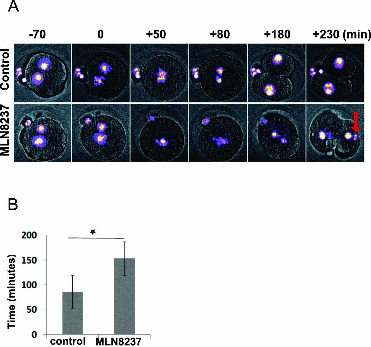
We monitored chromosome dynamics and segregation during the first mitosis in zygotes from H2B–GFP transgenic mice. Time of NEBD was arbitrarily set up as time 0 for further mitotic analysis (Fig. 5A and Video S1). Statistical analysis confirmed prolongation of the NEBD-anaphase period in all AURKA-inhibited embryos, as shown in the figure (Fig. 5B ). MLN8237-treated embryos entered anaphase significantly later (153 ± 30 min in MLN8237 vs. 86 ± 9 min in the control) and exhibited chromosome segregation errors. The newly created 2-cell embryos revealed misalignment of chromatin (micronuclei) when daughter nuclei were fully formed (Fig. 5A , arrow). These data suggest that AURKA is important for the timely normal anaphase onset and correct chromosome segregation. The delay in anaphase onset in MLN8237 embryos raises the question whether AURKA supports spindle formation.
Still images from a time-lapse recording of 1-cell mouse embryos expressing H2B–GFP during the first mitosis (A). Chromosomes are presented in pseudocolour mode to provide better contrast. Time-defined NEBD in both control and inhibited embryos was assigned as time 0. Misalignment of chromatin is visible in nuclei of 2-cell embryos (arrow). The delay of cytokinesis onset in inhibited embryos (control +80 versus MLN8237 +180) is shown in the graph (B). The asterisk indicates P < 0.001.

AURKA supports effective spindle formation in zygote
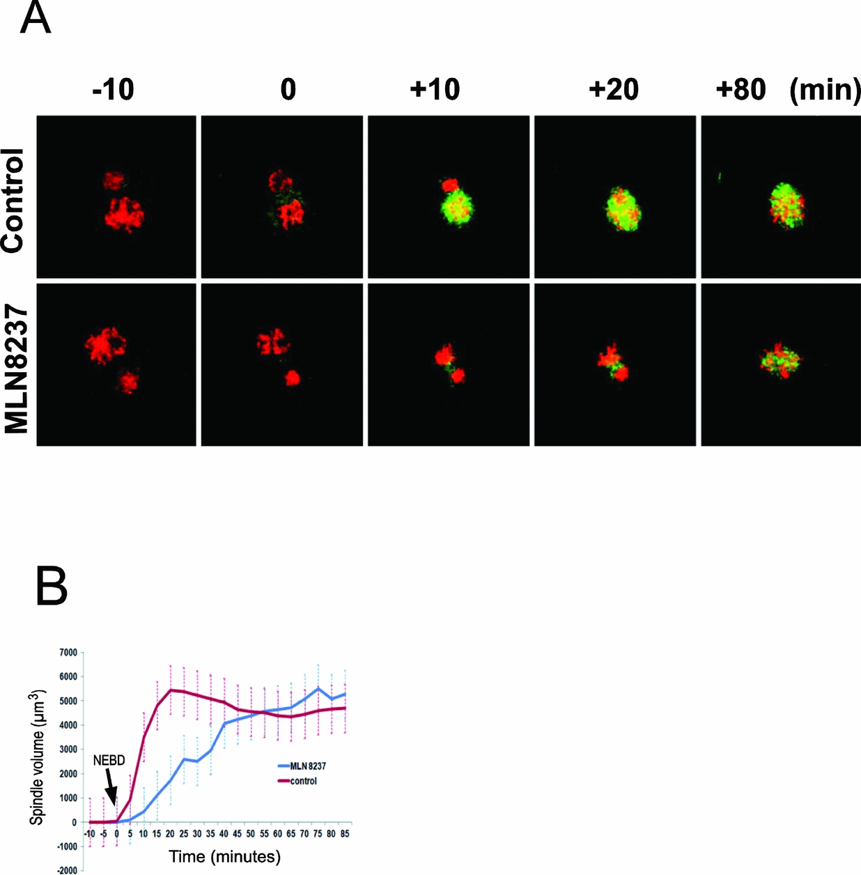
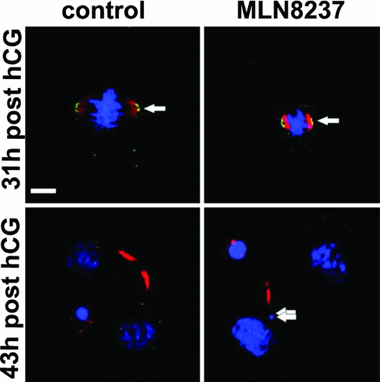
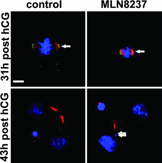
To monitor spindle assembly and chromosome segregation we expressed MAP4–EGFP and H2B–mCherry in live zygotes. The progress of spindle assembly is presented in Fig. 6A and in a real-time movie (Supporting data – Video S2). In control embryos, the spindles formed rapidly after NEBD. It was assigned as time 0 (Fig. 6A – red signal). In MLN8237-treated embryos, onset of the spindle formation was delayed but finally the spindle reached a similar volume as in the control. The spindle volume was measured after 3D reconstruction of MAP4–EGFP signals (Fig. 6A – green signal) recorded at individual time-lapse intervals. In the control embryos, the mean volume of the fully formed spindle was 5446 μm3, which was reached during a short period – at an average of 20 min (Fig. 6B – red line). Slower growth in spindle volume was recorded in embryos treated with MLN8237. In the AURKA-inhibited embryos, final spindle volume was reached during a longer developmental period – at an average of 80 min (Fig. 6B – blue line). The spindle volume was stabilized at a mean volume of 5498 μm3. The 4D image analysis (3D + time) revealed that the spindle-assembly process was slower in the AURKA-inhibited 1-cell embryos. Finally, the mitotic spindles were examined using immunofluorescence in fixed embryo controls and AURKA-inhibited 1-cell embryos (Fig. 7). In the inhibited embryos, double immunostaining for acetylated α-tubulin (Fig. 7 – red) and γ-tubulin (Fig. 7 – green, arrows) confirmed formation of a bipolar spindle with γ-tubulin accumulated on the poles of the spindle body. Chromatin staining with DAPI confirmed the presence of chromatin fragments (micronuclei) in 2-cell embryos (Fig. 7 – blue, double arrow). The same spindle phenotype was observed at both 500 nM and 1000 nM concentrations of MLN8237.
Still images from time-lapse recording of control and MLN8237-treated 1-cell mouse embryos expressing MAP4–EGFP (microtubules, green) and H2B–mCherry (chromosomes, red) during the first mitosis (A). Time-defined NEBD in both control and inhibited embryos was assigned as time 0. See also Video S2. The delay of acentriolar bipolar spindle establishment in inhibited embryos is expressed in the figure (B).

Immunofluorescence of γ-tubulin (green, arrows) and acetylated α-tubulin (red) in the control and MLN8237-treated embryo. DNA staining with DAPI (blue) shows chromatin fragments (micronuclei) in MLN8237-inhibited 2-cell embryos (double arrow). Scale bar = 10 μm.
Discussion
It was documented earlier that activation of AURKA precedes GVBD during resumption of meiosis (Saskova et al., Reference Saskova, Solc, Baran, Kubelka, Schultz and Motlik2008) and that kinase is restricted to MTOCs, because no significant changes in the amounts of phosphorylated AURKA were detected in the cytoplasm (Solc et al. Reference Solc, Baran, Mayer, Bohmova, Havlova-Panenkova, Saskova, Schultz and Motlik2012). The present study confirms the association of AURKA with MTOCs consistent with previous data for mammalian oocytes. In this study, we observed that pAURKA Thr288 is not recruited to the MTOCs of 1-cell mouse embryos treated with the AURKA inhibitor MNL8237. Despite the disrupted targeting of pAURKA Thr288 to the centrosomal structures, a bipolar spindle formed during the first mitosis of the inhibitor-treated embryos. The localization of pAURKA Thr288 on MTOCs was confirmed by double staining with pericentrin. Our study demonstrates that the activity of AURKA kinase is not essential for establishment of the bipolar acentrosomal spindle in the mouse 1-cell embryo. Conversely, inhibition of AURKA activity causes disorders in chromosome segregation during the early two embryonic divisions and arrest of cleavage ability before the 8-cell stage. We report that AURKA-inhibited cleaving of embryos promotes blastomeres to undergo apoptosis. In this context, Cowley et al. (Reference Cowley, Rivera-Pérez, Schliekelman, He, Oliver, Lu, O'Quinn, Salmon, Magnuson and Van Dyke2009) have observed that AURKA deletion in mid-gestation embryos causes an increased mitotic index and increased apoptosis. Several studies on tumour cells also showed that the action of Aurora kinase inhibitors is at least in part mediated through cell-cycle inhibition and apoptosis induction as a consequence of AURKA kinase inhibition (Kelly et al., Reference Kelly, Ecsedy, Medina, Mahalingam, Padmanabhan, Nawrocki, Giles and Carew2011; Qi et al., Reference Qi, Cooke, Liu, Rimsza, Roe, Manziolli, Persky, Miller and Mahadevan2011; Michaelis et al., Reference Michaelis, Selt, Rothweiler, Löschmann, Nüsse, Dirks, Zehner and Cinatl2014; Zhou et al., Reference Zhou, Singh, Mir, Parker, Lindner, Dreicer, Ecsedy, Zhang, Teh, Almasan and Hansel2013).
The mechanism of the first three embryonic divisions is largely shared with meiotic division, during which the spindle is self-assembled from randomly distributed MTOCs without centrioles. During cleavage from the 8-cell stage until the blastocyst stage, multiple MTOCs are focused into a sharp bipolar spindle and become full-valued centrosomes. The divisions after blastocyst (in which two centrosomes assemble a typical mitotic spindle) are the third phase of preimplantation mouse embryo development (Courtois et al., Reference Courtois, Schuh, Ellenberg and Hiiragi2012). Here we have shown that AURKA is not absolutely essential for bipolar spindle assembly but supports its correct formation. The fact that MLN8237-treated embryos exhibited chromosome segregation problems may be due to possible involvement of AURKA in microtubule–kinetochore attachment during anaphase. Alternatively microtubule–kinetochore attachment may be defective or weak already in metaphase but AURKA inhibition may weaken SAC activity. Consequently, development of 1-cell embryos was imaged over time, from the S-phase to completion of the first embryonic mitosis. Time-lapse recording at 5-min intervals pinpointed the timing of the individual phases of the first mitosis (NEBD, metaphase, cytokinesis). In this way we were able to detect prolongation of the NEBD-anaphase period in AURKA-inhibited embryos during the first cleavage stage. Relatively long G2 phase of the mouse 1-cell embryo is followed by entry into mitosis during which the pronuclei undergo NEBD. Sikora-Polaczek et al. (Reference Sikora-Polaczek, Hupalowka, Polanski, Kubiak and Ciemerych2006) showed that the first embryonic mitosis is prolonged by a transient metaphase arrest that is independent of the SAC. The authors postulated that the arrest is independent of the SAC machinery. However, the molecular mechanism of this transient arrest remains to be elucidated. In spite of the prolonged NEBD-anaphase period observed in the present study, the SAC does not block entry into anaphase because AURKA-inhibited embryos were able to finish the first mitosis by cleaving into 2-cell cells. It was presented earlier that AUKRA and PLK1 are directly involved in centrosome maturation, during which time centrosomal proteins are recruited to the PCM (Meraldi & Nigg, Reference Meraldi and Nigg2002) – the centrosome region responsible for microtubule nucleation by resident γ-tubulin (Khodjakov & Rieder, Reference Khodjakov and Rieder1999; Moser et al., Reference Moser, Fritzler, Ou and Rattner2010). Because mature mammalian oocytes have completely degraded centrioles and rodent spermatozoa lose PCM and spermatid centrioles during spermiogenesis (Manandhar et al., Reference Manandhar, Schatten and Sutovsky2005), the first three mouse embryo cleavages are centriole independent. Conversely, in non-rodent species, the sperm centriole duplicates after pronuclear apposition and the daughter centrioles separate toward the opposite poles of the pronuclei as a prelude to the formation of spindle poles (Schatten & Sun, Reference Schatten and Sun2009). Similar to somatic cells, maturation of embryonic MTOCs requires a cascade of phosphorylations that allows for the association of regulatory proteins with centrosomes to form a division-competent centrosome (Schatten & Sun, Reference Schatten and Sun2010). In this context, we have shown earlier that inhibition of PLK1 kinase activity resulted in γ-tubulin-free bipolar spindle formation without reduced microtubule-organizing activity. Although PLK1 kinase activity is not essential for mouse embryo spindle formation and bipolarity, kinase activity is essential for the metaphase–anaphase transition, independently of the SAC (Baran et al., Reference Baran, Solc, Kovarikova, Rehak and Sutovsky2013). In the present study, a bipolar spindle containing γ-tubulin accumulated on poles was formed when AURKA activity was inhibited, but microtubules failed to create a bipolar spindle when both AURKA and PLK1 were inhibited in the same embryos (not shown). In this case, γ-tubulin did not accumulate at the spindle poles. In the case of PLK1 inhibition, the arrest was persistent (Baran et al., Reference Baran, Solc, Kovarikova, Rehak and Sutovsky2013), but for AURKA inhibition, arrest was transient and 1-cell embryos completed the first division. It seems that activity of AURKA and PLK1 might co-regulate some processes during first mitosis of the early embryo. However, these kinases can antagonize each other. For example, the mitotic arrest that is induced by the inhibition of PLK1 can be overridden by the inhibition of Aurora B. Aurora B-inhibited cells exit mitosis in the presence of misattached chromosomes because the checkpoint response is ablated by inhibition of Aurora B activity. Conversely, mitotic arrest induced by inhibition of PLK1 is abrogated on inhibition of Aurora B function (for review see Lens et al., Reference Lens, Voest and Medena2010). Is a similar inactivation of the mitotic checkpoint induced by the inhibition of AURKA?
Based on our previous study (Baran et al., Reference Baran, Solc, Kovarikova, Rehak and Sutovsky2013) and the recent results it seems that inhibition of PLK1 or AURKA has different effects on mitotic progression in early embryo development. Inhibition of PLK1 activity in the oocyte (Solc et al. Reference Solc, Kitajima, Yoshida, Brzakova, Kaido, Baran, Mayer, Samalova, Motlik and Ellenberg2015), and the zygote (paper submitted) leads to SAC-independent metaphase arrest due to prevention of securin degradation before anaphase, whereas AURKA-inhibited early embryos finished the second mitosis displaying aberrant mitosis up to triggering cell death. More experiments will be required to test the exact contribution of AURKA (and other associated protein kinases) to the mitotic checkpoints and probably also in events of MTOCs transformation up to full-value centrosomes.
Taken together, our results suggest that AURKA activity is not essential for establishment of an acentrosomal bipolar spindle in the mouse 1-cell embryo but AURKA is essential for MTOC transformation up to full-value centrosomes, and for correct chromosome segregation in the first mitosis as a prerequisite for normal later development after first cleavage.
Supplementary material
To view supplementary material for this article, please visit http://dx.doi.org/10.1017/S0967199415000222
Acknowledgements
This study was supported by grant APVV-0237–10 from the Slovak Grant Agency. AB and PS was supported by ExAM CZ.1.05/2.1.00/03.0124.