Introduction
Reproductive biotechniques used for in vitro embryo production depend on fertilizable oocytes, which are present in limited numbers in the ovary and are enclosed in preovulatory follicles. In this context, the development of an in vitro culture system capable of promoting the growth and development of preantral follicles for the retrieval of competent mature oocytes could enable the production of a vast number of embryos from a single ovary. This fact would cause a profound impact on fundamental research through a better comprehension of the processes comprising folliculogenesis and could also be practically applicable in clinics. This efficient system could be employed for the treatment of infertility in humans, avoiding intensive ovarian stimulations, as well as in domestic animals in order to maximize the reproductive potential of genetically valuable individuals (Demeestere et al., Reference Demeestere, Simon, Englert and Delbaere2003; Haidari et al., Reference Haidari, Salehnia and Valojerdi2008).
Since methods for isolation and in vitro culture of preantral follicles from mammalian ovaries were established, several culture systems have been proposed for the study of the complex mechanisms regulating follicular growth and oocyte maturation in vitro. The effects of some factors apparently essential for the success of in vitro development have been evaluated, such as the culture of follicles in group or individually (Spears et al., Reference Spears, De Bruin and Gosden1996; Wu et al., Reference Wu, Emery and Carrell2001; Gupta et al., Reference Gupta, Nandi, Ravindranatha and Sarma2002) and culture medium composition, which has been supplemented with antioxidants, growth factors and hormones (Eppig & O'Brien, Reference Eppig and O'Brien1996; O'Brien et al., Reference O'Brien, Pendola and Eppig2003; Mao et al., Reference Mao, Smith, Rucker, Wu, Mccauley, Cantley, Prather, Didion and Day2004; Demeestere et al., Reference Demeestere, Centner, Gervy, Englert and Delbaere2005; Han et al., Reference Han, Lan, Wu, Han, Feng, Wang and Tan2006; Gao et al., Reference Gao, Wang and Wu2007; Gupta et al., Reference Gupta, Ramesh, Manjunatha, Nandi and Ravindra2008).
Some authors have demonstrated the importance of grouping preantral follicles retrieved from domestic animals for the in vitro culture, which increases growth rates in comparison with the culture of single follicles (Wu et al., Reference Wu, Emery and Carrell2001; Gupta et al., Reference Gupta, Nandi, Ravindranatha and Sarma2002). On the other hand, Spears et al. (1998) assessed the influence of interactions between follicles on in vitro growth until the antral stage, and demonstrated the occurrence of dominance in pairs of follicles cultured in contact. These authors hypothesized a specific interaction between adjacent follicles which determines the success of dominant follicle development.
Another important factor for the success of preantral follicles culture is the medium composition in which some key components, such as follicle stimulating hormone (FSH), play crucial roles. The in vitro development of preantral follicles is a complex process that comprises interactions of regulatory factors and hormonal signals. Ovarian function is critically regulated by FSH, which controls steroidogenesis, the proliferation and differentiation of granulosa cells, the development of antral follicles and the selection of dominant follicles (Baker & Spears, Reference Baker and Spears1999; Demeestere et al., Reference Demeestere, Centner, Gervy, Englert and Delbaere2005). Due to its pivotal functions, FSH has been included in the culture of preantral follicles of mice and other mammals (Nayadu & Osborn Reference Nayudu and Osborn1992; Cortvrindt et al., Reference Cortvrindt, Smitz and Van Steirteghem1996; Mao et al., Reference Mao, Wu, Smith, Mccauley, Cantley, Prather, Didion and Day2002).
Few studies have been reported regarding the in vitro development of isolated caprine preantral follicles. Moreover, the factors required in the production of competent oocytes for fertilization and in vitro embryo development are not fully defined. In this context, the aim of this study was to evaluate the influence of number of follicles per drop (one or three) and antral follicles on in vitro development of isolated goat preantral follicles.
Materials and methods
Chemicals
Unless mentioned otherwise, culture media, supplements and chemicals used in the present study were purchased from Sigma Chemical Co.
Source of ovaries
Caprine ovaries (n = 32) were obtained at a local slaughterhouse from adult cross-breed goats (n = 16). Immediately postmortem, ovaries were washed once in 70% alcohol for 10 s and then twice in minimum essential medium (MEM) containing HEPES and antibiotics (100 µg/ml penicillin and 100 µg/ml streptomycin) and transported within 1 h to the laboratory in MEM at 4°C.
Culture medium
The culture medium was alpha-MEM supplemented with 1.25 mg/ml bovine serum albumin (BSA), ITS (10 µg/ml insulin, 5.5 µg/ml transferrin and 5 ng/ml selenium), 2 mM glutamine, 2 mM hypoxanthine and 50 µg/ml ascorbic acid. For Experiments 2 and 3, the same medium was supplemented with recombinant FSH (Nanocore, Brazil) at the concentration of 1000 ng/ml.
Isolation and selection of preantral follicles
At the laboratory, the ovaries were stripped of surrounding fat tissue and ligaments, and thin (1 mm) slices from the ovarian surface were made using a surgical blade and washed in fragmentation medium (MEM HEPES) with antibiotics. Preantral follicles larger than 150 µm in diameter with no antral cavities were visualized under a dissecting microscope and isolated using 26G needles attached to a syringe, and then transferred to the culture medium for quality evaluation. Isolated follicles from different ovaries were pooled and only preantral follicles with a centrally located oocyte surrounded by various compact layers of granulosa cells and no damage in the basal membrane were selected for culture.
Experimental design
Experiment 1 was carried out to verify the effect of individual or group culturing of isolated follicles in medium without FSH. Selected preantral follicles were randomly distributed in 40 µl microdroplets of cultured medium covered with mineral oil in culture dishes (60 × 15 mm) at 39°C in a humidified atmosphere of 5% CO2 in air. Individually cultured follicles (one follicle per drop) represented the control, and groups of three follicles per drop were considered as the treatment group. The culture media were prepared daily and kept for equilibration in a CO2 incubator for at least 2 h before using for culture. Every alternate day, medium was partially replaced (20 µl per drop). The day on which the follicles were placed in culture was taken as day 0 (D0) and subsequent days as day 2 (D2), day four (D4), and so on.
In Experiment 2, to determine the effect of individual (one follicle per drop) and group (three follicles per drop) culture of isolated follicles in medium with FSH, preantral follicles were cultured under the same conditions as described above except for the addition of FSH (1000 ng/ml) to the culture medium.
In Experiment 3, to evaluate the effect of follicular dominance of early antral follicles over preantral follicles in vitro, the latter were cultured individually or in groups (three follicles per drop) in the presence or absence of an early antral follicle with a diameter of approximately 330 µm in medium containing 1000 ng/ml FSH. Early antral follicles and preantral follicles were dissected individually as described in Experiments 1 and 2. Isolated follicles were distributed in 40 µl microdroplets of the same medium used in Experiment 2. Treatment 1 consisted of one follicle per drop; treatment 2, one follicle per drop in co-culture with an early antral follicle; treatment 3 was carried out with three follicles per drop; treatment 4, group of three follicles co-cultured with an early antral follicle.
Morphological evaluation of follicle development
The viability of all of the preantral follicles was assessed every 6 days using a precalibrated ocular micrometer in a stereomicroscope (SMZ 645, Nikon) at ×20 magnification. Follicle degeneration was recognized through observation of rupture of the basement membrane, opacity of granulosa cells and/or extrusion of the oocyte from the follicle.
Follicle diameter and growth rate were recorded every 6 days by means of two perpendicular measures of each preantral follicle. Growth rate was calculated as the diameter variation during the culture period. The interval of 6 days of culture was chosen for diameter measurements because preliminary studies in our laboratory showed that no significant changes in follicle size occurred over a period shorter than 6 days. Antrum formation was considered as soon as small patches of antral cavities appeared.
Retrieval of oocytes from in vitro grown preantral follicles and viability assessment
At the end of culture, oocytes were dissected out from follicles with fine needles (26G) under a stereomicroscope from all of the surviving follicles. Previous studies demonstrated that caprine oocytes smaller than 100 µm were not able to resume meiosis (Crozet et al., Reference Crozet, Dahirel and Gall2000). Based on that finding, only oocytes larger than 110 µm were used for the in vitro maturation (IVM) procedure in our study. Oocytes with homogenous ooplasm and at least one layer of compact cumulus cells were considered acceptable for IVM. The recovery percentage of oocytes was calculated as the number of acceptable quality oocytes recovered out from the total number of cultured follicles. Collected oocytes were washed in IVM medium comprising TCM199 supplemented with 100 µg/ml luteinizing hormone (LH), 10% fetal bovine serum, 5 µg/ml FSH, 10 ng/ml epidermal growth factor, 22 µg/ml pyruvate and 1 µg/ml estradiol and placed in groups into 50 µl droplets of the same medium, previously equilibrated at 39°C under a humidified atmosphere of 5% CO2 in air. These droplets were covered with mineral oil and the oocytes were incubated under the same conditions for 26 to 30 hours.
After IVM, oocytes were denuded and live/dead fluorescence labelling was performed in 100 µl droplets of TCM-HEPES containing 4 µM calcein-AM, 2 µM ethidium homodimer-1 (Molecular Probes, Invitrogen), and 10 µl of Hoechst 33342 stain followed by an incubation at 37°C for 15 min. Finally, oocytes were mounted on glass slides and examined using a DMLB fluorescence microscope (Leica). The emitted fluorescent signals of Hoechst stain, calcein-AM and ethidium homodimer were collected at 350, 488 and 568 nm, respectively. While the first probe detected intracellular esterase activity of viable cells, the latter labelled the nucleic acids of non-viable cells with plasma membrane disruption. Oocytes were considered live if the cytoplasm was stained positively with calcein-AM (green) and if chromatin was not labelled with ethidium homodimer (red).
Statistical analysis
The percentages of follicular survival, antrum formation and maturation rates were compared by chi-squared test (StatView for Windows). Follicular diameter and growth rate after culture were compared by ANOVA and Kruskal–Wallis test. Values were considered statistically significant when p < 0.05.
Results
Experiment 1: The effect of individually (one follicle per drop) and group (three follicles per drop) cultured isolated preantral follicles in medium without FSH
Viability
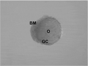
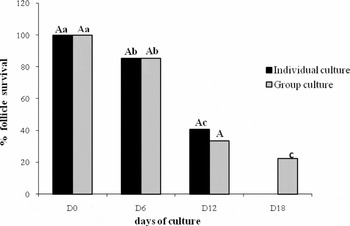
A total of 54 follicles were cultured for 22 days in Experiment 1. Immediately after isolation, preantral follicle granulosa cells surrounded a spherical oocyte, and the granulosa cells were limited by an intact basement membrane (Fig. 1). The follicular survival decreased significantly during the culture period (Fig. 2). There was no significant difference in the survival rate of the follicles when they were cultured in groups or alone in the microdrop until 12 days of culture. However, on D18, there were no viable follicles in the control group while 22.2% of the follicles cultured in group survived.
Isolated caprine preantral follicle on D0. BM, basement membrane; GC, granulosa cells; O, oocyte.

Percentages of follicular survival during 18 days of culture of preantral follicles cultured individually (control) or in groups (treatment) in medium without FSH (Experiment 1). A,B – indicates the significance between treatments on each day of culture; a,b,c – indicates the significance between the days of culture within each treatment.

Follicle diameter and growth rate
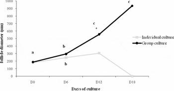
The mean diameters of the preantral follicles at the onset of the culture (D0) were 178.76 ± 40.9 µm and 180.02 ± 36.6 µm in the control and test group, respectively, which were similar to each other (p < 0.05) until D6 of culture. However, the diameter of follicles cultured in groups was significantly higher than that of the control from D12 of culture onwards (Fig. 3).
Diameter of follicles cultured individually (control) or in groups of three (treatment) in the absence of FSH (Experiment 1). * Indicates the significance between treatments from this day of culture onwards. a,b,c – indicates the significance between the days of culture within each treatment.

Growth rates of follicles cultured in groups were also significantly higher (21.4 ± 11.6 µm) than those of individually cultured follicles (13.6 ± 8.1 µm, p < 0.05).
Antral cavity formation
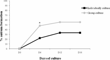
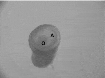
Antrum formation was detected through the visualization of translucent cavities using a stereomicroscope, and began on D2 of culture in follicles cultured in group and on D4 in the control (Fig. 4). A significantly higher percentage of antral cavity formation was observed in follicles cultured in groups (51.85%) in comparison with the control (22.2%) on D6 of culture. Nevertheless, from D12 of culture onwards, no significant difference was observed between the control (29%) and test group (55.6%, p < 0.05, Fig. 5).
Antrum formation in follicles cultured individually (control) or in groups of three (treatment) in the absence of FSH (Experiment 1). *Indicates the significance between treatments on this day of culture.

Antrum formation in a preantral follicle. A, antral cavity; O, oocyte.

Oocyte recovery after culture
In this experiment, oocytes did not reach the minimum diameter (110 µm) for IVM.
Experiment 2: The effect of individually (one follicle per drop) and group (three follicles per drop) cultured isolated preantral follicles in medium with FSH added
Viability
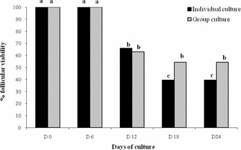
Percentages of viable follicles after individual (control) or group culture (treatment 2) were similar. In contrast to the results of Experiment 1 (culture without FSH), in which a significant decrease of the percentages of viable follicles was observed from D6 of culture onwards in both treatments, the use of FSH sustained viability until the same day of culture. Viability was significantly decreased in individually cultured follicles from D12 to D18 of culture, while percentages of viable follicles in groups were maintained from D12 until D24 of culture (Fig. 6).
Percentages of follicular survival during the 18 days of culture of preantral follicles cultured individually (control) or in groups (treatment) in medium with FSH (Experiment 2). a,b,c – indicates the significance between the days of culture within each treatment.

Follicle diameter and growth rate
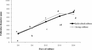
The mean diameters of follicles in both treatments were similar throughout the in vitro culture. Follicle growth rates in the control and test group were 19.29 ± 9.51 µm/day and 20.07 ± 9.52 µm/day, respectively, and did not differ statistically. With regard to the period of culture, follicle diameters increased significantly from D6 to D12 of culture in both treatments, but only follicles cultured in groups showed a significant growth from D12 to D18 (Fig. 7).
Follicular diameters of preantral follicles cultured individually (control) or in groups (treatment) in medium with FSH (Experiment 2) over 24 days. a,b,c,d – indicates the significance between the days of culture within each treatment.

Antral cavity formation
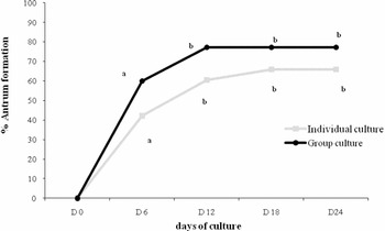
Percentages of follicles showing antrum formation were similar between the individual (control) and group culture (p < 0.05) during the entire experiment (Fig. 8).
Antrum formation in follicles cultured individually (control) or in groups of three in medium with FSH (Experiment 2). a,b – indicates the significance between the days of culture within each treatment.

Oocyte recovery after culture
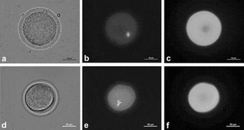
After 24 days of culture, a total of 33 oocytes reached the minimum diameter for IVM (≥110 µm) in both treatments. In the group of follicles cultured individually (n = 38), oocytes were recovered from 11 follicles (28.95%), while group cultured follicles yielded a higher rate of oocyte recovery (62.86%, 22 oocytes, p < 0.05). After IVM, 9.09% of the oocytes retrieved from follicles cultured in groups resumed meiosis and reached the metaphase I and anaphase I stages (Fig. 9A–C).
Meiosis resumption in oocytes recovered from preantral follicles cultured in groups in medium containing FSH (Experiment 2). A viable metaphase I oocyte (a–c) and a viable anaphase I oocyte (d–f) labelled with Hoechst stain (b, e) and calcein-AM (c, f). Scale bars represent 50 µm.

Experiment 3: The effect of early antral follicle co-culture with individually (one follicle per drop) and group (three follicles per drop) cultured isolated preantral follicles in medium with FSH
Viability
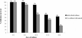
Individual × antral. After 12 and 24 days of culture, a higher percentage of viable single preantral follicles was observed in the group cultured without antral follicles (control group) (p < 0.05). Viability was sustained in the control until D12 of culture, while co-culture with antral follicles significantly reduced the percentages of viable follicles from D6 to D12, and at the end of the culture (D24), viability in this group was only 23% (Fig. 10).
Percentages of follicular survival during the 24 days of culture of preantral follicles cultured individually (control) or with antral follicles (treatment) (Experiment 3). A,B – indicates the significance between treatments on each day of culture; a,b,c – indicates the significance between the days of culture within each treatment.

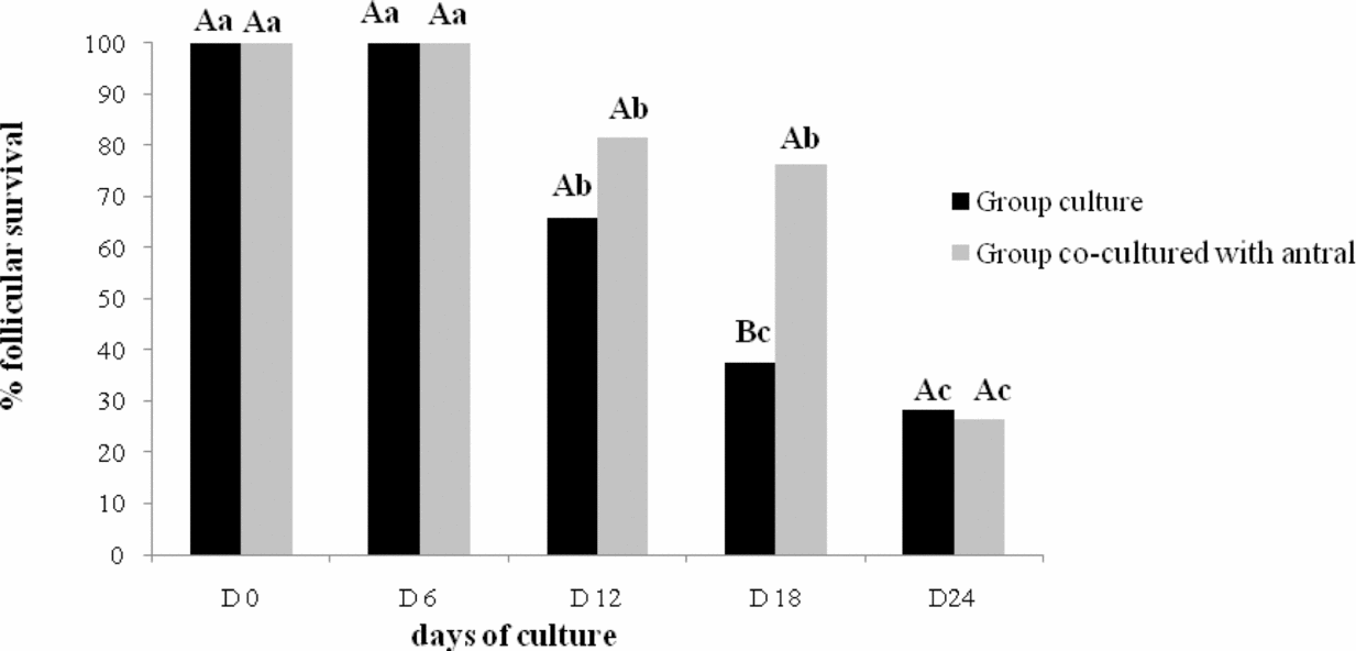
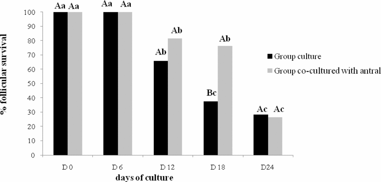
Group × antral. The presence of an antral follicle positively influenced the viability of preantral follicles cultured in groups, which was significantly higher (76%) on D18 of culture in comparison with the treatment without antral follicles (37.5%, p < 0.05). The latter group showed a significant reduction of viability from D12 to D18 of culture, while in the former treatment, percentages of viable follicles were decreased only on D24 (Fig. 11).
Percentages of follicular survival during the 24 days of culture of preantral follicles cultured in groups (control) or in groups co-cultured with antral follicles (treatment) (Experiment 3). A,B – indicates the significance between treatments on each day of culture; a,b,c – indicates the significance between the days of culture within each treatment.
Follicle diameter and growth rate
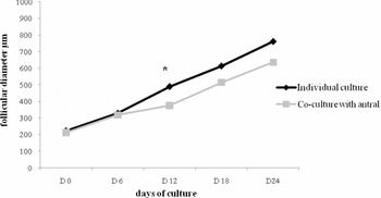
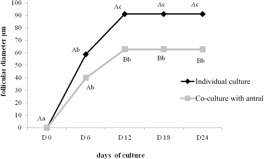
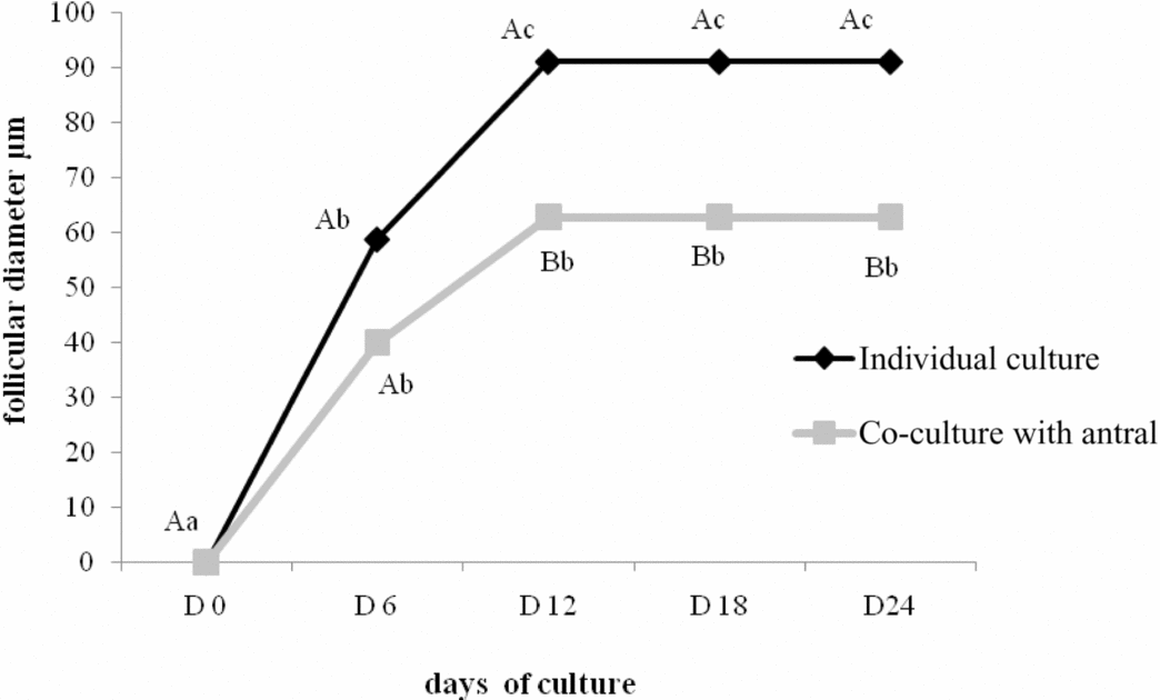
Individual × antral. The diameters of follicles cultured individually were similar to those co-cultured with an antral follicle throughout the experiment, except on D12, when single follicles presented significantly higher diameters (Fig. 12, p < 0.05).
Follicle diameters during the 24 days of culture of preantral follicles cultured individually without (control) or with antral follicles (treatment) (Experiment 3). *Indicates the significance between treatments on this day of culture.

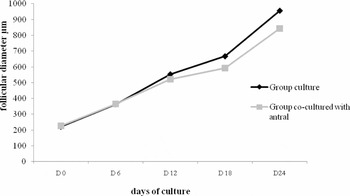
Group × antral. Although no significant difference in follicle diameter was observed between the treatments, a significantly higher growth rate was achieved in preantral follicles cultured without an antral follicle (control × group = 24.9 ± 9 µm/day) in comparison with the co-culture group (group × antral = 19.4 ± 6.6 µm/day) (Fig. 13).
Follicle diameter during the 24 days of culture of preantral follicles cultured in groups without (control) or with antral follicles (treatment) (Experiment 3).

Antrum formation
Individual × antral. The percentage of antrum formation was higher on D12 onwards in follicles cultured without an antral follicle compared with those co-cultured with an antral follicle (p < 0.05). During the culture period, the control group showed a significant increase in the percentage of antral cavity formation from D6 of culture, and this percentage became higher on D12. However, in the group co-cultured with the antral follicle, the significant increase was observed on D6 but was sustained until the end of culture (Fig. 14).
Antrum formation during the 24 days of culture of preantral follicles cultured individually without (control) or with antral follicles (treatment) (Experiment 3). A,B indicates the significance between treatments on each day of culture. a,b,c indicates the significance between the days of culture within each treatment.
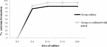
Group × antral. Antrum formation rate was not affected by the presence of an early antral follicle during in vitro culture of follicles in groups (Fig. 15).
Antrum formation during the 24 days of culture of preantral follicles cultured in groups without (control) or with antral follicles (treatment) (Experiment 3).

Oocyte recovery after culture
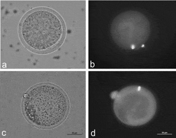
Individual × antral. The culture of follicles individually allowed the retrieval of a significantly higher number of adequate oocytes for IVM (21 oocytes measuring at least 110 µm, 61.8% of the total of cultured preantral follicles) than co-culture with an early antral follicle (12 oocytes, 34.3% of the total of cultured preantral follicles, p < 0.05). After IVM, one oocyte recovered from follicles cultured individually (control) reached the metaphase II stage (Fig. 16E,F), while one oocyte from the treatment group that was co-cultured with an antral follicle resumed meiosis until the metaphase I stage (Fig. 16A,B).
Meiosis resumption of oocytes recovered from preantral follicles cultured individually or in presence of an antral follicle in medium containing follicle stimulating hormone (FSH) (Experiment 3). MII oocyte from preantral follicle cultured for 24 days individually (a); and labelled with Hoechst stain (b); MI oocyte from preantral follicle after 24 days of in group culture in presence of an antral follicle (c); and labelled with Hoechst stain (d). Scale bars represent 50 µm.

Group × antral. With respect to the number of oocytes recovered for IVM, no statistical difference was found between follicles cultured in groups with or without an antral follicle (three oocytes in the control, 46.8%; three oocytes in the test group, 44.7%). Resumption of meiosis (metaphase I) was observed with oocytes in both treatment groups.
Discussion
The present study evaluated, for the first time, the effects of interactions between caprine preantral follicles on their in vitro growth, through the comparison of individual follicle and group culture. The influence of FSH and co-culture with early antral follicles on these interactions was assessed. The described model is a valuable tool to study the interactions and dominance between follicles in vitro.
The results of Experiment 1 showed that grouping caprine preantral follicles for in vitro culture without FSH enhances viability maintenance, growth and antrum formation after 12 days of culture. Several studies have demonstrated that culturing embryos in group increases cleavage and blastocyst rates, which may be due to the production of autocrine and paracrine factors that stimulate embryo development in vitro (Gardner & Lane, Reference Gardner and Lane1993; Pugh et al., 1993; Trounson et al., Reference Trounson, Pushett, Maclellan, Lewis and Gardner1994; Fukui et al., Reference Fukui, Lee and Araki1996). Some works have also shown that grouping preantral follicles for in vitro culture results in higher growth rates, and an ideal number of follicles in each culture drop stimulates their development through a paracrine interfollicular regulation involving secretion of growth factors (Wu et al., Reference Wu, Emery and Carrell2001; Gupta et al., Reference Gupta, Nandi, Ravindranatha and Sarma2002). In addition to the importance of co-cultured follicles, the volume of medium may also influence paracrine regulation. Secreted factors which stimulate growth can be diluted if large medium amounts are used, so it is necessary to balance numbers of follicles and medium volume. Low volumes of medium for group culturing of mouse, ovine and bovine embryos are beneficial for the paracrine interactions that stimulate development (Fukui et al., Reference Fukui, Lee and Araki1996). Follicular culture in groups may also be detrimental as excessive numbers of follicles can lead aggregation in single masses which can inhibit growth and development. Wu et al. (Reference Wu, Emery and Carrell2001) observed that the culture of five or more follicles per drop results in contact and subsequent growth suppression.
In Experiment 2, group culturing enhanced viability maintenance in the presence of FSH from day 18 onwards, while percentages of viable follicles cultured individually decreased significantly after culture for 12 days. The addition of FSH to the medium also to sustain viability in both treatments, since 39.5% and 54% viable follicles were observed after 18 days in the control and group culture treatment, respectively, while in Experiment 1 (culture without FSH), a viability of only 22% was observed in the follicles cultured in groups, and not a single viable follicle could be found in the control at the same culture period. These results are in accordance with other studies, in which only 10–17% of murine preantral follicles survived after in vitro culture without FSH, and a complete differentiation occurred only when this hormone was added to the culture during the advanced preantral stage (Cortvrindt et al., Reference Cortvrindt, Smitz and Van Steirteghem1997; Mitchell et al., Reference Mitchell, Kennedy and Hartshorne2002; Adriaens et al., Reference Adriaens, Cortvrindt and Smitz2004).
The role of FSH in folliculogenesis is well known. In vivo, this hormone is essential for steroidogenesis through the stimulation of the enzyme aromatase, differentiation of granulosa cells and the induction of the expression of LH receptors. Regulation of intercellular junctions between the oocytes and surrounding granulosa cells is also performed by FSH (Albertini et al., Reference Albertini, Combelles, Benecchi and Carabatsos2001). Moreover, gonadotrophins activate the expression of anti-apoptotic proteins in granulosa cells in vivo and in vitro (Wang et al., Reference Wang, Rippstein and Tsang2003).
Under action of FSH (Experiment 2), group culture significantly increased follicle diameter until day 18 of culture. Several authors have reported that co-culture of isolated follicles is beneficial for in vitro growth (Gutierrez et al., Reference Gutierrez, Ralph, Telfer, Wilmut and Webb2000; Gupta et al., Reference Gupta, Nandi, Ravindranatha and Sarma2002). Wu and et al. (Reference Wu, Emery and Carrell2001) demonstrated that culturing porcine preantral follicles in groups of three follicles promoted the highest growth rates in comparison with the incubation of one or five follicles, which suggests an ideal number of follicles in a culture drop for an optimum development by means of paracrine regulation. Spears et al. (Reference Spears, De Bruin and Gosden1996) reported that dominant follicles from mice can suppress the growth of contacting follicles, but group culturing these follicles without contact is favourable for in vitro growth. Zhou & Zhang (Reference Zhou and Zhang2006) cultured caprine follicles in microdrops and observed the aggregation of follicles which could grow within the resulting masses. This condition proved to be beneficial for oocyte growth probably because a close contact among granulosa cells is achieved, which may be essential for the growth of oocytes in vitro. Such tight association might assist gap junctions in the transport of small molecules from granulosa cells to the oocytes (Eppig, Reference Eppig1991).
The competence to resume meiosis is progressively acquired in caprine oocytes during the late phase of follicular growth, together with the capability to be fertilized and to sustain early embryo development. Only a small proportion of oocytes retrieved from caprine antral follicles is able to support in vitro embryo development (Crozet et al., Reference Crozet, Ahmed-Ali and Dubos1995). In the present study, culture of preantral follicles in groups positively influenced the in vitro growth of oocytes with the use of FSH (Experiment 2), making it possible to obtain oocytes with an adequate diameter (>110 µm) for IVM, which were able to resume meiosis. In this context, the development of an in vitro culture system capable of sustaining viability and promoting the growth of preantral follicles for longer periods will enable the retrieval of larger numbers of competent oocytes for the in vitro production of caprine embryos. Despite the fact that oocytes recovered in Experiment 1 were larger than 110 µm, a complete maturation (metaphase II) was not accomplished after IVM. Similarly to their counterparts in mice, cows and sows, caprine oocytes also need to acquire meiotic competence during the final phase of growth. Thus, a culture system capable of promoting higher growth rates for longer periods is required for adequate development and differentiation of oocytes, which need to express mRNA and synthesize specific proteins at a similar level as oocytes grown in vivo (De Smedt et al., Reference De Smedt, Crozet and Gall1994; Crozet et al., Reference Crozet, Dahirel and Gall2000).
In Experiment 3, co-culture of single preantral follicles with an early antral follicle had a detrimental effect on viability, growth, antrum formation and the production of oocytes with the minimum diameter for IVM. Nevertheless, preantral follicles cultured in groups benefited from the addition of an early antral follicle and presented higher viability, but no effect on antral cavity formation and retrieval of oocytes for IVM was observed. Conversely, the growth of these follicles was impaired, and significantly smaller diameters were found compared with groups of preantral follicles cultured without antral follicles.
Barker et al. (Reference Baker, Srsen, Lapping and Spears2001) observed that early antral follicles negatively influence the growth of mouse preantral follicles during in vitro culture, which demonstrates that dominance also occurs in vitro when follicles are co-cultured with others at different stages, and that larger follicles become dominant at the beginning of the culture and inhibit the growth of the others.
The process of follicle selection and dominance comprises complex interactions between a series of factors which are still not fully deciphered. The establishment of dominance and its maintenance are distinct events which can be studied separately. Interfollicular communication mediated through contact may act in the selection process by an inhibition of the development exerted over smaller follicles, and possibly the induction of a higher sensitivity to oscillations in FSH concentration (Baker & Spears, Reference Baker and Spears1999; Baker et al., Reference Baker, Srsen, Lapping and Spears2001). Follicular dominance is controlled by a number of mechanisms acting together, including the production of local factors which can inhibit the growth of subordinate follicles without affecting viability. These inhibitory factors can act by systemically suppressing follicular growth in the contralateral ovary and can also exert local effects on the proliferation of granulosa cells and aromatase activity stimulated by FSH (Armstrong & Webb, Reference Armstrong and Webb1997).
The development of subordinate follicles is suppressed through an endocrine mechanism in which inhibin and estradiol are secreted in increasing concentrations and downregulate the hypothalamus–hypophysis system. This downregulation leads to a decrease of circulating FSH, which in turn results in atresia of non-dominant follicles. In addition to this pathway, an intra-ovarian regulation of follicular dominance based on local inhibitory factors produced by follicles also has an important role. Such follicular interactions were evidenced by the results of the present study, in which the co-culture of preantral follicles in the same stage and an early antral follicle affected the in vitro development of preantral follicles.
Previous studies have shown that follicles can secrete factors which inhibit several aspects of the development of other follicles. Some works demonstrated a negative influence of antral cavity fluid on follicular development (Cahill et al., Reference Cahill, Driancourt, Chamely and Findlay1985). More recently, it was shown that low concentrations of follicular fluid from dominant follicles may contain beneficial factors for the growth of porcine preantral follicles, while fluid from smaller follicles can include compounds which inhibit the development of cultured preantral follicles (Metoki et al., Reference Metoki, Iwata, Itoh, Kasai, Takajyo, Suzuki, Kuwayama and Monji2008).
The findings of the present study (Experiment 3) indicate the existence of inhibitory factors produced by antral follicles which significantly slow down the growth of preantral follicles but do not induce atresia. In addition, this influence depends on the number of co-cultured follicles, since a negative effect on viability was observed only when a single preantral follicle was incubated with an antral follicle. Moreover, we hypothesize that a positive paracrine influence established among co-cultured preantral follicles counteracted the deleterious effect of the dominant follicle on viability observed in single preantral follicles co-incubated with an antral follicle.
Some elements, such as FSH concentration, can induce follicles to secrete stimulatory factors for other follicles in culture at the same developmental stage. Furthermore, FSH can suppress the inhibitory effect of factors produced by dominant follicles over in vitro co-cultured follicles in groups. During normal ovulatory cycles, the number of developing follicles can possibly be determined by a balance of local inhibitory and stimulatory factors and circulating levels of FSH at a given moment (Spears et al., Reference Spears, Baker, Srsen, Lapping, Mullan, Nelson and Allison2002). Therefore, the presence of FSH or its concentration in the in vitro culture of follicles at different stages might influence such follicular interactions.
In conclusion, in vitro culture of caprine preantral follicles in groups enhances development in comparison with incubation of single follicles, and increases the production of competent oocytes for IVM. Furthermore, follicles at more advanced developmental stages can affect the in vitro growth of preantral follicles through interactions which depend on the number of co-cultured follicles. Future investigations are required to characterize the factors which determine dominance and other follicular interactions.
Acknowledgements
We thank Anderson Almeida for fluorescence microscopy and micrograph assistance. This work was supported by CAPES via FUNCAP.