I. INTRODUCTION
Cytochrome c (cyt c) is a water-soluble containing 104-amino-acid globular protein with a covalently bound heme active center,Reference Shang, Nuffer, Muñiz-Papandrea, Colón, Siegel and Dordick1 which acts as a key protein to complete the electron transfer in the mitochondrial respiratory chain.Reference Li, Sun, Hao, He, Zhang and Wang2 Recent studies indicated the release of cyt c into the cytoplasm can induce the cell apoptosis to cause some disease.Reference Salehnia, Hosseini and Ganjali3 Therefore, cyt c was identified as an important indicator in the apoptotic process and as an early signs of cell apoptosis.Reference Yin, Cai, Feng, Wu, Zou and Cai4 Hence, researchers have tried their best to study and detect cyt c to better understand cell apoptosis and certain disease. Recently, various methods have been used to detect cyt c, including electrochemistry,Reference Pandiaraj, Madasamy, Gollavilli, Balamurugan, Kotamraju, Rao, Bhargava and Karunakaran5,Reference Yang, Shin, Choi, Lee and Shin6 mass spectrometry,Reference Liu and Yan7 fluorescent sensorReference Cao, Li, Qin, Kang and Li8 liquid chromatography,Reference Crouser, Gadd, Julian, Huff, Broekemeier, Robbins and Pfeiffer9 etc. During the detection process, the enrichment and purification of cyt c is very essential for the analysis of cyt c because of the minute quantity in the cytoplasm. Therefore, a simple, economical, and fast method is necessary for the detection of cyt c in clinic and research applications. As we all know, adsorption-based method is widely considered as a high efficiency, simple, and ecofriendly method.Reference Hao, Song, Zhang, Wan, Tang and Lv10 Additionally, research studies showed that adsorption can make the proteins retain the original structure and their activity.Reference Vinu, Murugesan, Tangermann and Hartmann11,Reference Washmon-Kriel, Jimenez and Balkus12 Therefore, great efforts have been made to develop the excellent adsorbents.
In recent years, more attention has been paid to nanostructured materials,Reference Patila, Pavlidis, Diamanti, Katapodis, Gournis and Stamatis13 especially carbon-based materials, including carbon nanotubes and graphene-based materials due to their large surface area, easy modification, and good biocompatibility.Reference Patila, Pavlidis, Diamanti, Katapodis, Gournis and Stamatis13,Reference Feng and Ji14 These unique physicochemical properties were conducive to ions,Reference Song, Hao, Tian, Wan, Zhang and Lv15 protein,Reference Zhang, Liang, Han, Wan and Ding16 and biomolecule loading.Reference Lu, Yang, Zhu, Chen and Chen17 Since its historical discovery by Novoselov KS et al. in 2004, graphene has been a rising star in the field of biomaterial.Reference Novoselov, Geim, Morozov, Jiang, Zhang, Dubonos, Grigorieva and Firsov18 Research studies on the functions of graphene-based biomaterials relying on biomolecular interaction have bloomed in the recent decades, such as cellular imaging,Reference Mei, Chen, Zhao, Yang, Liu, Liu and Zhang19 drug delivering,Reference Wang, Hao, Zhang, Chen and Zhang20 molecular diagnostics,Reference Gollavelli and Ling21 biosensor,Reference Wisitsoraat, Mensing, Karuwan, Sriprachuabwong, Jaruwongrungsee, Phokharatkul, Daniels, Liewhiran and Tuantranont22 etc. Besides, studies showed that graphene oxide (GO) was easy to aggregate in solution because of its thin thickness and highly toxic to mammalian cells compared with reduced graphene oxide (rGO).Reference Hao, Wang, Huang, Liu, Zhang, Chen, Zhang and Cai23 Moreover, rGO showed more active sites than pure graphene and GO derived from the residual oxygenic groups.Reference Hao, Gai, Huang, Liu, Sun, Fu and Lin24 Therefore, much more attention has been paid to prepare the excellent rGO.
A brand new material called tea polyphenol (TP) functionalized and reduced graphene oxide (TPG) has drawn much attention owing to its superior properties and therapy potential.Reference Huang, Hao, Xie, Gong, Liao and Lin25 There, TPs act as a biocompatible reducing agents as well as a biodegradable stabilizer. Also, research studies have revealed that TPs showed high anti-inflammatory,Reference Hisanaga, Ishida, Sakao, Sogo, Kumamoto, Hashimoto and Hou26 antioxidant,Reference Forester and Lambert27 and anti-mutagenicReference Butt and Sultan28 properties in various biological systems and can modulate signaling pathways involved in cell proliferation, angiogenesis, and apoptosis.Reference Cho, Kim, Lee, Park, Kim and Chun29–Reference Syed, Afaq, Kweon, Hadi, Bhatia, Spiegelman and Mukhtar31 Furthermore, the addition of TPs can reduce the agglomeration of the material and increase the surface area of TPG, thus providing more adsorptive active sites. However, few articles have quantified the exact adsorption of proteins on the surface of TPG. Needless to say, factors which could influence the adsorption such as pH, concentration of both material and analytes, time, etc. are hardly investigated in detail.
In the present work, a novel, friendly, and high-efficiency adsorptivity material, named TPG, was first used as the adsorbent for cyt c as a model protein. The adsorption behaviors were analyzed by using UV-Vis spectroscopy. Moreover, the effects of adsorption, including pH, contact time, and the initial concentration of cyt c, and adsorption kinetic as well as isotherms were discussed in detail. It was interesting to find that TPG showed the fast adsorption and superior adsorption capacity toward cyt c, which will provide a novel and effective material to construct a biosensor of proteins.
II. EXPERIMENT SECTION
A. Materials
Chemicals used in the experiments including graphic powder, sodium hydroxide (NaOH), nitric acid (HNO3), phosphate-buffered saline (PBS) solutions (pH = 7.800–8.000), and cytochrome c (cyt c) were all of analytical grade and used without further purification. Deionized water from ULUPURE Water Purification System (Chengdu, China) was used in all the experiments. Biological reagents used in this paper were RPMI 1640 medium and fetal bovine serum (FBS), obtained from Gibco Life Technologies (Grand Island, New York). Mouse fibroblast cell line (L929) was obtained from the State Key Laboratory of Oral Diseases of Sichuan University (Chengdu, China). The Cell Counting Kit-8 (CCK-8) was obtained from Dojindo Laboratory.
B. Preparation of TPG
TPG was prepared according to our previous report.Reference Song, Hao, Tian, Wan, Zhang and Lv15 Briefly, 0.5 mg/mL GO was sonicated for 2 h to form complete exfoliation and then centrifuged for 30 min (3500 rpm) to remove nonexfoliated material. TPG was prepared using hydrothermal method: 3 mg/mL TPs solution (TP, 20 mL) was dispersed with GO (20 mL) under vigorous stirring to form a homogeneous solution. Finally, the mixed solution was transferred into a 50 mL Teflon-lined stainless-steel autoclave at 80 °C for 8 h. The materials were collected and washed repeatedly with deionized water by centrifugation at 11,000 rpm for 30 min, then dialyzed to remove excess TP molecules.
C. Characterization and apparatus
The surface morphology of the materials was examined by field emission scanning electron microscopy (FE-SEM; Inspect F) and atomic force microscopy (AFM; Shimadzu SPM-9700). X-ray diffraction (XRD) measurements (5–90°) were taken using an X’Pert Pro X-ray diffractometer (Philips) with Cu Kα radiation (λ = 1.5406 Å). Also, the surface properties were detected with Fourier transform infrared (FTIR, Shimadzu Corp., Tokyo, Japan), spectroscopy (Thermo Nicolet IS10, Thermo Fisher Scientific, Waltham, Massachusetts) in the 4000–500 cm−1 frequency range, using samples diluted in KBr pellets.
D. Batch adsorption experiment
Batch adsorption tests for cyt c were carried out at the real environment of human bodies (37 °C) as the incubation temperature to study the effects of various parameters as follows: a certain amount of TPG (1 mL) were added to cyt c with different concentrations (1 mL). Then the common incubation conditions were shaken at a speed of 220 rpm for 30 min expect the kinetic experiments. Finally, the solution mixtures were centrifuged at 10,000 rpm for 10 min to collect the supernatant, and the concentrations of the residual cyt c were analyzed by using a U-2910 UV-Vis spectrophotometer. In this study, the effects of dose of TPG, pH values, contact time, and the concentration of cyt c on the adsorption were investigated. And the adsorption efficiency (E, %) and the adsorption capacity (Q, mg/g) were calculated according to the following equations:
 $$E = {{{C_0} - {C_{\rm{e}}}} \over {{C_0}}} \times 100\% \quad ,$$
 $$Q = {{\left( {{C_0} - {C_{\rm{e}}}} \right) \cdot V} \over m}\quad ,$$
where C 0 and C e (mg/L) are the initial and equilibrium concentrations of cyt c in aqueous phase, V is the volume of the solution (L), and m is the mass of adsorbent TPG used (g).
E. Biocompatibility experiment
The biocompatibility experiments include the water and humoral stability, and cytotoxicity. As for the first property, we have studied in our previous report.Reference Hao, Wang, Huang, Liu, Zhang, Chen, Zhang and Cai23 For the cytotoxicity, mouse fibroblast cell line (L929) was incubated with different concentrations of TPG. Briefly, the L929 cell line was cultured at 104 cells per well in 96-well plates overnight before treatment. Then, the different final concentrations of TPG (5, 25, 50, 100, 150, and 250 µg/mL) were incubated with the above cells from the 96-well plates for 24, 48, and 72 h, respectively. At last, CCK-8 method was used to monitor the cytotoxicity at a wavelength of 450 nm using a microplate reader (VariOskanFlas 3001, Thermo Fisher Scientific).
III. RESULTS AND DISCUSSION
A. Biocompatibility of TPG
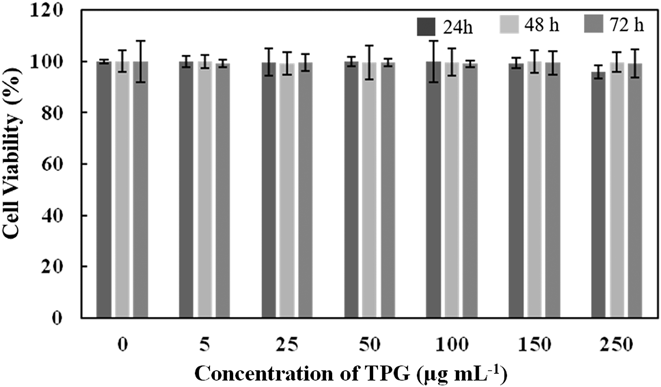
The biocompatibility of materials was the one of the important biological evaluation systems. In general, the biocompatibility follows biological stability and biosafety guidelines. For the biological stability, the previous study has been reported, indicating that TPG was stable in water and physiological environment, which was important for potential biological applications.Reference Hao, Wang, Huang, Liu, Zhang, Chen, Zhang and Cai23 For the biosafety, the different concentrations of TPG incubated with the normal cells (L929) for 24, 48, and 72 h. As shown in Fig. 1, obvious cell proliferation inhibition was not found even at the high concentration and long time, which indicated TPG was friendly to the normal cells. To sum up, the TPG possessed good biocompatibility.
Biocompatibility of TPG with normal cells (L929 cell line). The cytotoxicity of TPG with different concentrations (0, 5, 25, 50, 100, 150, and 250 µg/mL) to L929 cell line. Cells were incubated with TPG at different concentrations for 24, 48, and 72 h, followed by measurement using the Cell Counting Kit-8 (CCK-8).

B. Effect of dose of TPG for adsorption cyt c
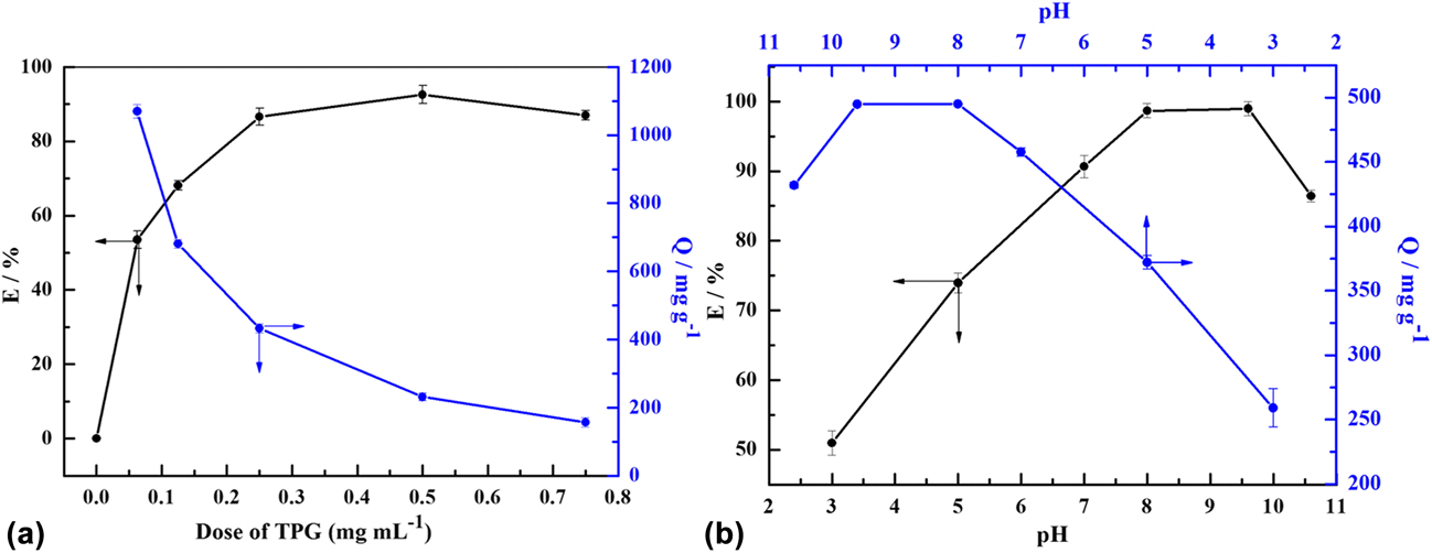
TPG solution was diluted with PBS, reaching a concentration of 0.125 mg/mL, 0.250 mg/mL, 0.500 mg/mL, 1.00 mg/mL, 1.50 mg/mL, respectively. Then the different concentrations of TPG solutions were added into cyt c solution (0.250 mg/mL) separately with the volume ratio of 1:1. After 1 h, the mixtures were centrifuged at a speed of 10,000 rpm for 25 min. Finally, the supernatant was collected for further tests. As shown in Fig. 2(a), as the concentration of TPG increased, the adsorption efficiency increased except at the concentration of 0.750 mg/mL, at which there were so many TPG that affect cyt c quantification because of the difficulty of the separation of TPG and cyt c. And the adsorption efficiency reached the highest at 0.500 mg/mL as 92.55%. While the adsorption capacity decreased with the concentration of TPG increased, which was attributed to the excess active sites from superfluous TPG. According to the above results, the optimum dose of TPG was chosen as 1.00 mg, namely the final concentration was 0.500 mg/mL in the following experiments.
Effect of the dose of TPG (a) and pH (b) for the adsorption efficiency (left) and capacity (right) of cyt c: adsorption time, 1 h; cyt c concentration, 0.25 mg/mL; adsorbent dose, 0.5 mg/mL for (b).

C. Effect of pH for adsorption cyt c
The pH of the solution plays a great effect on the adsorption target protein. Too acid or too alkaline will affect the structure of protein to affect the determination of protein.Reference Vinu, Murugesan, Tangermann and Hartmann11 In this study, PBS solution with different pH values was prepared with NaOH solution (0.1 M) and HNO3 solution (0.1 M) to form the final pH values as 3.00, 5.00, 7.00, 8.00, 9.60, and 10.6, which was used to prepare TPG (0.500 mg/mL) and cyt c (0.250 mg/mL) solutions at different pH values. The following procedures were as before. Cyt c is not stable below 3.00 and above 11.0; therefore, we chose the range of pH was 3.00–10.0, which was not outside this pH range. According to Fig. 2(b), it was clearly observed that the adsorption efficiency and capacity gradually raised from 3.00 to 9.60 because of the electrostatic interaction between cyt c and TPG. As we all know, the isoelectric point (pI) of cyt c is 9.80.Reference Vinu, Murugesan, Tangermann and Hartmann11 Therefore, cyt c is positively charged at pH below pI and opposite above pI. Thus, cyt c was positively charged at pH below 9.80. Under acidic conditions, the surface of TPG may be partially protonated. Therefore, as shown in Fig. 2(b), the adsorption efficiency and capacity were low at acidic condition and had a growing trend below 10.6. At above 9.80, cyt c and TPG were negatively charged at pH 10.6, which may generate electrostatic repulsion to affect the adsorption of cyt c. Consequently, the optimal adsorption pH was chosen as 9.60, which was close to the pI of cyt c (9.80).
D. Effect of time for adsorption cyt c and adsorption kinetics
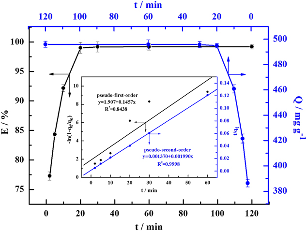
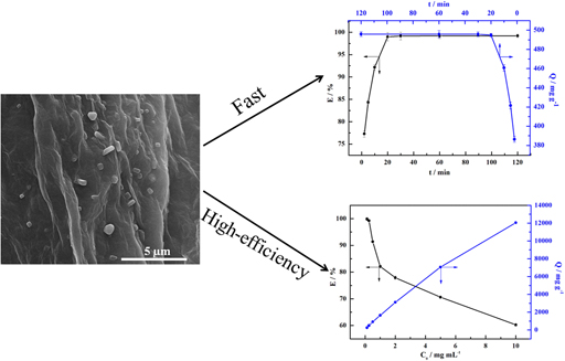
The study on the effect of time is important to ensure the high efficiency adsorption as soon as possible under TPG solution (0.500 mg/mL) and cyt c solution (0.250 mg/mL) at pH = 9.60. All the reaction conditions were kept the same except for the reaction time. Lastly, the mixtures were centrifuged after incubations of 2, 5, 10, 20, 30, 60, and 120 min, respectively. According to Fig. 3, the adsorption efficiency and capacity were increased sharply within 20 min with the adsorption efficiency reaching 98.99%, and the adsorption process reached equilibrium about after 30 min, which was attributed the laminated structure and large surface area of TPG. Hence, 30 min was chosen as the optimum reaction time to ensure as high as possible adsorption efficiency at short time. Meanwhile, two kinetic models were used to understand the adsorption behavior and mechanism using Lagergren pseudo-first-order and pseudo-second-order models. The kinetic equations were expressed as (3) and (4) as follows,Reference Hao, Song, Zhang, Wan, Tang and Lv10 respectively:
 $$- \ln \left( {1 - {{{q_t}} \over {{q_{\rm{e}}}}}} \right) = {k_1}t\quad ,$$
 $${t \over {{q_t}}} = {1 \over {{k_2}q_{\rm{e}}^2}} + {t \over {{q_{\rm{e}}}}}\quad ,$$
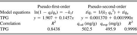
where q t and q e are the amounts of cyt c adsorbed on TPG at time t and at equilibrium (mg/g), respectively. k 1 (min−1) and k 2 [g/(mg min)] represent the pseudo-first-order and second-order rate constants for the adsorption process, which can be obtained by a plot of −ln(1 − q t/q e) and t/q t against t, respectively. As shown in the inset of Fig. 3, the adsorption process was in accordance with the pseudo-second-order model, which had a higher correlation coefficient of 0.9998 than that of pseudo-first-order model (0.8438). Moreover, it was concluded from Table I that the experimental adsorption capacity (q exp) and calculated adsorption capacity (q cal) was very close, which further confirmed the conformance of pseudo-second-order model. The results suggested the adsorption process may be the rate-limiting step through electronic sharing or exchange between TPG and cyt c.Reference Wang, Hao, Zhang, Chen and Zhang20
Effect of contact time for the adsorption efficiency (Left) and capacity (Right) of cyt c; inset: adsorption kinetics models: pH, 9.8; cyt c concentration, 0.25 mg/mL; adsorbent dose, 0.5 mg/mL.

Kinetics models for the adsorption of cyt c on TPG.

E. Effect of concentration of cyt c and adsorption isotherms
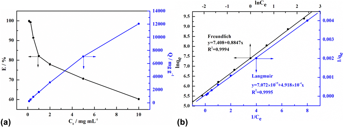
The initial cyt c concentration was also studied in various ranges of 0.125–10.0 mg/mL at pH 9.60 for 30 min. As shown in Fig. 4(a), the adsorption capacity increased with the concentration of cyt c, while the adsorption efficiency decreased markedly. At low initial concentration, there were large surface area and more reactive sites to adsorb more cyt c. However, at higher initial concentration, the residual reactive sites became less, thus resulting in the decrease of adsorption efficiency. To understand the adsorption behaviors, the adsorption isotherms models were proposed, including Langmuir and Freundlich isotherms models [Fig. 4(b)].
Effect of initial cyt c concentration (a) and Langmuir and Freundlich isotherms (b) for the adsorption of cyt c onto TPG: pH, 9.8; adsorption time, 30 min; cyt c concentration, 0.25 mg/mL; adsorbent dose, 0.5 mg/mL.

The Langmuir isotherm is expressed as Eq. (5), which describes monolayer adsorption on adsorbent with equal adsorbate affinity.Reference Song, Hao, Tian, Wan, Zhang and Lv15
 $${1 \over {{q_{\rm{e}}}}} = {1 \over {{q_{\rm{m}}}}} + {1 \over {{K_{\rm{L}}} \cdot {q_{\rm{m}}} \cdot {C_{\rm{e}}}}}\quad .$$
On the other hand, the Freundlich isotherm model assumes the multilayer adsorption with heterogeneity and is described as followsReference Song, Hao, Tian, Wan, Zhang and Lv15:
 $$\ln {q_{\rm{e}}} = \ln {K_{\rm{F}}} + {1 \over n} \cdot \ln {C_{\rm{e}}}\quad ,$$
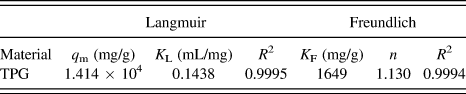
where q e and C e are the adsorption capacity (mg/g) and the equilibrium concentration of cyt c in solution (mg/mL), respectively. K L (mL/mg) is the constant related to the free energy of adsorption and q m (mg/g) is the maximum adsorption capacity, which is calculated according to Langmuir model. K F (mg/g) and n, as the Freundlich constants, represent the adsorption capacity and the adsorption strength, which can be obtained from intercept and the slope of the linear plot of ln q e versus ln C e, respectively. As shown in Fig. 4(b) and the data presented in Table II, the adsorption process was in accordance with Langmuir and Freundlich isotherm models, which had high correlation coefficients, suggesting that the adsorption behavior took place by monolayer as well as multilayer on homogeneous sites of the surface of TPG.Reference Li, Zhang, Du, Peng, Liu, Wang, Xia, Zhang, Wang, Zhu and Wu32 According to Langmuir isotherm model, the theoretical maximum adsorption capacity (q m) for cyt c was calculated as 1.414 × 10⁴ mg/g, close to the experimental maximum adsorption capacity value of 1.205 × 10⁴ mg/g. The results showed TPG has excellent absorbability for cyt c because of the large surface area and the functional groups.
Langmuir and Freundlich adsorption isotherm parameters for cyt c on TPG.

F. Adsorption mechanism
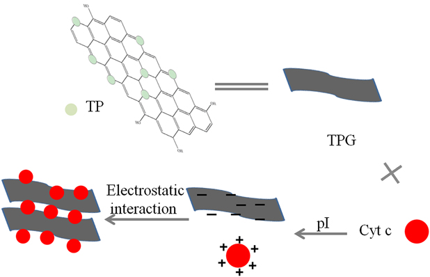
In general, the possible adsorption mechanisms can be summarized as the following two points: (i) The large surface area.Reference Hao, Wang, Huang, Liu, Zhang, Chen, Zhang and Cai23 The addition of TP was helpful to reduce the agglomeration of graphene to provide more reactive sites and introduce some active oxygen-containing functional groups, which is in favor of the adsorption of protein. (ii) Electrostatic interaction.Reference Wang, Hao, Zhang, Chen and Zhang20,Reference Hao, Wang, Huang, Liu, Zhang, Chen, Zhang and Cai23 According to the pH results, the electrostatic repulsion was strong below pI of cyt c, at which TPG was easy to be protonated. And above pI of cyt c, cyt c and TPG was both negatively charged to the existence of electrostatic repulsion force. The repulsion force was minimal at near isoelectric point, and the adsorption efficiency and capacity were high. So the electrostatic interaction between cyt c and TPG was one of the most important adsorption mechanisms. The above adsorption mechanism is shown in Fig. 5. However, the more accurate mechanisms should be further studied.
The schematic diagram of adsorption mechanism between TPG and cyt c.

G. Characterization of TPG-cyt c
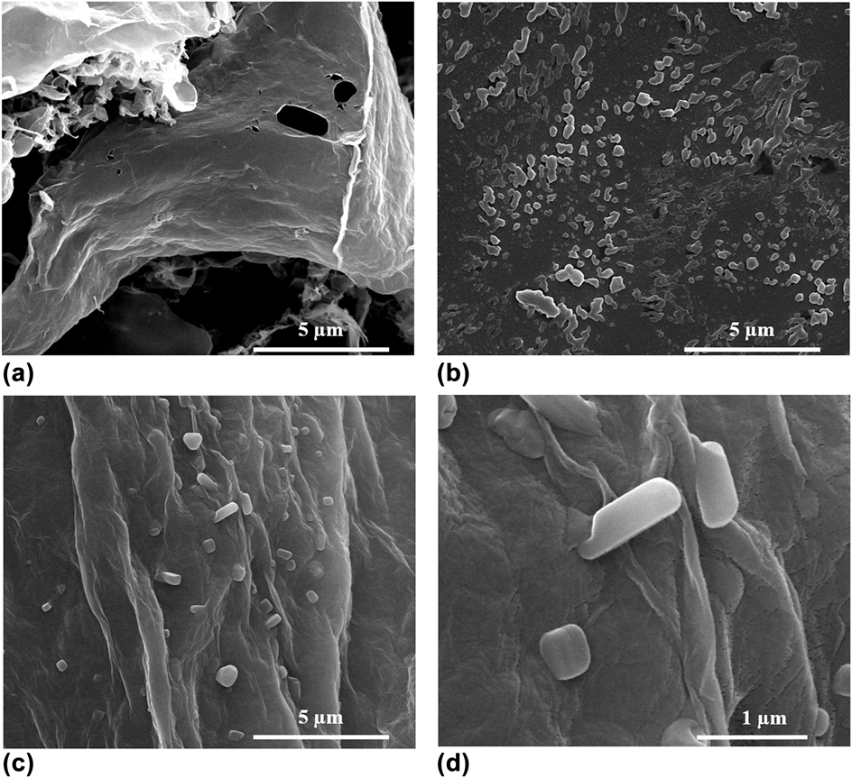
Some necessary characterizations were performed to verify the loading of cyt c on TPG. First, the visual morphologies of pure TPG, cyt c, and TPG-cyt c were observed by SEM and tapping-mode atomic force microscopy (AFM). TPG nanosheets with large surface area and visible defects and a few wrinkles are observed in Fig. 6(a), which indicated more reactive sites have been produced after the addition of TP. Figure 6(b) shows the SEM image of pure cyt c with long strips about 300 nm. From Fig. 6(c), the layer structure of TPG was well observed, and long strips cyt c were dispersed the surfaces of TPG to form the “sandwich” structure, resulting in the high adsorption capacity of cyt c.
SEM images of TPG (a), free cyt c (b), TPG-cyt c (c), and high magnification image of (c and d).

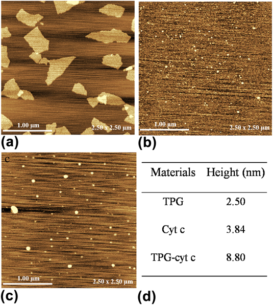
Also, AFM was used to further verify the successful adsorption of cyt c on TPG. As shown in Figs. 7(a)–7(d), TPG showed the thin layer structure, and the pure cyt c presented small particle-structure. After the adsorption, cyt c was aggregated and observed on the surface of TPG, which was proved by the AFM height analysis. The cross section analysis in Fig. 7(d) indicated the TPG nanosheets were about 2.50 nm, and the height of cyt c was about 3.84 nm. And the height of TPG-cyt c increased to 8.80 nm, which was attributed to the adsorption of cyt c on the surface of TPG. The SEM and AFM images, together with the height data, indicated cyt c was successfully adsorbed on the surface of TPG forming a sandwich-like structure.Reference Hao, Wang, Huang, Liu, Zhang, Chen, Zhang and Cai23
AFM images of TPG (a), free cyt c (b), TPG-cyt c (c), and the summary of the height of these materials (d).

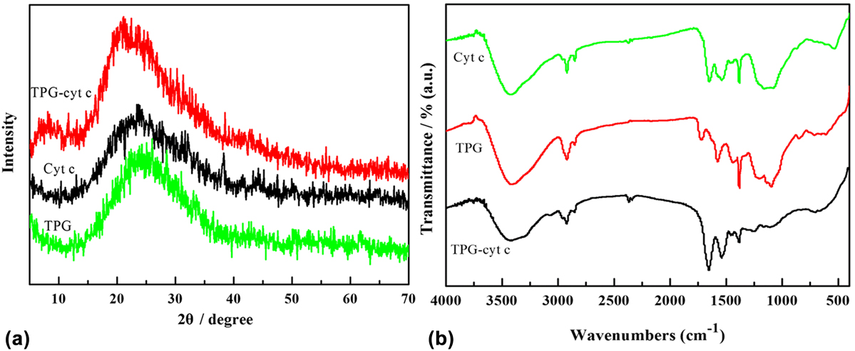
In addition to the observation of morphology, the crystalline phase, structure, and chemical functional groups were characterized by XRD and FTIR. As shown in Fig. 8(a), the characteristic peaks of pure TPG and cyt c were clearly observed at 2θ = 25.6° for cyt c and 26.0° for TPG.Reference Song, Hao, Tian, Wan, Zhang and Lv15,Reference Huang, Hao, Xie, Gong, Liao and Lin25 Additionally, the peak of TPG-cyt c was blue-shifted to 23.6°, which was attributed to the adsorption of cyt c on the surface of TPG resulted in the increase of interlayer spacing of TPG. Moreover, FTIR was used to verify the successful binging of cyt c to the surface of TPG by the chemical functional groups in Fig. 8(b). In the spectrum of cyt c, the main characteristic peaks were observed at 3430 cm−1 with stretching vibrations O–H from adsorbed water and/or the sample; 1655 cm−1 was arising from the amide Ι band, attributed to C=O stretch; 1543 cm−1 was mainly attributed to the amide II band from C–N stretch with N–H bending mode; and 673 cm−1 was derived from the stretching vibrations of C–S.Reference Park, Park, Selvaraj, Kim and Ha33,Reference Šimšíková and Antalík34 As for the spectrum of TPG, the dominant peaks at 1718 cm−1 for the stretching vibrations of C=O of the carboxylic group; 1097 cm−1 corresponding to the C–O groups in residual epoxide moieties.Reference Huang, Hao, Xie, Gong, Liao and Lin25 As for the TPG-cyt c, the characteristic peaks of cyt c and C–O at 1097 cm−1 from TPG were clearly observed, suggesting the successful loading of cyt c on the surface of TPG.
XRD patterns (a) and FTIR spectra (b) of TPG, free cyt c, and TPG-cyt c.

IV. CONCLUSIONS
A simple, effective method for immobilizing cyt c on the novel carbonaceous material-TPG nanosheets was designed using the adsorption approach for improving the loading capacity of cyt c. The effects of adsorption including the dose of TPG, pH, contact time, and the initial concentration were discussed in detail as well as the adsorption kinetics and isotherms. The results indicated the adsorption method was fast and high effective to adsorb cyt c with the maximum adsorption capacity as high as 1.414 × 10⁴ mg/g, which was higher than that of the other method and materials. Moreover, the possible adsorption mechanism was proposed as the large surface area and electrostatic interaction. Additionally, the TPG after the adsorption of cyt c was characterized by SEM, AFM, XRD, and FTIR. This development was useful in the enrichment of protein for widespread applications, and the as-obtained TPG nanosheets with large surface area were a novel adsorbent for the high-efficient adsorption cyt c. Moreover, TPG with good biocompatibility had potential application in biomedical fields.
ACKNOWLEDGMENT
This work was supported by National Natural Science Foundation of China (81400534).