Implications
This work has shown that the intestinal microbiota in pigs can metabolize even readily available nutrients such as supplemented amino acids, hence affecting there availability to the host. Significant amount of literature has demonstrated that dl-2-hydroxy-4-methylthiobutanoic acid (MHA-FA) has lower bioefficacy as a methionine source than dl-methionine (MET). We provide evidence that in swine diets the lower bioefficacy of MHA-FA is, in part, due to its high microbial metabolism relative to MET. However, we did not find any effect of diet composition on the degree of microbial metabolism of MET or MHA-FA, indicating that changes in microbial composition do not necessarily reflect a significant change in the level of metabolism of supplemented amino acid source.
Introduction
There is increasing realization that along with a nutritional contribution mainly by the hindgut communities, the upper gut microflora competes with animal cells for a wide variety of nutrients, including the amino acids (Savage, Reference Savage1986). Conventional microflora were reported to catabolize ascorbic acid and chlorogenic acid (Cobb et al., Reference Cobb, Holloway and Rivers1991; Gonthier et al., Reference Gonthier, Verny, Besson, Remesy and Scalbert2003) and can also take up and incorporate amino acids into microbial protein (Salter et al., Reference Salter, Coates and Hewitt1974) or utilize the carbon skeleton as an energy source. Liu et al. (Reference Liu, Holland and Crow2003) concluded that the various Lactobacillus species, which predominate in the pig small intestine (SI) (Hill et al., Reference Hill, Hemmingsen, Blair, Dumonceaux, Klassen, Zijlstra, Goh and Van Kessel2005) show a wide diversity in their abilities to metabolize amino acids in in vitro experiments.
Crystalline amino acids are commonly supplemented in livestock diets to provide optimum essential amino acid balance with minimum nitrogen excretion. dl-Methionine (dl-MET) and hydroxy-analogue of methionine, dl-2-hydroxy-4-methylthiobutanoic acid (MHA-FA) are alternate sources for supplementation of methionine. The Dutch Central Bureau for Livestock Feeding (Jansman et al., Reference Jansman, Kan and Wiebenga2003) summarized the available literature and reported bio-efficacy values of dl-MHA-FA as 77% in broiler chickens, 83% in layers and 82% in piglets on equimolar basis, compared to dl-MET.
The mechanisms responsible for the lower bio-efficacy of MHA-FA could involve efficiency of intestinal transport, efficiency of conversion of MHA-FA to l-MET and microbial metabolism. Studies in poultry have reported lower apparent absorption of MHA-FA associated with low affinity, low velocity brush border transport mechanisms compared to methionine (Maenz and Engele Schaan, Reference Maenz and Engele Schaan1996b). Earlier results from our laboratory, using the germ-free chicken, suggested that the more slowly absorbed MHA-FA may be metabolized by the intestinal microbiota, reducing host availability (Drew et al., Reference Drew, Van Kessel and Maenz2003). Therefore, the present study was designed to examine whether gastrointestinal microbiota similarly affect the apparent absorption of methionine and MHA-FA in conventional (CON) and gnotobiotic pigs. Since diet composition, and specifically cereal grain variety, affects microbial composition in the pig SI (Hill et al., Reference Hill, Hemmingsen, Blair, Dumonceaux, Klassen, Zijlstra, Goh and Van Kessel2005; Pieper et al., Reference Pieper, Jha, Rossnagel, Van Kessel, Souffrant and Leterme2008), we also investigated whether diets formulated on corn or barley and wheat affected apparent absorption of methionine sources.
Material and methods
Source and synthesis of radiotracers
l-[methyl-3H] MET (93% pure) was purchased from Amersham Biosciences (Oakville, ON, Canada). l-[methyl-3H] MHA-FA was synthesized by nitrous deamination of l-[methyl-3H] MET according to the procedure of Winitz et al. (Reference Winitz, Bloch-Frankenthal, Izumiya, Birnabaum, Barker and Greenstein1956) except that sodium nitrite was used at 5.68 mol/l to improve yield. The method for the purification and separation of l-[methyl-3H] MET and MHA-FA was first developed and validated with non radioactive l-MET (Sigma Chemical Co., St. Louis, MO, USA) and MHA-FA (Novus International Inc., St. Charles, MO, USA) on HPLC using an ultraviolet post column detection system, at 210 nm. For HPLC analysis, a C-18 column (Luna 3μ, 150 × 4.6 mm; Phenomenex, Torrance, CA, USA) with a mobile phase of 17.5% methanol (pH 3.8) at a flow rate of 0.8 ml/min was used. l-[methyl-3H] MET was isolated from contaminant radioactivity by single peak elution on HPLC. Elution profiles of l-[methyl-3H] MET (Amersham Biosciences) and synthesized l-[methyl-3H] MHA-FA were determined by scintillation counting (Beckman LS6000 TA liquid scintillation counter; Beckman Instruments, Fullerton, CA, USA). Aliquots of purified l-[methyl-3H] MET (99%) and l-[methyl-3H] MHA-FA (99%) were stored in liquid nitrogen and used within 3 weeks.
Experimental diets
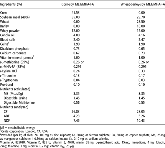
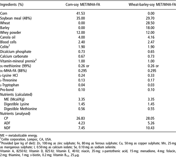
Dietary treatments included diets based on corn or wheat and barley and supplemented with dl-MET or its hydroxy-analogue (MHA-FA) as the methionine source. Diet formulations, calculated and analysed nutrient composition are presented in Table 1. Diets were formulated to meet or exceed nutrient requirements (National Research Council, 1998). Methionine was added as either dl-MET (0.26% of 99% pure product) or MHA-FA (0.295% of 88% pure product) on an equimolar basis generating four experimental diets. For gnotobiotic experiments, diets were sterilized by gamma-irradiation at 5 Mrads (MDS Nordion, Canadian Irradiation Centre, Laval, QC, Canada). All protocols were approved by the University of Saskatchewan Animal Care and Use Committee and Radiation Safety Committee.
Diet formulations used in the experiment
ME = metabolizable energy.
1Celite corporation, Lompoc, CA, USA.
2Provided (per kg of diet): Zn, 100 mg as zinc sulphate; Fe, 80 mg as ferrous sulphate; Cu, 50 mg as copper sulphate; Mn, 25 mg as manganous sulphate; I, 0.50 mg as calcium iodate; Se, 0.10 mg as sodium selenite.
Vitamin A, 8250 IU; Vitamin D, 825 IU; Vitamin E, 40 IU; niacin, 35 mg; d-pantothenic acid, 15 mg; menadione, 4 mg; folacin, 2 mg; thiamine, 1 mg; d-biotin, 0.2 mg; Vitamin B12, 25 μg.
Conventional pig experimental design
All experiments were conducted using Large White × White Duroc pigs sourced from Prairie Swine Centre, Inc. (Saskatoon, SK, Canada). In two replicate experiments, a total of 32 vaginally delivered, sow-reared pigs were weaned at 14 days of age and randomly assigned to one of four experimental diets (four pigs per diet in each experiment), balanced for litter of origin, sex and body weight. At 10 days after weaning, and following a 10 h overnight fast, pigs were offered a test meal (20 g/kg BW) of the corresponding experimental diet with added chromic oxide (0.5%) and 107 Bq of HPLC purified l-[methyl-3H] MET or l-[methyl-3H] MHA-FA per kilogram feed. At 3 h following the meal, pigs were killed by asphyxiation with carbon dioxide and exsanguinated.
Preparation of gnotobiotic isolators
Gnotobiotic isolators (Class Biologically Clean Ltd., Madison, WI, USA) were maintained under positive pressure, and ventilation occurred through high efficiency particulate air (HEPA) filters to maintain sterility. All components of the isolators and experimental materials placed inside were sterilized by autoclaving at 121°C for 30 min or by spraying with 1 : 10 : 1 (base : water : activator) clidox (Pharmacal Research laboratories, Naugatuck, CT, USA). The isolators were further sterilized with a 5% solution of peracetic acid (35%; FMC Corp., Philadelphia, PA, USA), sealed for 24 h, and vented for a minimum of at least 36 h prior to pig placement.
Gnotobiotic pig derivation and maintenance
Pigs were delivered by caesarian section and aseptically passed through a betadine filled (10% povidone-iodine; Purdue Pharma, Stanford, CT, USA) dip tank into a sterile HEPA-filtered transfer unit. Pigs from two sows were revived in the transfer unit, dried and then transferred aseptically to gnotobiotic isolators (four pigs/isolator), balancing for litter of origin, sex and body weight. Each of the piglets were bottle-fed 100 ml of γ-irradiated colostrum within the first 24 h and thereafter fed infant formula (Similac, Abbott Laboratories, Abbott Park, IL, USA) as similac : water in the ratio of 2 : 1 (vol/vol) containing 3.13 g protein, 8.13 g lipid and 16.2 g carbohydrate per 100 ml, as fed. Pigs were fed ad libitum in three equal feedings at 8 h intervals until weaning at 14 days of age. Room temperature was maintained at 34°C on day 1 and reduced by 1°C every 2 days until 30°C was reached.
Gnotobiotic pig experimental design
In each of the two gnotobiotic trials, 16 pigs were assigned to four gnotobiotic isolators, each with a capacity of rearing four pigs. At 14 days of age, one pig in each isolator received one of the four sterilized experimental diets such that there were four pigs per treatment in each experiment. Sterile water was available ad libitum. At 10 days post weaning, pigs were fasted overnight and fed the corresponding experimental diets containing a 3H-labelled methionine source as a meal, using an identical protocol as described for CON pigs.
Sample collection
Immediately following exsanguination, an incision was made along the ventral midline of abdomen and the SI was dissected from mesentery, after clamping at the pylorus and ileo-caecal junction. The stomach and caecum were removed separately. The length of the SI was measured and regions corresponding to 5%, 25%, 50%, 75% and 95% of length beginning at the pyloric sphincter were identified. Digesta was collected from the stomach and a 30 cm segment at each small intestinal location, snap-frozen in liquid nitrogen and frozen at −80°C for subsequent analysis. Approximately 10 cm tissue segment was also collected from 5% of SI length, snap-frozen in liquid nitrogen and frozen at −80°C for analysis of 3H activity.
Microbial identification and enumeration
Digesta was sampled (100 mg) aseptically from 25% and 75% of SI location, placed in 15 ml sterile plastic tubes containing 1 ml of 0.1% sterile peptone buffer with 5 g/l of cysteine hydrochloride (Sigma Chemical Co., St. Louis, MO, USA) and kept on ice until diluted for culture within 3 h of collection. For CON pigs, total aerobes and anaerobes were enumerated on BBL blood agar base (VWR Int., Mississauga, ON, Canada) containing 5% sheep blood, using an automated spiral plater (Autoplate; Spiral Biotech Inc., Bethesda, MD, USA) and incubated aerobically and anaerobically (10% CO2, 10% H2 and 80% N2), respectively, for 24 h at 37°C. Lactobacilli were enumerated by culture on de Man, Rogosa and Sharpe agar (Becton, Dickinson and Co., Sparks, MD, USA) anaerobically for 48 h at 37°C. Digesta samples were also cultured aerobically on MacConkey’s agar (Becton, Dickinson and Co.) and Bile esculin agar (Becton, Dickinson and Co.) for the enumeration of enterobacteria and enterococci, respectively. For gnotobiotic pigs, only total aerobes and anaerobes were enumerated. All dilutions were plated using an automated spiral plater (Autoplate; Spiral Biotech Inc.). Results were expressed as log10 colony forming units per gram of wet intestinal contents.
For gnotobiotic experiments, colonies cultured from digesta samples were isolated and further cultured in trypticase soy (TS) broth (Difco, Becton Dickinson and Co., Sparks, MD, USA) followed by freezing in 15% glycerol at −80°C. Colony taxonomic identification was by cpn60 universal target (UT) sequencing (Hill et al., Reference Hill, Penny, Crowell, Goh and Hemmingsen2004). Briefly, after overnight culture from frozen stocks in TS broth, bacterial cells were harvested by centrifugation and genomic DNA was extracted with phenol–chloroform–isoamyl alcohol according to previously described methods (Dumonceaux et al., Reference Dumonceaux, Hill, Briggs, Amoako, Hemmingsen and Van Kessel2006). The cpn60 UT was amplified by PCR using 1 μl of extracted DNA, 0.5 U of Taq polymerase, 50 mM MgCl2, 10 mM of dNTPs (deoxynucleotide triphosphates) and 0.375 μM for each of the degenerate primers H729 and H730 (Hill et al., Reference Hill, Seipp, Betts, Hawkins, Van Kessel, Crosby and Hemmingsen2002). Reactions were subject to 95°C for 3 min, followed by 40 cycles of 1 min at 95°C, 1 min at 46°C, 1 min at 72°C and a terminal 5 min extension at 72°C. PCR products were purified using QIAquick Gel extraction Kit (Qiagen, Mississauga, ON, Canada) and sequenced directly (National Research Council, Plant Biotechnology Institute, Saskatoon, SK, Canada). Nucleotide sequences were trimmed of plasmid and primer sequence and identified by comparison to typed strain sequences in cpnDB (http://cpndb.cbr.nrc.ca) using BlastN (Hill et al., Reference Hill, Penny, Crowell, Goh and Hemmingsen2004).
Measurement of apparent retention of l-[methyl-3H] MET and l-[methyl-3H] MHA-FA in digesta
Chromic oxide (Cr2O3) was estimated in diets and freeze-dried digesta according to the procedure of Fenton and Fenton (Reference Fenton and Fenton1979) adapted for small sample sizes. Approximately 100 mg of freeze-dried digesta or 400 mg of feed was digested with the digestion mixture (Fenton and Fenton, Reference Fenton and Fenton1979) following overnight ashing in muffle furnace at 450°C, further dilution to 10 ml with distilled water and centrifugation (3000 × g, 10 min). Absorbance was measured at 440 nm in a spectrophotometer (Spectramax; Molecular Devices Corp., Sunnyvale, CA, USA). Chromic oxide concentration was calculated from the regression equation developed from a standard curve ranging from 250 μg to 3 mg chromic oxide (Anachemia Canada Inc., Lachine, QC, Canada). 3H activity in freeze dried digesta and feed samples was determined according to methods described previously (Drew et al., Reference Drew, Van Kessel and Maenz2003). Briefly, 20 mg of freeze-dried digesta or approximately 50 mg of diet was mixed with 1 ml of BTS-450 tissue solubilizer (Beckman Coulter Inc., Mississauga, ON, Canada) in 18 ml scintillation vials, and the mixture was incubated for 2 h at 40°C followed by addition of 0.5 ml isopropanol and 0.2 ml hydrogen peroxide. After 10 min at room temperature and a further 2 h at 40°C, the mixture was diluted with 5 ml deionized water, combined with 10 ml scintillation cocktail (Beckman Coulter Inc.) and counts per minute (cpm) recorded using a Beckman LS600 TA liquid scintillation counter (Beckman Coulter Inc.). Apparent retention of methionine and MHA-FA in the digesta was determined using chromic oxide as indigestible marker according to following equation:

Analysis of 3H activity in intestinal tissue
To measure 3H activity in intestinal tissue, frozen samples were coarsely ground under liquid nitrogen with mortar and pestle. An 80 to 100 mg homogenous sub-sample was weighed and mixed with 1 ml of BTS-450 tissue solubilizer (Beckman Coulter Inc.). The tissue was incubated at 40°C for 3 to 4 h with frequent vortexing. Thereafter, 10 ml of scintillation cocktail and 70 μl of glacial acetic acid was added. Radioactivity was determined using a Beckman LS600 TA liquid scintillation counter and reported as cpm/mg wet tissue.
Statistical analyses
Monoassociation of isolator-reared pigs with either of two bacterial species required a reconsideration of data analysis. Analysis of data from monoassociated (MA) pigs by one-way ANOVA using a GLM (SPSS Inc., Chicago, IL, USA) indicated no effect of monoassociated species on any parameter. Similarly, a separate one-way ANOVA indicated no replication effect in the CON pig experiments. Consequently, data was analysed as 2 × 2 × 2 factorial using a GLM procedure with fixed main effects of cereal grain (corn v. wheat and barley), methionine source (dl-MET v. dl-MHA-FA) and microbial status (conventional v. monoassociated), plus interactions as sources of variation. Because of significant (P < 0.07) methionine source by microbial status interaction at 75% location and absence of data at 95% SI length, data were analysed separately for conventional and monoassociated pig experiments as a 2 × 2 factorial with cereal grain and methionine source as main effects.
Results
Health and microbial status
In the conventional pig experiment, one pig was euthanized at 21 days of age due to anorexia without other clinical signs. Three isolator-reared pigs were emaciated after failing to consume the post-weaning diet and were euthanized at 24 days of age. All other CON and MA pigs appeared healthy based on visual appearance and appetite.
Microbial enumeration: conventional pigs
Standard plate counts on selective agars revealed significant differences only for digesta collected at the 25% SI location. Total aerobic counts (log colony-forming unit (cfu)/g digesta) in digesta collected at 25% of SI length for pigs fed wheat-barley diets (5.48 ± 0.14) was higher (P < 0.05) compared to corn-based diets (4.81 ± 0.23). Total aerobe count was also significantly (P < 0.05) higher with MHA-FA supplementation (5.42 ± 0.19) compared to with methionine supplementation (4.90 ± 0.20). Similarly, lactobacilli counts increased significantly in digesta from the 25% SI location for the MHA-FA group (6.22 ± 0.15) compared to the MET group (5.63 ± 0.19). Cereal grain type or source of methionine did not affect total anaerobes, enterobacteria or enterococci at 25% or 75% SI location.
Microbial enumeration and identification: monoassociated pigs
Culture of small intestinal contents at 25% and 75% locations showed bacterial contamination of isolator-reared pigs. Morphologic examination of the colonies on aerobic and anaerobic blood agar suggested the presence of a single bacterial species. Identification by cpn60 UT sequence analysis of two to three colonies from each pig confirmed that pigs in two isolators were monoassociated with a Gram-negative Providencia like species (98% identity to Providencia rettgeri). Pigs in the remaining isolators were monoassociated with Gram-positive Enterococcus faecium (100% identity).
Among Gram-negative MA pigs, total aerobic counts were 7.24 ± 0.21 and 8.31 ± 0.17 log cfu/g at 25% and 75% of SI length, respectively. Anaerobic plate counts were 7.10 ± 0.21 at 25% and 8.26 ± 0.20 log cfu/g at 25% and 75% SI locations, respectively. For the Gram-positive MA pigs, total aerobe counts (log cfu/g) were 7.66 ± 0.24 and 8.76 ± 0.09, whereas total anaerobic counts were 7.98 ± 0.31 and 8.88 ± 0.07 at 25% and 75% of SI length, respectively.
Apparent retention of l-[methyl-3H] MET and l-[methyl-3H] MHA-FA in digesta
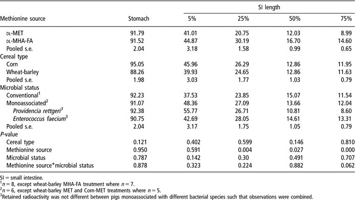
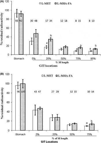
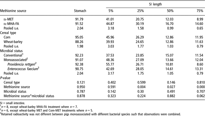
Residual l-[methyl-3H] MET and l-[methyl-3H] MHA-FA activity in contents of conventionally raised and MA pigs is shown in Table 2. In the stomach of CON and MA pigs, both methionine sources were present at similar high levels (92% to 99%) which were not affected by cereal source or microbial status. Both methionine sources disappeared rapidly from proximal SI such that less than 50% residual activity was observed at the 5% location. Approximately 12% to 14% residual activity was observed in the distal SI. Cereal grain did not affect the residual amounts of either methionine source. At 75% of SI length an interaction (P = 0.06) between methionine source and microbial status was observed. Furthermore, no data could be determined for the 95% of SI length in MA pigs because chromic oxide concentration was below detection at this location at 3 h after feeding. Separate analysis of residual activity for CON pigs revealed that residual l-MHA-FA was significantly (P < 0.01) higher (34%, 15% and 13%) compared to l-MET (16%, 8% and 7.8%) at 25%, 75% and 95% of SI length, respectively (Figure 1A). In MA pigs, the apparent retention of MET and MHA-FA was similar at all small intestinal locations, with an exception at the 75% SI location where a trend (P < 0.07) to increase residual MHA-FA was observed (Figure 1B).
Percent retained radioactivity in gastrointestinal tracts of conventional and monoassociated pigs fed corn and wheat-barley based diets supplemented with dl-methionine or dl-MHA-FA on equimolar basis
SI = small intestine.
1n = 8, except wheat-barley MHA-FA treatment where n = 7.
2n = 6, except wheat-barley MET and Corn-MET treatments where n = 5.
3Retained radioactivity was not different between pigs monoassociated with different bacterial species such that observations were combined.
Apparent retention of l-[methyl-3H] MET and l-[methyl-3H] MHA-FA in digesta in conventional (A) and monoassociated pigs (B) fed corn or wheat-barley based post-weaning diets. Values over each bar are mean percent retained radioactivity. The vertical bars depict s.e. Bars with different letters are significantly different at P < 0.05 (A) or P < 0.07 (B). SI = small intestine; GIT = gastrointestinal tract.

Apparent tissue associated l-[methyl-3H] MET and l-[methyl-3H] MHA-FA activity
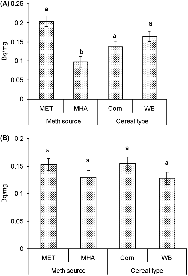
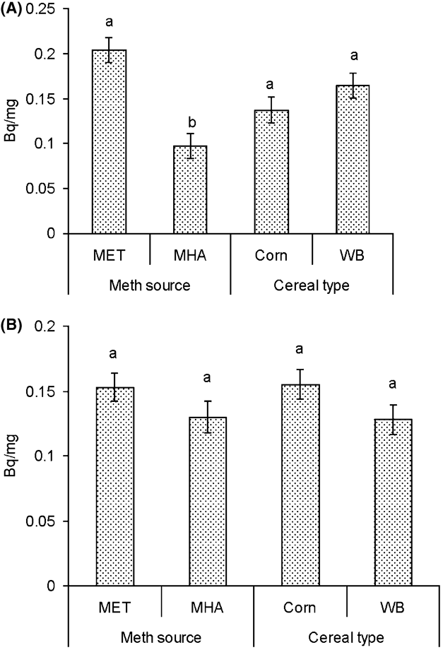
Radioactivity in small intestinal tissue associated with feeding l-[methyl-3H] MET and l-[methyl-3H] MHA-FA was detectable only at the 5% SI location (Figure 2 A and B). A significant (P < 0.01) methionine source by microbial status interaction was observed for tissue radioactivity (Bq/mg wet weight) such that at the 5% SI location activity associated with l-[methyl-3H] MET feeding (8.56 ± 0.47) was significantly (P < 0.05) higher as compared to l-[methyl-3H] MHA-FA feeding (5.45 ± 0.50) in CON pigs only (Figure 2A). Cereal grain type did not affect tissue radioactivity levels.
Apparent tissue associated l-[methyl-3H] MET and l-[methyl-3H] MHA-FA activity in conventional (A) and monoassociated pigs (B) fed corn or wheat-barley (WB) based post-weaning diets. The vertical bars depict s.e. Within methionine source or cereal type, bars with a different letters are significantly different (P < 0.05).
Discussion
The highly complex enteric microbiota play an important role in modifying the structure, biochemistry and physiology of the gastrointestinal tract, impacting nutrition and health of the host (Falk et al., Reference Falk, Hooper, Midvedt and Gordon1998). Gnotobiotic animals, where gastrointestinal bacterial populations are clearly defined, offer an indispensable tool to study a broad range of specific host–bacterial and host–nutrient–bacterial interactions. Here, we utilized the gnotobiotic model to establish the role of the intestinal microbiota, in the pig, in determining the availability for absorption of supplemented amino acids, particularly supplemented methionine and MHA-FA. Since different cereal grains are known to affect the composition of intestinal microbiota in conventionally reared animals (Hill et al., Reference Hill, Hemmingsen, Blair, Dumonceaux, Klassen, Zijlstra, Goh and Van Kessel2005), the study compared corn and wheat/barley based diets.
In the present study, residual 3H-MHA-FA activity observed in intestinal contents was significantly higher than 3H-MET activity along the entire SI in the conventionally reared pigs. In contrast, in MA pigs, residual 3H-MHA-FA and 3H-MET activity in small intestinal contents was similar. We hypothesize that the increase in residual MHA-FA activity in digesta of CON pigs, but not MA pigs, reflects a preferential metabolism of MHA-FA, relative to methionine, by the complex consortia of bacteria present in the intestine. This increase in microbial metabolism of MHA-FA could reduce the amount of MHA-FA available for absorption and contribute to the lower bio-efficacy of MHA-FA (Jansman et al., Reference Jansman, Kan and Wiebenga2003). Our results in pigs are in agreement with an earlier report by our laboratory in chickens, where conventional chickens had increased residual 3H-MHA-FA in intestinal contents compared with the germ-free chicken (Drew et al., Reference Drew, Van Kessel and Maenz2003).
MHA-FA is transported by non-stereospecific intermediate affinity H+-dependent system whereas methionine is transported by Na+-dependent system B transporter with higher substrate affinity and maximal velocity compared to H+-dependent system (Maenz and Engele Schaan, Reference Maenz and Engele Schaan1996b). We have previously suggested that the slower rate of transport of MHA-FA may prolong the opportunity for microbial metabolism relative to methionine (Drew et al., Reference Drew, Van Kessel and Maenz2003).
Many studies in chicken (Esteve Garcia and Austic, Reference Esteve Garcia and Austic1993; Lingens and Molnar, Reference Lingens and Molnar1996; Maenz and Engele Schaan, Reference Maenz and Engele Schaan1996a) used radiolabelled methionine and MHA-FA and estimated intestinal absorption based on residual radioactivity in intestinal contents. This method is not affected by bacterial metabolism of methionine and MHA-FA since the radiotracer would remain within the intestinal contents even following metabolism. All three studies reported that 10% to 20% of the original radiolabelled MHA-FA activity in the feed was present in the distal sections of the SI compared to 3.5% to 5% for methionine, thus in agreement with our results. Moreover, HPLC analysis of gut contents from the distal ileum showed that only 10% of the residual radioactivity was associated with MHA-FA, suggesting that the compound had been metabolized during intestinal transit (Maenz and Engele Schaan, Reference Maenz and Engele Schaan1996a) and further supporting metabolism by gut microbiota.
We found that MHA-FA supplementation increased the number of total aerobes and lactobacilli in the upper SI, a response that may have contributed to the increased residual MHA-FA activity in upper small intestinal contents. Among lactic acid bacteria, which are dominant in the upper SI, there is significant inter- and intra-species differences in their utilization of various amino acids (Liu et al., Reference Liu, Holland and Crow2003). Hegedus et al. (Reference Hegedus, Andrasofszky, Brydll, Veresegyhazy and Tamas1993) reported that of the three lactobacilli species (Lactobacillus plantarum, L. casei and Leuconostoc mesenteroides) studied, none could utilize MHA-FA though all could use methionine. In this regard, further characterization of the proximal gut microbiota and species variation in metabolizing various amino acids is required.
Although the methionine sources disappeared rapidly from the upper SI in the pig, interestingly, the residual activity in intestinal contents of both methionine sources was much lower (9% to 15%) in the upper SI of chicken (Drew et al., Reference Drew, Van Kessel and Maenz2003) as compared to the pig (35% to 45%). The rate of absorption of methionine sources may be slower in the upper SI of pig compared to chicken. Also, both methionine and MHA-FA were essentially completely retained in the pig stomach in contrast to a report by Richards et al. (Reference Richards, Atwell, Vazquez-Anon and Dibner2005) in the chicken, in which only 15% of supplemented MHA-FA could be detected by HPLC in digesta recovered just prior to the duodenum. The authors suggested 85% of supplemented MHA-FA was passively absorbed from the upper tract of the chicken (crop and proventriculus), a phenomenon that does not appear to occur appreciably in pig stomach. Alternatively, it is possible that the low recovery of MHA-FA by HPLC in chicken proventriculus could be partly explained by microbial metabolism to alternate products not detected by HPLC.
Wester et al. (Reference Wester, Vazquez-Anon, Dibner, Parker, Calder and Lobley2006) reported that in lambs, small intestinal tissues are capable of converting MHA-FA to methionine and further that MHA-FA-derived methionine is preferentially retained in the intestine to support anabolism, sparing absorbed methionine from dietary protein for transport to the liver. This raised the possibility that, in the current study, increased radioactivity observed in digesta in the MHA-FA supplemented pigs may be due to preferential in situ metabolism which could contribute to higher radioactivity associated with endogenous losses. In the current study, however, increased upper small intestinal tissue radioactivity was associated with the methionine-supplemented diet compared to MHA-FA. This is consistent with greater apparent absorption of methionine from the upper SI (lower residual radioactivity) and suggests, although not conclusively, that the increased residual radioactivity in the digesta observed for MHA-FA supplementation could not be accounted for by increased radioactivity in endogenous losses.
Chromic oxide content in digesta from 95% of SI length was below the assay detection limit for MA pigs, preventing determination of residual radioactivity in digesta. In contrast, chromic oxide content in CON pigs was always highest at 95% length of SI. This could be due to a slower transit rate from stomach to distal SI in MA pigs resulting in negligible chromic oxide content. Previous reports indicate that the commensal microbiota decrease the intestinal transit time in rats (Riottot et al., Reference Riottot, Sacquet, Vila and Leprince1980).
Gut microbiota is influenced by many factors including age, weaning and diet. Nutrients in the gut affect the microflora by providing preferential substrates for bacterial growth (Jensen and Jorgensen, Reference Jensen and Jorgensen1994; Hill et al., Reference Hill, Hemmingsen, Blair, Dumonceaux, Klassen, Zijlstra, Goh and Van Kessel2005). Although we did not conduct an extensive molecular examination of the composition of the intestinal microbiota in conventional animals in the current study, limited culture-based analysis detected an effect of cereal grain type. Interestingly, wheat-barley based diets increased total aerobes in the upper gastrointestinal tract, a location for the greatest opportunity for microbial metabolism of supplemented methionine source, given the rapid absorption. Nevertheless, no effect of cereal grain source on residual activity of MHA-FA or methionine was observed.
Our results support the hypothesis that intestinal microbiota compete with the host for metabolism of readily available nutrients such as supplemented amino acids. Our previous results of increased residual 3H-MHA-FA in intestinal contents of conventional, but not germ-free, chicken and current similar results in CON and MA pigs suggest that microbial metabolism of MHA-FA reduces the availability of MHA-FA for absorption relative to methionine. Whether MHA-FA is a preferred substrate for intestinal bacteria or the slower absorption kinetics relative to methionine provides greater opportunity for microbial metabolism, is not clear. Microbial metabolism of MHA-FA is, however, a likely contributor to reduced bio-efficacy of MHA-FA relative to methionine.
Acknowledgements
This research was funded by the Natural Sciences and Engineering Research Council of Canada and Evonik-Degussa GmbH. We are grateful for the skilful technical assistance provided by Jason Marshall.