Introduction
Vascular anomalies comprise 1 per cent of congenital cardiac anomalies.Reference Park and Park1 They rarely present with cardiovascular manifestations, however, being more likely to present with many of the common paediatric upper aero-digestive tract symptoms, such as wheeze, stridor, cough and dysphagia. The pattern of presentation is dependent on the particular type of anomaly, with varying degrees of tracheal and/or oesophageal compression being present. Most cases present in the first year of life, with subsequent surgical intervention providing safe and effective relief of presenting symptoms.Reference Shah, Mora, Bacha, Sena, Buonomo and Del Nido2
We present a rare case of a vascular ring which was diagnosed following the removal of a foreign body from the oesophagus. Despite foreign body removal, symptoms persisted, leading to repeat endoscopy and subsequent diagnosis of a double aortic arch. To our knowledge, this is only the second reported case of a double aortic arch presenting in a patient following removal of an oesophageal foreign body.Reference Currarino and Nikaidoh3
Case report
An eight-month-old male infant presented with a two-day history of deteriorating respiratory symptoms of wheeze, biphasic stridor and cough. A recent choking episode was reported in the days prior to admission. No swallowing difficulties were reported, and the infant had been tolerating fluids orally prior to admission. On closer questioning, baseline respiratory symptoms had been present for a period of six weeks.
Rigid bronchoscopy was performed, revealing significant tracheomalacia with posterior tracheal compression. Rigid oesophagoscopy identified a metallic foreign body in the upper oesophagus. Significant ulceration of the oesophageal mucosa was noted following removal of the foreign body.
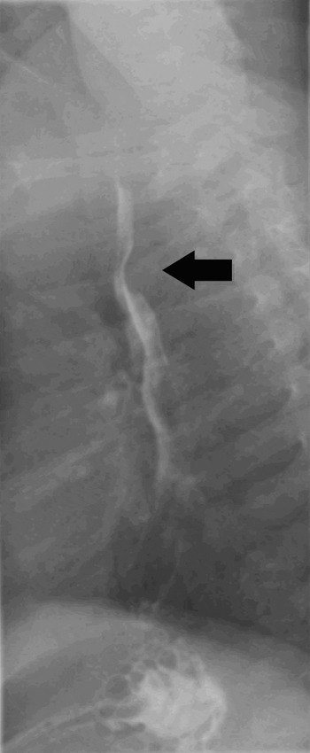
A barium swallow examination on the first post-operative day confirmed no oesophageal perforation; however, note was made during this examination of an indentation into the posterior oesophageal wall (Figure 1).
Barium swallow radiograph performed following removal of the oesophageal foreign body, showing an extrinsic defect in the posterior wall of the upper oesophagus at the level of the aortic arch, consistent with a compressive vascular ring.

The infant's respiratory symptoms responded immediately to the removal of the foreign body, and he was discharged home on the second post-operative day with follow up arranged.
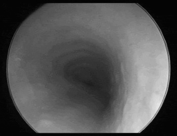
However, the infant's respiratory symptoms returned shortly after discharge, and a repeat bronchoscopy and oesophagoscopy were performed. These showed significant residual, non-pulsatile tracheal compression, arising posteriorly and laterally from the right side (Figure 2). Rigid oesophagoscopy was normal.
Bronchoscopy image showing extrinsic compression of the trachea.

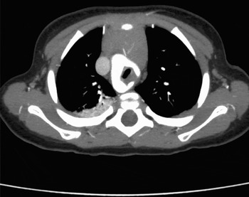
In view of these findings and the previously abnormal barium study, a computed tomography (CT) scan of the thorax was performed. This identified a double aortic arch encircling and compressing the trachea and oesophagus, at a level which corresponded to the site of foreign body impaction in the oesophagus at the initial presentation (Figure 3).
Axial, contrast-enhanced computed tomography image of the thorax, showing a double aortic arch encircling and compressing the trachea.

A subsequent echocardiogram revealed no other associated cardiac anomalies.
The patient was referred to the cardiothoracic surgery department, and subsequently underwent a thoracotomy and division of a non-dominant left aortic arch. No significant post-operative complications were reported.
Discussion
This case was a rare instance of a foreign body lodging in the oesophagus above the level of compression caused by a vascular ring. It emphasises the importance of observing basic principles in the investigation and diagnosis of persistent respiratory symptoms. In our case of persistent respiratory symptoms, an abnormal barium swallow and the finding of residual tracheomalacia at bronchoscopy lead to the diagnosis of a vascular ring.
In many cases, the diagnosis of a foreign body in either the oesophagus or the airway is obvious due to a strong history from the child's parents and corresponding physical and/or radiological findings. However, there may often be other, coexisting symptoms, often present for many days or even weeks, which can potentially lead to difficulties or delays in diagnosis. It is our view that any child in whom the presence of a foreign body in the upper aero-digestive tract has been suggested or is suspected should undergo formal bronchoscopy and oesophagoscopy in a timely fashion, in order to avoid the potentially serious consequences of a missed foreign body.
Congenital vascular anomalies occur due to failure of the normal embryological development of the great vessels. The aortic arch system develops from six pairs of arches connecting a dorsal and ventral aorta. A vascular anomaly occurs when there is persistence or failure of regression of portions of the primitive circulatory system. A double aortic arch occurs when both fourth arches persist.Reference Kaiser, Kron and Spray4, Reference Shields, LoCicero and Ponn5
The most common types of anomalies of the great vessels are (in decreasing order of frequency): (1) double aortic arch, (2) right-sided aortic arch with an aberrant left subclavian artery and ligamentum arteriosum, (3) innominate artery compression, (4) aberrant right subclavian artery, (5) pulmonary artery sling, and (6) aberrant left subclavian artery.Reference Woods, Sharp and Holcomb6
Vascular anomalies may be classified as vascular rings, which encircle major structures such as the trachea and oesophagus, and vascular slings, which cause symptoms by compressing either structure without encircling it. Vascular rings include the double aortic arch and the right-sided aortic arch with aberrant left subclavian artery and ligamentum arteriosum. A double aortic arch is the only true vascular ring. Incomplete vascular rings include innominate artery compression and a pulmonary artery sling.
Symptoms at presentation vary depending on which structures are compressed by the vascular anomaly and the degree of compression. Vascular rings, such as the double aortic arch, tend to present with mainly respiratory symptoms due to tracheal compression, while oesophageal symptoms predominate with other anomalies such as innominate artery compression.Reference Turner, Gavel and Coutts7
• A rare case is presented of an oesophageal foreign body lodging above the level of oesophageal compression by a vascular ring
• Symptoms persisted despite removal of the oesophageal foreign body, leading to the diagnosis of tracheal and oesophageal compression by a double aortic arch
• The literature surrounding congenital anomalies of the major vessels is reviewed
A variety of investigations may lead to the diagnosis of a vascular anomaly, depending on the primary symptoms at presentation. Barium swallow is a simple, useful investigation, which will typically identify an indentation in the oesophagus on a lateral view. Bronchoscopy may reveal tracheal compression which may or may not be pulsatile in nature. Bronchoscopy is useful also to document normal vocal fold movement prior to any planned cardiothoracic surgery, and also to detect the presence of any coexisting airway pathology such as laryngomalacia. Some debate exists on the most appropriate imaging modality for diagnosis of the various types of vascular anomaly. While magnetic resonance imaging and magnetic resonance angiography have previously been considered superior, newer generation CT scanning with contrast now provides comparable accuracy and diagnostic information necessary for planning surgical repair.Reference Turner, Gavel and Coutts7 Echocardiography should be performed on all patients diagnosed with a cardiac vascular anomaly, due to the high incidence of associated congenital cardiac abnormalities.Reference Lillehei and Colan8
Management involves thoracic surgery, which may be performed by thoracotomy or as open surgery via a formal sternotomy. Overall survival figures are excellent. The principal complications are vocal fold paralysis, pneumothorax and chylothorax. Persistent residual tracheomalacia is commonly reported following surgery; despite removal of the compressive vascular anomaly, tracheomalacia may persist, often for many months.Reference Shah, Mora, Bacha, Sena, Buonomo and Del Nido2
Conclusion
Vascular anomalies are rare but important causes of both respiratory and swallowing symptoms in the paediatric population. The presented patient was a rare case of a foreign body lodging above the site of oesophageal compression by a vascular ring. This case highlights the usefulness of basic diagnostic tests such as barium studies in the paediatric patient. Our patient also emphasises the importance of repeating diagnostic examinations such as bronchoscopy, in cases in which the clinical history and physical signs fail to agree.