Introduction
The anhydrous sodium–copper sulfates are of interest for mineralogists, geochemists, physicists and materials scientists because of their genetic significance in postvolcanic fumarolic systems and perspective magnetic and non-linear optic properties (Kovrugin et al., Reference Kovrugin, Nekrasova, Siidra, Mentre, Masquelier, Stefanovich and Colmont2019; Borisov et al., Reference Borisov, Siidra, Kovrugin, Golovin, Depmeier, Nazarchuk and Holzheid2021; Siidra et al., Reference Siidra, Borisov, Charkin, Depmeier and Platonova2021a, Reference Siidra, Charkin, Kovrugin and Borisov2021b, Reference Siidra, Nekrasova, Charkin, Zaitsev, Borisov, Colmont, Mentre and Spiridonova2021c; Singh et al., Reference Singh, Neveu, Jayanthi, Das, Chakraborty, Navrotsky, Pralong and Barpanda2022). These papers have shown, for example, that the eldfellite-type phase NaFe(SO4)2 is characterised by 8 mAh/g of reversible capacity with a discharge voltage of 3.0 V and that the synthetic analogue of saranchinaite can be considered as a high voltage cathode (4.84 V vs Li+ /Li0). However, natural samples of such sulfates and their synthetic counterparts tend to have narrow fields of thermodynamic stability and are affected by different transformations including hydration. Thus, each novel natural Na–Cu sulfate sheds a light on formation conditions and crystal chemistry of these types of compounds.
All anhydrous Na–Cu sulfates discovered recently in Nature are endemics of active fumaroles of the Tolbachik volcano (Kamchatka, Russia), namely puninite Na2Cu3O(SO4)3 (Siidra et al., Reference Siidra, Nazarchuk, Zaitsev, Lukina, Avdontseva, Vergasova, Vlasenko, Filatov, Turner and Karpov2017), saranchinaite Na2Cu(SO4)2 (Siidra et al., Reference Siidra, Lukina, Nazarchuk, Depmeier, Bubnova, Agakhanov, Avdontseva, Filatov and Kovrugin2018), petrovite Na12Cu2(SO4)8 (Filatov et al., Reference Filatov, Shablinskii, Krivovichev, Vergasova and Moskaleva2020), and the mineral described here, cuprodobrovolskyite Na4Cu(SO4)3. This latter mineral differs from others in stoichiometry, symmetry, crystal structure and physical properties.
Cuprodobrovolskyite (Cyrillic: купродобровольскиит) is named as an analogue of dobrovolskyite Na4Ca(SO4)3 (Shablinskii et al., Reference Shablinskii, Filatov, Krivovichev, Vergasova, Moskaleva, Avdontseva, Knyazev and Bubnova2021) with copper prevailing over calcium. Both the new mineral and its name (symbol Cdvo) have been approved by the Commission on New Minerals, Nomenclature and Classification of the International Mineralogical Association (IMA No. 2022-061, Shchipalkina et al., Reference Shchipalkina, Pekov, Koshlyakova, Belakovskiy, Zubkova, Agakhanov, Britvin and Nazarova2023b). The type specimen is deposited in the systematic collection of the Fersman Mineralogical Museum of the Russian Academy of Sciences, Moscow, Russia with catalogue number 98046.
Occurrence, morphology and physical properties
The specimens with the new mineral were collected by us in July 2021 at the Arsenatnaya fumarole located at the summit of the Second scoria cone of the Northern Breakthrough of the Great Tolbachik Fissure Eruption 1975–1976, Tolbachik volcano, Kamchatka. This active, high-temperature oxidising-type fumarole is famous as the type locality of more than sixty new mineral species. Its general description is given by Pekov et al. (Reference Pekov, Koshlyakova, Zubkova, Lykova, Britvin, Yapaskurt, Agakhanov, Schipalkina, Turchkova and Sidorov2018) and Shchipalkina et al. (Reference Shchipalkina, Pekov, Koshlyakova, Britvin, Zubkova, Varlamov and Sidorov2020b). The specimens with cuprodobrovolskyite were collected in fumarolic pockets with temperatures of 350–400°C (the temperature was measured with a chromel–alumel thermocouple during sampling).
Cuprodobrovolskyite is a typical fumarolic mineral. We believe that it was deposited directly from the gas phase as a volcanic sublimate at temperatures not lower than 400°C. Minerals associated closely with cuprodobrovolskyite are petrovite, saranchinaite, euchlorine, krasheninnikovite, langbeinite, calciolangbeinite, anhydrite, sanidine, tenorite and hematite.
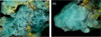
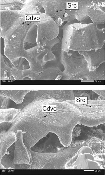
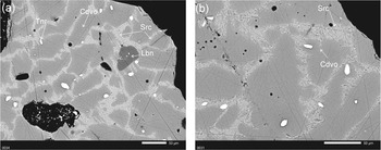
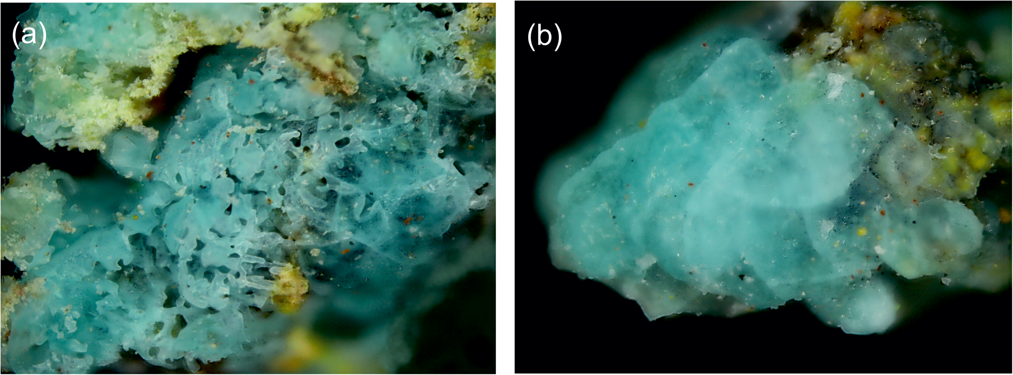
Cuprodobrovolskyite occurs as two morphological varieties: (1) coarse hexagonal tabular or equant, typically skeletal crystals up to 1 mm and their clusters or open-work crusts (Figs 1a and 2) up to 5 mm across; and (2) massive crusts, typically interrupted, up to 1.5 cm × 2.5 cm in area and up to 1 mm thick, with a ‘glazed’ surface (Fig. 1b) which cover basalt scoria altered by fumarolic gas. Cuprodobrovolskyite crystals and crusts contain abundant lamellar and irregular shaped ingrowths of saranchinaite; on the surface, these ingrowths look like grooves (Fig. 2). The relationship between saranchinaite and the host cuprodobrovolskyite is shown in Fig. 3. Other sulfates (petrovite, langbeinite and calciolangbeinite) also form intimate intergrowths with cuprodobrovolskyite.
Light blue crusts of cuprodobrovolskyite: (a) open-work crust consisting of coarse skeletal crystals, (b) massive crust. Field of view, width – 1 mm (both images). Holotype specimen, # 98046.
Sulfate crusts consisting of coarse skeletal cuprodobrovolskyite (Cdvo) crystals with ingrowths of saranchinaite (Src). The mineral symbols are given after Warr (Reference Warr2021). Scanning electron microscopy (SEM) images, SE mode. Holotype specimen, # 98046.

Crystals of cuprodobrovolskyite (Cdvo) with ingrowths (probably relics) of saranchinaite (Src); Tnr – tenorite and Lbn – langbeinite. Polished sections, SEM images, BSE mode. Holotype specimen, # 98046.

Cuprodobrovolskyite is transparent, light blue or greenish-bluish to almost colourless. The streak is white. The lustre is vitreous. The Mohs’ hardness is ca. 3. The mineral is brittle, cleavage or parting was not observed. The density could not be determined correctly owing to several intergrown minerals, represented mainly by saranchinaite. The density calculated using the empirical formula is 2.783 g cm–3.
Cuprodobrovolskyite is optically uniaxial (+) with ω = 1.509(3) and ɛ = 1.528(3) (589 nm). Under the microscope in plane polarised transmitted light cuprodobrovolskyite is colourless and non-pleochroic. The Gladstone-Dale compatibility index is –0.043 (good).
Methods and results
Chemical composition
The chemical composition of cuprodobrovolskyite was studied using an electron microprobe. The analyses were carried out with a JEOL JXA-8230 (WDS mode) at the Laboratory of Analytical Techniques of High Spatial Resolution, Dept. of Petrology, Moscow State University. The operating conditions included an accelerating voltage of 20 kV and beam current of 20 nA; the beam was rastered on an area 4 μm × 4 μm. The data reduction was carried out using the INCA Energy 300 software package. The following standards were used for quantitative analysis: diopside (Mg), metal Cu (Cu), ZnS (Zn), anorthite (Ca and Al), PbTe (Pb), SrSO4 (Sr) and pyrite (S). Contents of other elements with atomic numbers higher than carbon are below detection limits.
The chemical composition of cuprodobrovolskyite is given in Table 1. The averaged empirical formula of the holotype specimen calculated on the basis of 12 O atoms per formula unit (apfu) is (Na3.64K0.09Pb0.03)Σ3.76(Cu0.51Ca0.22Mg0.16Zn0.07Al0.01Mn0.01)Σ0.98S3.04O12. The simplified formula is Na4(Cu,Ca,Mg,Zn)(SO4)3. The ideal formula Na4Cu(SO4)3 requires Na2O 27.88, CuO 17.94, SO3 54.18, total 100 wt.%.
Chemical composition of cuprodobrovolskyite from the Arsenatnaya fumarole: holotype (#1) and other specimens (##2, 3) and cuprodobrovolskyite samples after annealing (##4, 5).
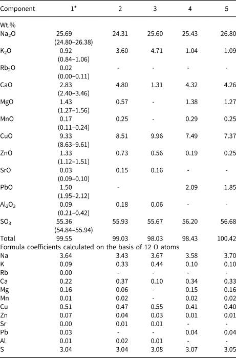
* Averaged for 10 spot analyses, ranges are in parentheses; ‘–’ means the content was below the detection limit.
Raman spectroscopy
The Raman spectra of cuprodobrovolskyite and, for comparison, dobrovolskyite, saranchinaite and petrovite (Fig. 4) were recorded using an EnSpectr R532 spectrometer with a green laser (532 nm) at room temperature. The output power of the laser beam was ~7 mW. The spectrum was processed using the EnSpectr expert mode program in the range from 100 to 4000 cm–1 with the use of a holographic diffraction grating with 1800 lines/cm and a resolution of ~6 cm–1. The diameter of the focal spot on the sample was ~10 μm. The Raman spectra were acquired on polycrystalline samples previously checked by powder X-ray diffraction (XRD).
The Raman spectra of (a) saranchinaite, (b) petrovite, (c) dobrovolskyite and (d) cuprodobrovolskyite. For Raman shift values of bands see Table 2.
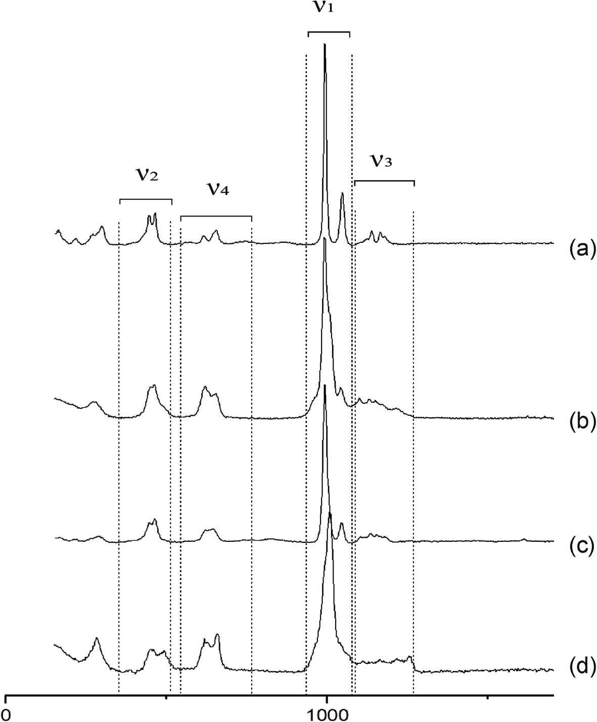
Three main regions are usually distinguished in the Raman spectra of anhydrous sulfates with bivalent cations: (1) 800–1300 cm−1, SO4 stretching vibrations (ν1 and ν3 modes); (2) 800–400 cm−1, SO4 bending vibrations (ν2 and ν4 modes); (3) 400–100 cm−1, M–O (M = Cu, Mg, Fe, Ca, Zn and Na) vibrations and lattice modes (Nakamoto, Reference Nakamoto1986; Kosek et al., Reference Kosek, Culka and Jehlicka2018 and references therein). Comparison of the Raman spectra of the above-listed minerals is presented in Fig. 4, Raman shift values and assignment of bands are given in Table 2.
Vibration modes of SO4 tetrahedra in the Raman spectra of saranchinaite, petrovite, dobrovolskyite and cuprodobrovolskyite.

* ‘No.’ = Number of non-equivalent S sites (per unit cell)
Notes: sh – shoulder, w – weak band; strong bands are highlighted in bold type, bands with medium intensity have no marks.
Despite the common features, the Raman spectra of these sulfates are different from each other in numbers, intensities and positions of bands (Table 2). Symmetric bending vibrations of SO4 tetrahedra in saranchinaite, petrovite and dobrovolskyite spectra appear in the range 447–464 cm−1, whereas in cuprodobrovolskyite the positions of these bands are shifted to higher frequencies: 452–492 cm–1. The band of asymmetric stretching vibrations of SO4 tetrahedra in cuprodobrovolskyite has the highest frequency among these minerals: 1258 cm–1 (Table 2). Another distinctive feature of the spectrum of cuprodobrovolskyite is a single strong band at 1009 cm–1 (S–O stretching symmetric vibrations) with shoulders (1044 and 1064 cm–1), whereas in the spectra of saranchinaite, petrovite and dobrovolskyite there are distinct doublets: strong band at 991–993 cm–1 and weaker band at 1042–1047 cm–1.
The absence of bands with frequencies higher than 1300 cm–1 indicates the absence of groups with O–H, C–H, C–O, N–H and N–O bonds in cuprodobrovolskyite and other studied sulfates.
Powder X-ray diffraction data and crystal structure
The attempts of single-crystal XRD studies of cuprodobrovolskyite were unsuccessful because of the imperfectness (microblocky character) of crystals. Powder XRD data for the new mineral (on a sample polluted with petrovite and saranchinaite) were collected using a Rigaku R-AXIS Rapid II diffractometer equipped with image plate detector and rotating anode with the microfocus optics, CoKα radiation, 40 kV, 15 mA, Debye-Scherrer geometry, d = 127.4 mm and exposure time of 15 min. The raw data to profile conversion was performed with the osc2xrd program (Britvin et al., Reference Britvin, Dolivo-Dobrovolsky and Krzhizhanovskaya2017). Powder data are reported in Table 3. On the basis of the data on dobrovolskyite (Shablinskii et al., Reference Shablinskii, Filatov, Krivovichev, Vergasova, Moskaleva, Avdontseva, Knyazev and Bubnova2021), we found that the mineral is trigonal, space group R3. The unit-cell parameters of cuprodobrovolskyite calculated from the powder data are: a = 15.702(2), c = 22.017(5) Å, V = 4701.0(2) Å3, and Z = 9.
Powder X-ray diffraction data (d in Å) for cuprodobrovolskyite.

* Reflections overlapped with reflections of admixed petrovite. The strongest reflections are marked in boldtype.
The crystal structure of cuprodobrovolskyite was studied on a powder sample using the Rietveld method. Data treatment and the Rietveld structure analysis were carried out using the Jana2006 software (Petříček et al., Reference Petriček, Duŝek and Palatinus2014). A total of 13,700 observed intensity envelope points were used in the refinement. The profiles of individual reflections were modelled using a Pseudo-Voigt function.
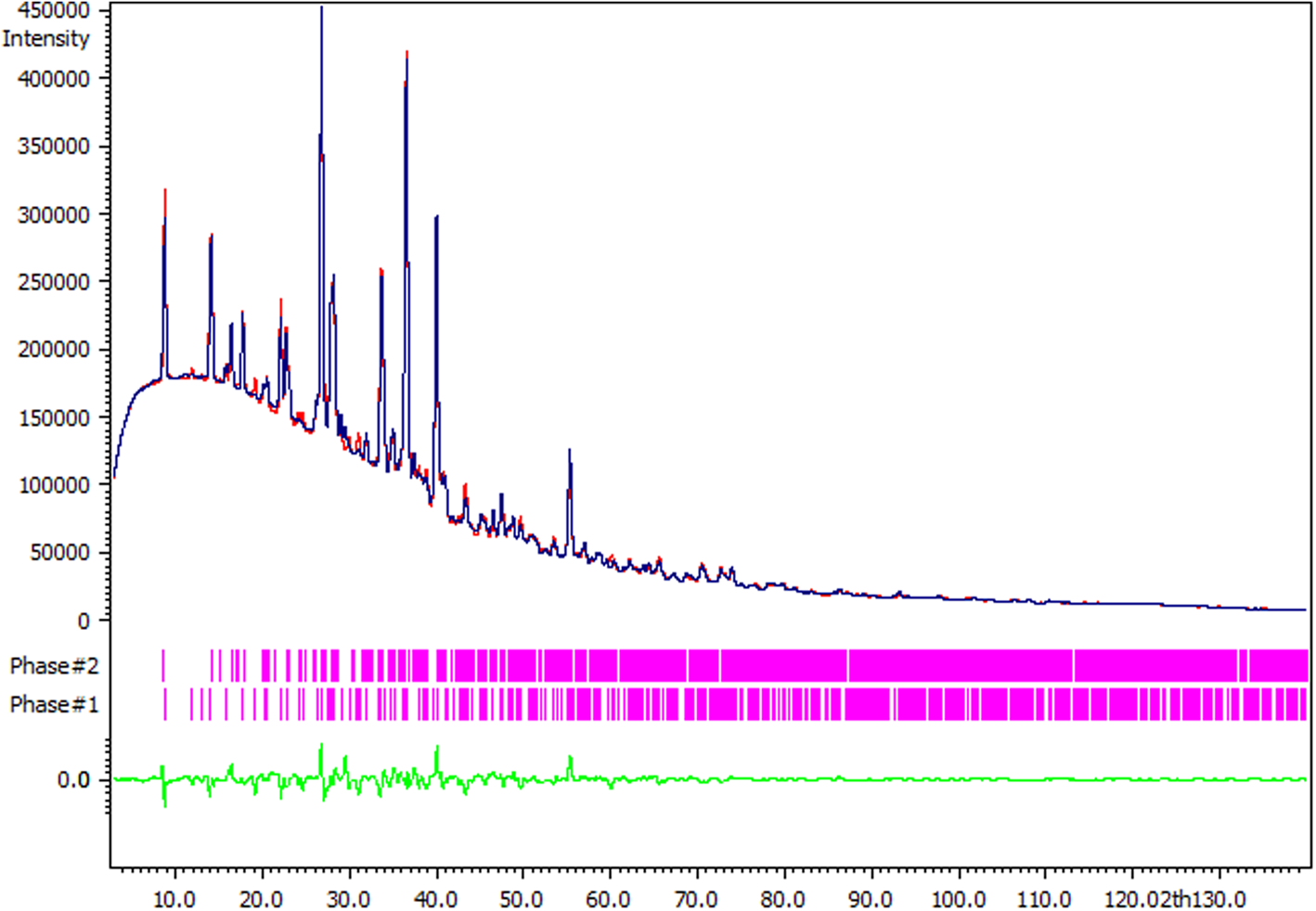
The refinement of the crystal structure of cuprodobrovolskyite was complicated by the presence of admixed petrovite and saranchinaite in the powder sample (Fig. 5). As shown in Table 4, cuprodobrovolskyite is well-distinguished from petrovite and saranchinaite by number and position of strong reflections in powder XRD pattern. Generally, the powder XRD diagram is a good diagnostic tool to distinguish cuprodobrovolskyite from other Na–Cu sulfates including petrovite though quite similar in chemistry.
Rietveld refinement plot of mixture of cuprodobrovolskyite and petrovite. The models of dobrovolskyite (Shablinskii et al., Reference Shablinskii, Filatov, Krivovichev, Vergasova, Moskaleva, Avdontseva, Knyazev and Bubnova2021) (Phase #1; see Table 4) and petrovite (Filatov et al., Reference Filatov, Shablinskii, Krivovichev, Vergasova and Moskaleva2020) (Phase #2) were used, Bragg positions of their reflections are coloured in magenta. The blue curve is the experimental pattern of the mixture consisting of cuprodobrovolskyite and petrovite, the calculated pattern is outlined in red, the difference curve is outlined in green.
Comparative data on cuprodobrovolskyite and related minerals.

* The formula of cuprodobrovolskyite is given for Z = 18 for better comparison with other presented minerals.
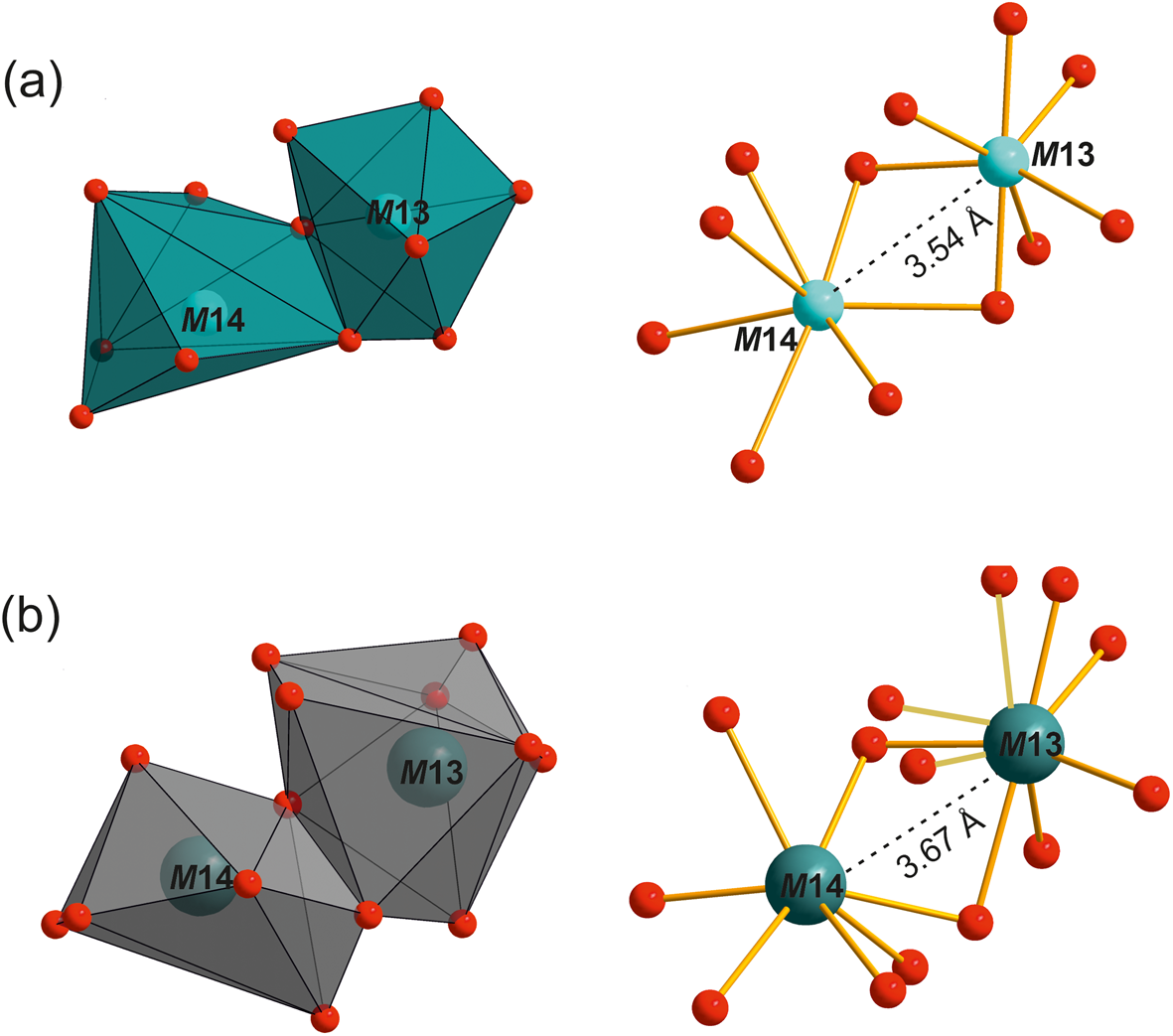
Data collection information and structure refinement details for cuprodobrovolskyite are given in Table 5. However, the pollution of the sample by these two minerals led to the insufficient description of the profile, which affected the refinement of anisotropic displacement parameters for O atoms and metals. The accuracy of calculations of the location of O atoms and, respectively, the interatomic M–O and S–O distances did not allow us to calculate the BVS. Atomic scattering factors together with anomalous dispersion corrections were taken from the International Tables for X-Ray Crystallography (Ibers and Hamilton, Reference Ibers and Hamilton1974). The final refinement cycles were finished with R p = 0.0246, R wp = 0.0325, R 1 = 0.0521, wR 2 = 0.0770 and GOF = 7.70 for all data. Fractional atomic coordinates, site occupancies based on refined electron numbers and equivalent atomic displacement parameters (U eq) are given in Table 6. The selected interatomic distances in the cuprodobrovolskyite structure are presented in Table 7. The crystallographic information file has been deposited with the Principal Editor of Mineralogical Magazine and is available as Supplementary material (see below). All SO4 tetrahedra are quite distorted with S–O distances from 1.37 to 1.62 Å. For two S sites (S2 and S3), coordination of S by O atoms includes additional variative O sites by analogy with dobrovolskyite. Sodium atoms centre different polyhedra, with coordination numbers from 6 to 9 (Table 7). These polyhedra, joining via edges, vertices and SO4 tetrahedra, form rods described by Shablinskii et al. (Reference Shablinskii, Filatov, Krivovichev, Vergasova, Moskaleva, Avdontseva, Knyazev and Bubnova2021) for dobrovolskyite. Cu2+ cations occupy two independent M sites coordinated by seven O atoms with average distances of 2.39 and 2.66 Å for M13 and M14, respectively. The refined numbers of electrons (e ref) are 26 for M13 and 25 for M14 whereas for other cation sites e ref were between 6 and 12. The only ‘heavy’ component, which is present in the mineral in a significant amount to cause e ref values for M13 = 26 and for M14 = 25, is Cu, whereas the amounts of other constituents with high atomic numbers, Pb and Zn, are minor: 0.04 and 0.07 apfu, respectively. Thus, it is undoubtedly Cu that is the prevailing cation in both M13 and M14 sites. The second component in M13 and M14 should be Ca. This consideration is supported by two arguments: (1) in the isostructural mineral dobrovolskyite, ideally Na4Ca(SO4)3, Ca occurs in these sites (Shablinskii et al., Reference Shablinskii, Filatov, Krivovichev, Vergasova, Moskaleva, Avdontseva, Knyazev and Bubnova2021); and (2) in synthetic analogue of the Cu-bearing variety of dobrovolskyite, Na4(Ca,Cu)(SO4)3, both Ca and Cu2+ occupy these sites together (Shorets, Reference Shorets2022).
Crystal data and Rietveld refinement details for cuprodobrovolskyite.

Coordinates and equivalent displacement parameters (Å2) of atoms and site occupancies in crystal structure of cuprodobrovolskyite.

Selected interatomic distances in crystal structure of cuprodobrovolskyite.

Heat treatment of cuprodobrovolskyite
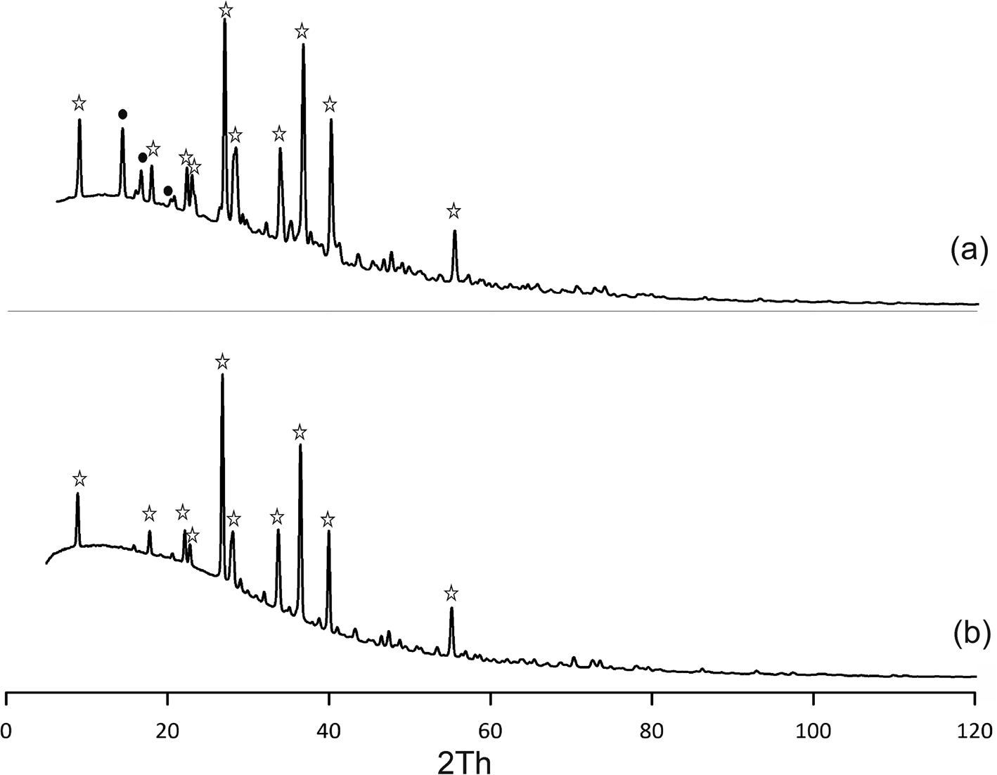
The heating of greenish-blue crusts composed of cuprodobrovolskyite with ingrowths of petrovite and saranchinaite at 600°C for 4 h was carried out using the muffle furnace. The experiment reveals that only cuprodobrovolskyite remains in the sample (verified by powder XRD: Fig. 6). The colour of the sample changed after heating from blue to light green. The chemical composition of the obtained crystals is close to that of cuprodobrovolskyite before the heating (Table 1). Notably, the studied mixture partly decomposed with segregation of CuO on the crucible walls.
Powder X-ray diffraction patterns (λ = 1.79021 Å) of cuprodobrovolskyite with (a) a petrovite admixture and (b) the same sample after heating to 600°C for 4 hours in a muffle furnace. The distinctive reflections of cuprodobrovolskyite and petrovite are marked as stars and filled circles, respectively.
Discussion
Comparative crystal chemistry of anhydrous sodium–copper sulfates
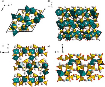
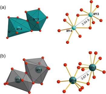
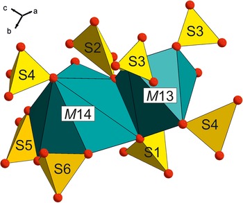
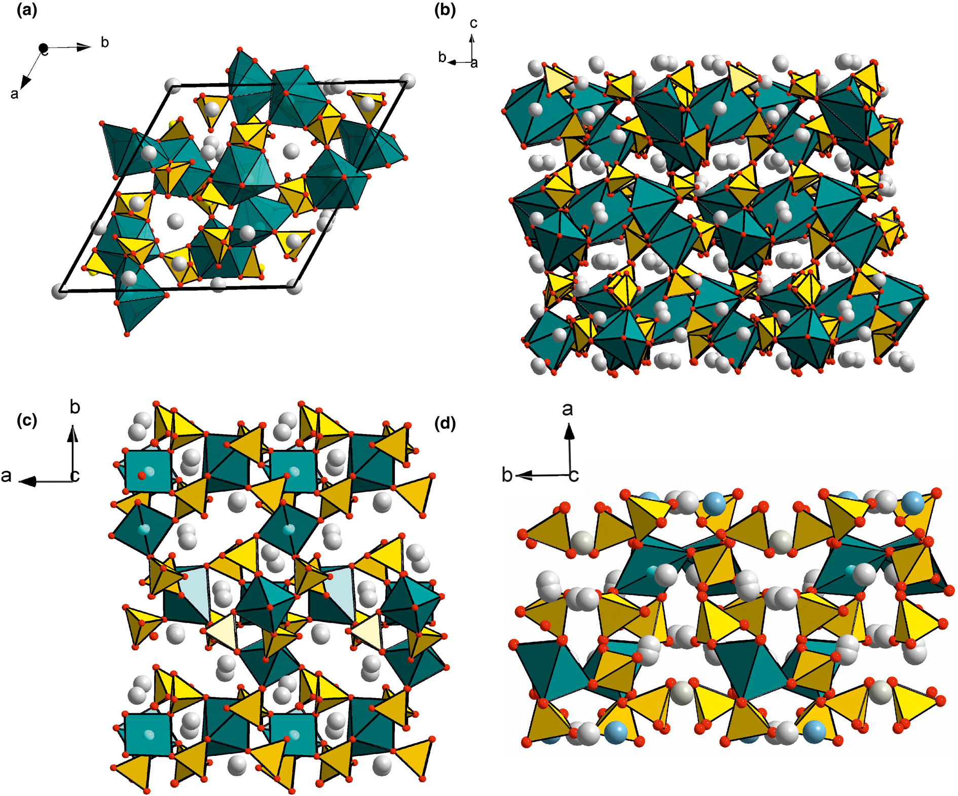
Three natural anhydrous Na–Cu sulfates without additional oxygen, saranchinaite Na2Cu(SO4)2, petrovite Na12Cu2(SO4)8, and cuprodobrovolskyite Na4Cu(SO4)3 belong to three different structural types. The main structural units in these minerals are based on motifs built by Cu2+-centred polyhedra and SO4 tetrahedra, however, there are significant differences (Fig. 7). Saranchinaite is considered as a specific compound with a three-dimensional [Cu4(SO4)8]8– framework (Siidra et al., Reference Siidra, Nazarchuk, Zaitsev, Lukina, Avdontseva, Vergasova, Vlasenko, Filatov, Turner and Karpov2017) whereas the crystal structure of petrovite includes isolated [Cu2(SO4)8]12– clusters in which CuO7 polyhedra are connected via vertices with SO4 tetrahedra (Filatov et al., Reference Filatov, Shablinskii, Krivovichev, Vergasova and Moskaleva2020). In all three discussed crystal structures, Cu2+ cations centre 7-fold polyhedra (in saranchinaite there are also CuO6 octahedra with Jahn–Teller distortion). In cuprodobrovolskyite, Cu-centred 7-fold polyhedra are linked to each other via common edges, the same as Ca polyhedra in dobrovolskyite (Fig. 8). The joint of SO4 tetrahedra with CuO7 polyhedra in cuprodobrovolskyite is illustrated in Fig. 9.
The fragments of crystal structures of (a and b) cuprodobrovolskyite, (c) saranchinaite (drawn after Siidra et al., Reference Siidra, Lukina, Nazarchuk, Depmeier, Bubnova, Agakhanov, Avdontseva, Filatov and Kovrugin2018) and (d) petrovite (drawn after Filatov et al., Reference Filatov, Shablinskii, Krivovichev, Vergasova and Moskaleva2020). SO4 tetrahedra are yellow, Cu-centred polyhedra are bluish-green, Na atoms are presented as greyish spheres. Drawn using Diamond 3.2 (Diamond - Crystal and Molecular Structure Visualization, Crystal Impact - Dr. H. Putz & Dr. K. Brandenburg GbR, Kreuzherrenstr. 102, 53227 Bonn, Germany, https://www.crystalimpact.de/diamond).
Coordination of Cu and Ca atoms and joint of Cu- and Ca-centred polyhedra (M13 and M14) in crystal structures of (a) cuprodobrovolskyite and (b) dobrovolskyite (drawn after Shablinskii et al., Reference Shablinskii, Filatov, Krivovichev, Vergasova, Moskaleva, Avdontseva, Knyazev and Bubnova2021).
The fragment of the crystal structure of cuprodobrovolskyite with polyhedra centred by bivalent cations.

It is noteworthy that the crystal structures with edge-shared Cu-polyhedra are not exotic. There are numerous compounds with such type of Cu polyhedra stacking, e.g. walls composed of CuO5 polyhedra are described for α-(Cu2−xZnx)V2O7 (Shi et al., Reference Shi, Sanson, Venier, Fan, Sun, Xing and Chen2020) and chains of edge-shared CuO6 octahedra for β-NaCuPO4 (Ulutagay-Kartin et al., Reference Ulutagay-Kartin, Etheredge, Schimek and Hwu2002). The experiments performed by Siidra et al. (2021) clearly show that the appearance of highly-coordinated Cu2+ (7-fold coordination by O atoms) is, in general, not unique for anhydrous alkali copper sulfates. Cuprodobrovolskyite is one more example which demonstrates this phenomenon.
Cuprodobrovolskyite belongs to the family of sulfates with structures derived from the archetype of hexagonal (space group P63/mmc) sodium sulfate with an aphthitalite-like structure known among synthetic compounds as Na2SO4(I) (Fischmeister, Reference Fischmeister1962; Rasmussen et al., Reference Rasmussen, Jorgensen and Lundtoft1996 and references therein) and in Nature as metathénardite (Pekov et al., Reference Pekov, Shchipalkina, Zubkova, Gurzhiy, Agakhanov, Belakovskiy, Chukanov, Lykova, Vigasina, Koshlyakova, Sidorov and Giester2019). Its trigonal derivatives with the same unit-cell metrics (a = 5.3–5.8 and c = 7.05–7.4 Å) are aphthitalite K3Na(SO4)2, natroaphthitalite KNa3(SO4)2 (both P $\bar{3}$ m1), and belomarinaite KNaSO4 (P3m1) (Filatov et al., Reference Filatov, Shablinskii, Vergasova, Saprikina, Bubnova, Moskaleva and Belousov2019; Shchipalkina et al., Reference Shchipalkina, Pekov, Chukanov, Belakovskiy, Zubkova, Koshlyakova, Britvin and Sidorov2020a). The derivatives with multiplied unit-cell parameters are bubnovaite K2Na8Ca(SO4)6, (P31c, a = 10.8 and c = 22.0 Å, Gorelova et al., Reference Gorelova, Vergasova, Krivovichev, Avdontseva, Moskaleva, Karpov and Filatov2016), dobrovolskyite Na4Ca(SO4)3 (Shablinskii et al., Reference Shablinskii, Filatov, Krivovichev, Vergasova, Moskaleva, Avdontseva, Knyazev and Bubnova2021), and cuprodobrovolskyite Na4Cu(SO4)3 (both R3, a = 15.7 and c = 22.0 Å: Table 4). All these minerals, except for aphthitalite, were described as new species in exhalations of the Tolbachik fumaroles. The relationship of the dobrovolskyite structure type with the archetype of metathénardite and relative superstructures such as bubnovaite K2Na8Ca(SO4)6 and hanksite Na22K(SO4)9(CO3)2Cl was described by Shablinskii et al. (Reference Shablinskii, Filatov, Krivovichev, Vergasova, Moskaleva, Avdontseva, Knyazev and Bubnova2021). They represented the series of aphthitalite-related crystal structures in terms of stacking cation layers and their arrangement in unit cells. The dobrovolskyite structure is considered as unique with a 3×3×3 superstructure and ordered vacant sites in the cation array (Shablinskii et al., Reference Shablinskii, Filatov, Krivovichev, Vergasova, Moskaleva, Avdontseva, Knyazev and Bubnova2021). It is interesting that the dobrovolskyite structure type is suitable for isomorphism of Cu2+ → Ca despite the discrepancy in their ionic radii: 0.72 and 1.06 Å for Cu2+ and Ca, respectively (Shannon and Prewitt, Reference Shannon and Prewitt1969). However, there are several proved examples of solid-solution series between isotypic Ca- and Cu-compounds. The crystal structure data on these compounds show that positions of Ca2+ and Cu2+ cations are not exactly the same though they are located closely. Thus, among relatively simple compounds, the synthetic solid-solution series Ca1–xCu2+xO3 is a promising example (Ruck et al., Reference Ruck, Wolf, Ruck, Eckert, Krabbes and Müller2001). In Nature for example, alluaudite-group minerals show such a phenomenon. The most demonstrative example here is the continuous solid-solution series johillerite NaCuMg3(AsO4)3 – nickenichite Na(Ca0.5Cu0.5)Mg3(AsO4)3 – calciojohillerite NaCaMg3(AsO4)3: in intermediate members, the closely located A(1) and A(1)' sites are occupied with Ca and Cu2+, respectively [Hatert, Reference Hatert2019, and references therein). Another interesting example of isomorphous substitution of Ca by Cu was stated for synthetic hydroxylapatite (Guo et al. Reference Guo, Liang, Song, Loh, Kherani, Wang, Kubel, Dai, Wang and Ozin2021). According to the crystal chemical data obtained in this work, a similar character of Ca–Cu2+ substitution in cuprodobrovolskyite and dobrovolskyite can be proposed, especially noting the unusual interatomic distances for Ca and Cu. Thus, these cations can occupy the closely located positions. The relatively high values of atom displacement parameters for the cation sites, especially M14 (Table 6), as well as significant distortion of polyhedra, could be a result of a splitting of the M13 and M14 sites to subsites statistically occupied with Cu or Ca. It is not excluded that these subsites could be described with different oxygen coordinations, however, the quality of the available samples does not allow us to prove this suggestion. It is noteworthy, that the recently IMA-approved mineral enricofrancoite (IMA No. 2023-002) with the ideal formula KNaCaSi4O10 from the Somma-Vesuvius volcanic complex (Naples, Italy) is an analogue of litidionite KNaCuSi4O10 with Ca instead of Cu (Balassone et al., Reference Balassone, Panikorovskii, Pellino, Bazai, Bocharov, Goychuk, Avdontseva, Yakovenchuk, Krivovichev, Petti, Cappelletti, Mondillo, Moliterni and Altomare2023). This find is more indirect evidence that post-volcanic processes can contribute to Ca–Cu2+ isomorphic substitution.
Genetic features of anhydrous Na–Cu sulfates: relationship between of saranchinaite Na2Cu(SO4)2, petrovite Na12Cu2(SO4)8 and cuprodobrovolskyite Na4Cu(SO4)3
As was shown by Siidra et al. (Reference Siidra, Lukina, Nazarchuk, Depmeier, Bubnova, Agakhanov, Avdontseva, Filatov and Kovrugin2018) for genesis of saranchinaite: (1) it can be a product of dehydration of kröhnkite Na2Cu(SO4)2(H2O)2 with complete transformation at 200°C; and (2) at temperatures higher than 475°C saranchinaite decomposes into tenorite, thénardite (metathénardite? – our note) and an unidentified phase. The latter can correspond to petrovite or cuprodobrovolskyite, but the data given in the cited paper are scarce and do not allow comparison of the reflections on the powder XRD patterns.
If we assume that petrovite and cuprodobrovolskyite can be not only the primary sulfates but also the products of transformations of several associated minerals (of which there are many in large amounts) in sulfate-rich zones of the Arsenatnaya fumarole, then the following reactions can be suggested:


Dobrovolskyite Na4Ca(SO4)3 and cuprodobrovolskyite from the Arsenatnaya fumarole are supposed to form a solid-solution series, which is most probably, limited (Fig. 10). The presence of admixed Ca in both petrovite and the studied cuprodobrovolskyite could be due to the participation of anhydrite CaSO4 in the reactions as suggested for petrovite by Filatov et al. (Reference Filatov, Shablinskii, Krivovichev, Vergasova and Moskaleva2020):

and, as we can now assume for the intermediate members of the dobrovolskyite Na4Ca(SO4)3 – cuprodobrovolskyite Na4Cu(SO4)3 series:

The Ca:Cu ratio (formula based on 12 O apfu) in cuprodobrovolskyite (diamonds) and the holotype dobrovolskyite (Shablinskii et al., Reference Shablinskii, Filatov, Krivovichev, Vergasova, Moskaleva, Avdontseva, Knyazev and Bubnova2021) (red circle). The composition of the cuprodobrovolskyite sample with the studied crystal structure is marked as a green diamond. The dotted line shows the formal border between dobrovolskyite and cuprodobrovolskyite.

The indirect evidence of the high-temperature origin of cuprodobrovolskyite (and probably dobrovolskyite) follows from the results of the experiments aimed at synthesis of petrovite reported by Shorets (Reference Shorets2022) who used the mixture of Na2SO4, CaSO4 and CuSO4 in ratios of 5:1:1, pressed this mixture in tablets and then heated the tablets at T = 600°C for 60 h. However, instead of the expected petrovite, the main phase formed in this experiment was a Cu-bearing variety of dobrovolskyite with tenorite admixture (Shorets, Reference Shorets2022).
Our heating experiment shows the sample studied, with the assemblage of cuprodobrovolskyite, petrovite and saranchinaite, partly decomposed with segregation of CuO on the crucible walls. Among the mentioned sulfates only cuprodobrovolskyite stays stable after heating, as was confirmed by the powder X-ray diffraction (Fig. 6). This demonstrates that cuprodobrovolskyite can be assumed as the most high-temperature phase in comparison with saranchinaite and petrovite. The appearance of cuprodobrovolskyite in the high-temperature fumarolic mineral association could be due to the heating of initial kröhnkite – a supergene mineral formed in winter in the upper part of this fumarole system and its subsequent transformations as a result of the interactions between abundant alkali-copper sulfates (euchlorine, wulffite, etc.) with atmospheric water and water vapour.
We believe, cuprodobrovolskyite forms at temperatures higher than 400°C whereas saranchinaite and petrovite seem to be more low-temperature sulfates which can appear, in particular, as products of transformations of cuprodobrovolskyite after cooling. As was shown by us (Shchipalkina et al., Reference Shchipalkina, Pekov, Britvin, Koshlyakova and Sidorov2021, Reference Shchipalkina, Koshlyakova, Pekov, Agakhanov, Britvin and Nazarova2023a), different types of exsolution and other solid-state transformations are typical for high-temperature Na-rich sulfates with aphthitalite-related structures in fumarolic systems. Generally, data on structure-superstructure relationships between aphthitalite-like crystal structures, the occurrence of cuprodobrovolskyite, and its relationship with other sulfates, allow us to propose cuprodobrovolskyite being a high-temperature phase (possibly quenched) with ordered univalent (Na) and bivalent (Cu, Ca) cations.
Acknowledgements
We are grateful to Andrey Yu. Bychkov for his help with heating experiments. We thank anonymous referees for their valuable comments. The mineralogical and structural studies of cuprodobrovolskyite and crystal chemical analysis by NVS, IVP and NVZ were supported by the Russian Science Foundation, grant no. 19-17-00050. The technical support by the SPbSU X-Ray Diffraction Resource Center in the powder XRD study is acknowledged.
Supplementary material
The supplementary material for this article can be found at https://doi.org/10.1180/mgm.2023.85.
Competing interests
The authors declare none.