In congenital aortic valve stenosis, left ventricular outflow obstruction during systole leads to an increase in left ventricular pressure with subsequent hypertrophy of the cardiac muscle.Reference Piorecka-Makula and Werner 1 The hypertrophied myocardium with abnormal micro architecture of myocytes and connective tissue becomes a potential source of ventricular cardiac arrhythmias that can lead to sudden death.Reference Piorecka-Makula and Werner 1 , Reference Kremer 2
At present, echocardiography at rest is the primary method used to diagnose and longitudinally assess congenital aortic valve stenosis, upon which the indication for intervention is largely based.Reference Taylor 3 Although different severity classifications co-exist,Reference Taylor 3 – Reference Baumgartner, Hung and Bermejo 6 the three main parameters that are favoured are aortic jet velocity (m/second), mean transvalvular pressure gradient (mmHg), and functional valve area (cm2).Reference Baumgartner, Hung and Bermejo 6
In the paediatric age group, mild aortic valve stenosis typically shows a slow progression during infancy, childhood, and adolescence, whereas significant mortality is reported in neonates with severe aortic valve stenosis.Reference Ten Harkel, Berkhout, Hop, Witsenburg and Helbing 7 This leaves children with moderate aortic valve stenosis in the grey zone. On one hand, QT dispersion (d-QT), a marker for myocardial electrical instabilityReference Guiraud, Gayda and Curnier 8 and inhomogeneity of myocardial re-polarisation,Reference Darbar, Cherry and Kerins 9 is also a predictor of arrhythmic events.Reference Guiraud, Gayda and Curnier 8 In aortic valve stenosis, resting d-QT is increased with significant positive correlations with left ventricular mass index and pressure gradient.Reference Piorecka-Makula and Werner 1 From the paediatric perspective, significantly increased d-QT is recorded in neonates with pressure overload due to isolated severe aortic coarctation but normal left ventricular geometry otherwise.Reference Nigro, Russo and Rago 10
On the other hand, exercise testing is useful for risk stratification and unmasking cardiac symptoms that are not detected at rest.Reference Das, Rimington and Chambers 11 Several studies in adults suggest that exercise testing may provide incremental prognostic value beyond what can be obtained from the clinical evaluation of the patient in resting condition,Reference Das, Rimington and Chambers 11 – Reference Lancellotti, Lebois, Simon, Tombeux, Chauvel and Pierard 14 which could be particularly helpful in valvular disease.Reference Pierard and Lancellotti 15 Based on these observations, the present study investigated the pattern of QT and d-QT response to exercise challenge in children with moderate aortic valve stenosis in order to evaluate the possibility of unmasking electrophysiological alteration, which cannot be evaluated at rest. We hypothesised that exercise induces QT modifications in congenital moderate aortic valve stenosis.
Materials and methods
Study population
This study is a retrospective analysis of data formerly obtained for clinical indications, carried out in our Division of Paediatric Cardiology. Institutional approval was obtained before the conduct of the study. Exercise tests included in our study were completed between January, 2009 and September, 2013. Exercise tests performed by isolated asymptomatic moderate aortic valve stenosis – left ventricle to aortic mean Doppler gradient between 25 and 40 mmHgReference Nishimura, Otto and Bonow 16 – patients and those who met the following criteria were included in our study: completion of the test until exhaustion as perceived by the patient; patient not under cardiovascular or pulmonary medication; patient with absent or mild aortic regurgitation; absence of or presence of trivial mitral, pulmonary, or tricuspid valve regurgitation; patient free of acute or chronic cardio-pulmonary ailments; electrocardiograph tracings where QT intervals could be reliably measured in a minimum of eight leadsReference Hnatkova, Malik, Kautzner, Gang and Camm 17 at rest, peak exercise, and 1 and 3 minutes of recovery. Mild aortic valve regurgitation was defined as a retrograde/anterograde velocity time integral ratio at the level of the thoracic aorta of 20–40%.Reference Beroukhim, Graham, Margossian, Brown, Geva and Colan 18 Finally, both native and previously dilated isolated aortic valve stenosis during infancy were accepted for the study.
The control group was age- and sex-matched to study patients among a clinical paediatric population referred to our division for non-specific chest pain or dyspnoea, who had normal physical examination, and echocardiography examination, as well as normal resting and exercise electrocardiography, no ischaemic changes or arrhythmia. Exclusion criteria consisted of pre-existing arrhythmia, metabolic disease, cardiopulmonary conditions limiting exercise capacity, cardiovascular medication, or medication affecting the circulatory system. Patients who experienced chest pain during the test and those who exhibited ST-segment change or significant arrhythmia were also excluded from the control group.
Electrocardiograph measurements
All exercise tests were conducted by experienced staff using the modified Bruce protocolReference Bruce, Kusumi and Hosmer 19 on a GE Case P2 series system (Milwaukee, Illinois, United States of America) for continuous 12-lead electrocardiograph monitoring, set at 25 mm/second paper speed. Selected electrocardiograph tracings were scanned with a resolution of 200 pixels/inch. Manual readings were made on a computer screen with magnified electrocardiographic images, using Adobe Photoshop software (Adobe System Inc., San Jose, CA, United States of America) by a single operator who was blinded to patient’s group category. All interval measurements were performed by a single trained observer at rest, peak exercise, and at 1 and 3 minutes into recovery. QT interval was measured from the onset of QRS to the apex of T wave (QTa), which is a recognised and reliable parameter for QT analysis upon exercise.Reference Tutar, Ocal, Imamoglu and Atalay 20 When noticeable, the U wave was not included in the QT interval. QT duration was disregarded in the leads where the T wave was isoelectric, or when preceded by the occasional ectopic beats. For each assessed QT, the preceding RR interval was measured to calculate the corrected QT intervals. We subsequently applied the Fridericia correction formulaReference Fridericia 21 to the various stages of exercise electrocardiograph tracings due to its superiority in exercise compared with Bazzet’s formula.Reference Davey 22 Therefore, the corrected QT dispersion – that is, the difference between the longest and the shortest QT among the measurable leads – using the Fridericia equation was applied to the various exercise stages.Reference Benatar and Decraene 23
The following are the resulting QT interval abbreviations: QT interval measured from the onset of QRS complex to the apex of the T-wave and corrected with the Fridericia’s correction formula (QTaF), sum of the QTa segments divided by the number of readable leads (MQTa interval) with mean QTaF measured in the 12 leads (MQTaF), and dispersion of QTa with Fridericia’s correction (d-QTaF).
Statistical analysis
The Mann–Whitney rank-sum test was used for comparison of baseline variables between groups. For most exercise variables, data distributions were significantly different from normal, as assessed by the Shapiro–Wilks’ test, with the variables displaying skewed distributions. A log-transformation was applied to those variables, effectively reducing the skewness of distributions. Exercise variables were then compared by computing repeated-measures analysis of variance. Greenhouse–Geisser’s adjustment of degrees of freedom was used when Mauchly’s test for sphericity of data was significant. When statistical significance among groups was identified by the analysis of variance, multiple comparisons were computed using Bonferroni’s adjustment for α. Continuous variables are reported as mean±standard deviation of untransformed data. Proportions are reported as count (percentage). All the statistical analyses were carried out using IBM SPSS software version 20. For all tests, a p-value<0.05 was considered significant.
Results
Characteristics of the study groups
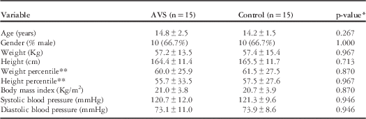
A total of 15 patients included in this study for whom the peak pressure gradient was 51.2±9.2 mmHg and mean pressure gradient was 27.7±4.8 mmHg. From those 15 patients, 1 had no aortic insufficiency and 14 had mild aortic insufficiency. Controls were 15 age- and sex-matched healthy patients. There were no significant differences between the groups in terms of weight and height percentilesReference Owen 24 , as well as body mass index. All the patients were Caucasians. None among both the groups had a resting QT abnormality according to paediatric reference values.Reference Tutar, Ocal, Imamoglu and Atalay 20 Similarly, resting QTc was comparable between the groups. Detailed basic characteristics are summarised in Table 1.
Baseline characteristics comparison between control and aortic valve stenosis (AVS) group.

Data are mean±SD or number (percentage)
* Mann–Whitney test for continuous variables and the χ 2 test for proportions
** Percentile ranks for sex and age according to reference values of the Center for Disease Control (1978)
Readability of the electrocardiograph tracings
QT and RR intervals could be reliably determined in all of the leads in the aortic valve stenosis group. There were >10 readable leads in 52/60 (86.7%) studied electrocardiograph tracings and >8 readable leads in the remaining 8/60 (13.3%). Therefore, all electrocardiographic studies were compatible for analysis.
Exercise response patterns
All the patients completed the test within the normal limits for their age;Reference Chatrath, Shenoy, Serratto and Thoele 25 endurance time was 12.6±2.8 in the study group compared with 12.4±2.2 in the control group (p=0.870). In contrast, the age-adjusted peak heart rate (% of theoretical maximum heart rate) was significantly lower in the study group (88.4±6.9) compared with controls (93.9±5.7) (p=0.037), despite achieving a normal endurance time in all. None of the patients exhibited significant ST-segment changes, T wave abnormalities, or clinical signs of ischaemia along the exercise challenge. There was no statistically significant difference in the RR interval between the study groups at rest or at any subsequent stage of the test, including recovery (repeated measures analysis of variance; p=0.596).
Re-polarisation parameters
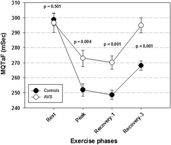
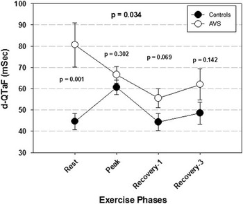
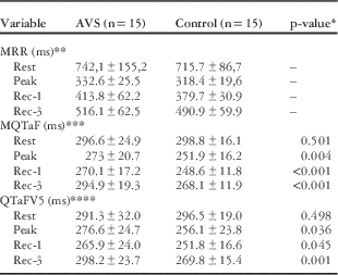
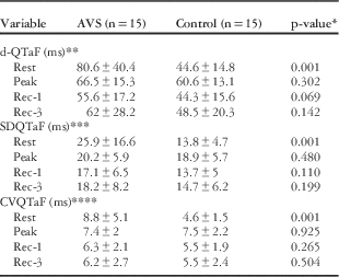
Exercise-induced MQTaF was statistically different between the two groups (repeated measures analysis of variance; p<0.001) (Table 2). Although baseline values were comparable at rest (p=0.501), MQTaF remained significantly elevated in the aortic valve stenosis group in the subsequent stages, including peak exercise (273.0±20.7 versus 251.9±16.2; p=0.004), 1 minute into recovery (270.1±17.2 versus 248.6±11.8, p<0.001), and 3 minutes into recovery (294.9±19.3 versus 268.1±11.9, p<0.001) (Fig 1). Similarly, QTaF in V5 was statistically different between the groups (repeated measures analysis of variance; p<0.001) (Table 2). Although baseline values were comparable at rest (p=0.498), QTaF in V5 remained significantly elevated in the aortic valve stenosis groups in the subsequent stages, including peak exercise (276.6±24.7 versus 256.1±23.8, p=0.036), 1 minute into recovery (265.9±24.0 versus 251.8±16.6, p=0.045), and 3 minutes into recovery (298.2±23.7 versus 269.8±15.4, p=0.001). The d-QTaF yielded a different pattern instead (Fig 2), despite a statistical association between groups for different exercise stages (repeated measures analysis of variance; p=0.034). In essence, the statistically significant difference between groups was obviated with a higher measurement in the aortic valve stenosis group at rest (p=0.001), a trend of higher values at 1 minute into recovery (p=0.069), but no significance at 3 minutes into recovery (p=0.142). Moreover, in the control group, d-QTaF was significantly increased at peak exercise compared with other stages (p<0.001 versus baseline, p=0.004 and 0.002 versus 1 and 3 minutes into recovery), whereas no significant differences in d-QTaF were observed among the different stages in the study group. QT dispersion analysed according to two other indices – the standard deviation of QTaF (SDQTaF) and the coefficient of variation of QTaF (CVQTaF) – yielded a similar pattern to d-QTaF – that is, higher values at rest in aortic valve stenosis and no significant differences between groups at peak exercise and during recovery (Table 3).
Mean QTaF (MQTaF) during the Bruce test. AVS=aortic valve stenosis; MQTaF=mean QTaF measured in the 12 leads; QTaF=QTa with Fridericia’s correction.

QTaF dispersion (d-QTaF) during the Bruce test. AVS=aortic valve stenosis; d-QTaF=dispersion of QTaF; QTaF=QTa with Fridericia’s correction.

MQTaF and RR intervals during the Bruce test.

MQTaF=mean QTaF measured in the 12 leads; MRR=mean of RR interval measured in all readable leads; QTaFV5=QT interval measured from QRS to the apex of T wave and corrected with Fridericia’s formula in lead V5
Data are mean±SD or number (percentage)
* p-value for pairwise comparisons between groups, only shown when the repeated-measures analysis of variance detected a significant interaction
** No significant interaction or group effect (p>0.05)
*** Significant interaction (p<0.001)
**** Significant interaction (p<0.001)
Dispersion, standard deviation, and coefficient of variation of QTaF interval during the Bruce test.

CVQTaF=coefficient of variation of QTaF; d-QTaF=dispersion of QTaF; QTaF=QTa with Fridericia’s correction; SDQTaF=standard deviation of QTaF
Data are mean±SD
* Pairwise comparisons between groups for a given exercise stage
** Significant interaction (p=0.034)
*** Significant interaction (p=0.023)
**** Significant interaction (p=0.004)
Discussion
We have validated our hypothesis that exercise induces QT modifications in congenital moderate aortic valve stenosis. In this study, we have measured electrocardiography parameters at rest and during an exercise stress test, which unmasked electrophysiological abnormalities of ventricular re-polarisation that were undetectable at rest. We have shown that in the moderate congenital aortic valve stenosis group, exercise induces increased corrected QT interval at peak effort and during recovery compared with the control group, whereas the QT interval is similar to the control group at rest. Few studies have used exercise electrocardiography in children with congenital aortic valve stenosis. The results we obtained are consistent with theirs.Reference Bastianon, Del Bolgia, Boscioni, Gobbi, Marzano and Collorid 26 , Reference Yilmaz, Ozme, Ozer, Tokel and Celiker 27 In addition, the range of moderate aortic valve stenosis could be equated to mild stenosis in the lower end and to severe stenosis in the upper end of the spectrum. Therefore, exercise electrocardiographic assessment for abnormal re-polarisation changes may improve risk stratification of these patients in the future.
To the best of our knowledge, two studiesReference Bastianon, Del Bolgia, Boscioni, Gobbi, Marzano and Collorid 26 , Reference Yilmaz, Ozme, Ozer, Tokel and Celiker 27 reported exercise stress testing with electrocardiographic assessment of dynamic QT changes in children with mild and moderate congenital aortic stenosis. Both studies showed no statistically significant differences in QT intervals between patients and controls at rest. In the first study,Reference Bastianon, Del Bolgia, Boscioni, Gobbi, Marzano and Collorid 26 the mean QT during exercise challenge was significantly longer in congenital aortic valve stenosis group (p<0.05), but the measures were not corrected for heart rate. In the second study,Reference Yilmaz, Ozme, Ozer, Tokel and Celiker 27 longer QT intervals were observed during exercise as well, calculated at various pre-determined heart rates of 140±5 bpm (p<0.001), 160±5 bpm (p<0.001), and 180±5 bpm (p<0.001). In our study, despite comparable QTc interval at rest, we were able to uncover statistically significant differences during exertion and upon recovery. In the two previous studies, RR and QT intervals were only measured in leads II and V6, as opposed to 12-lead assessment in our series. Our approach reduces the potential bias associated with a selected lead. In addition, 12-lead analyses are more representative of the whole ventricle’s re-polarisation pattern.
In aortic valve stenosis, the narrowing of the aortic valve induces pressure overload on the left ventricular walls, which becomes more intense during exercise. Such mechanism causes mechanical stress and stretching of the ventricular walls. The stretched myocardium induces electrical instability, which has been experimentally observed in terms of increased dispersion of refractoriness and re-polarisation.Reference Sarubbi, Calvanese, Cappelli Bigazzi, Santoro, Giovanna Russo and Calabro 28 This mechano-electric feedback mechanism, by which mechanical strain influences the electrophysiology of the myocardium, is now well-established at all levels from cell to man.Reference Lab 29 – Reference Kohl, Bollensdorff and Garny 34 This alteration of action potential duration and refractoriness is induced by mechanical stretch through stretch-activated channelsReference Lab 35 , Reference Taggart and Lab 36 or by influencing calcium cycling.Reference Taggart and Lab 36 , Reference Lab, Allen and Orchard 37 It has been suggested that such an effect on refractoriness could be an arrhythmogenic mechanism, particularly if regional variations in action potential duration manifest within the myocardium.Reference Taggart and Lab 36 The lengthening of action potential duration, and therefore of ventricular re-polarisation, is reflected by a prolonged QT interval, which may induce sudden death through serious arrhythmias.Reference Yilmaz, Ozme, Ozer, Tokel and Celiker 27 We used exercise testing, during which myocardial mechanical stress is higher due to a greater haemodynamic demand, in order to identify patients whose risk for arrhythmias may be enhanced by altered re-polarisation duration; however, although there are evidences of mechano-electric feedback in aortic valve stenosis,Reference Sarubbi, Calvanese, Cappelli Bigazzi, Santoro, Giovanna Russo and Calabro 28 there is still a leap of understanding between cellular findings and clinical arrhythmias. We cannot state beyond doubt that our results are due to this mechanism, but we can affirm that the increased ventricular re-polarisation duration in our patients during exercise testing suggests a greater electrical instability.
In addition to an increased QTc during exercise, we report an elevated resting QT dispersion in aortic valve stenosis. QTc dispersion is thought to reflect the autonomic regulation of cardiovascular function, with an increased dispersion as a response to higher sympathetic and lower parasympathetic input to the heart.Reference Wei, Dorian, Newman and Langer 38 Furthermore, our study identifies a blunted response of QTc dispersion to exercise in moderate aortic valve stenosis. This phenomenon was previously described in children following Kawasaki disease.Reference Gravel, Dahdah, Fournier, Mathieu and Curnier 39 QTc dispersion has been reported to increase during exercise in healthy adults and to decrease towards baseline values during recovery,Reference Akutsu, Li, Shinozuka, Kodama, Yamanaka and Katagiri 40 whereas the pathologic QTc response has been mostly studied in coronary artery disease and is still equivocal, as it has been shown either to increase or remain unchanged from rest to peak exercise.Reference Akutsu, Li, Shinozuka, Kodama, Yamanaka and Katagiri 40 , Reference Roukema, Singh, Meijs, Carvalho and Hart 41 Therefore, the significance of a blunted response of QTc dispersion to exercise remains to be elucidated.
Risk stratification in the aortic valve stenosis patients is still controversial.Reference Sawaya, Stewart and Babaliaros 42 – Reference Ennezat, Marechaux, Iung, Chauvel, LeJemtel and Pibarot 44 There have been a lot of investigations on the asymptomatic severe aortic valve stenosis in the adult and elderly population, but very few in the young. In the adult population, aortic valve replacement is recommended in patients with severe aortic valve stenosis and any symptoms related to the condition, level of recommendation 1B, and in asymptomatic patients with severe stenosis and abnormal exercise test inducing symptoms clearly related to the stenosis, level of recommendation 1C.Reference Taylor 3 Moreover, aortic valve replacement is not recommended in the early stages of aortic valve stenosis and before the onset of symptoms because the associated risk could outweigh the benefit.Reference Sawaya, Stewart and Babaliaros 42 In infants with aortic valve stenosis, progression of mild stenosis is slow, whereas significant mortality is recorded in severe cases;Reference Bartz, Driscoll and Keane 45 however, although the majority (65%) of children with moderate aortic valve stenosis showed no progression over time in the previous series, the remaining 35% showed rapid progression, ultimately needing intervention. There are little clinical tools to distinguish between the two subsets of moderate aortic valve stenosis, however. This emphasises the need to improve the indication of intervention parameters beyond what can be obtained with echocardiography at rest. On the other hand, conservative indications for aortic valve replacement may indicate the surgical intervention at a stage of the disease where myocardial impairment is, at least in part, irreversible.Reference Lund 46 , Reference Lund and Larsen 47
There is extensive literature on the usefulness of exercise testing in predicting the onset of symptoms. By increasing the haemodynamic demand, exercise testing induces symptoms otherwise unreported at rest and could, therefore, be a strong predictor of clinical events in patients who claim to be asymptomatic. Such patients unconsciously limit their physical activity secondarily to progressing aortic valve stenosis, and the exercise test could be of value in unmasking their symptoms.Reference Rafique, Biner, Ray, Forrester, Tolstrup and Siegel 43 Exercise testing is proven to be a good predictor of clinical outcome;Reference Das, Rimington and Chambers 11 however, there is uncertainty about which specific parameters are the most useful. In our study, none of our patients had a positive stress test. Nevertheless, we observed exercise-induced electrophysiological modification of re-polarisation in the aortic valve stenosis group solely. Accordingly, some have suggested that a longer QT interval of moderate aortic stenosis cases compared with normal children during exercise could be interpreted as the first sign of myocardial alteration and could lead to fatal ventricular arrhythmias and sudden death.Reference Yilmaz, Ozme, Ozer, Tokel and Celiker 27 At present, there is no clear indication that the risk of serious arrhythmias is increased in patients with mildly long QT intervals measured solely during exercise. Nevetheless, significantly increased QT dispersion in newborns, for instance, with isolated coarctation and normal left ventricular geometry had no other obvious reason for such anomalies beyond elevated myocardial pressure load.Reference Nigro, Russo and Rago 10 Based on our observations, we believe that there is added value of attributing the electrocardiograph alterations to the aortic valve stenosis in our study, and given their young age our patients have less co-morbidities as they are not afflicted by the chronicity of classical cardiovascular diseases and risk factors.
Study limitations
The small number of patients represents the first limitation of this study. It would have been sound to compare data from patients with severe aortic valve stenosis as well; nevertheless, there is little indication and a greater risk to perform a stress test in such population. Finally, based on our study, we cannot state that increased ventricular potential duration evaluated by QT interval during peak exercise and recovery reflects increased risks of serious arrhythmias at rest. In the future, we plan to combine 24-hour Holter monitoring for this purpose.
Conclusion
Exercise challenge uncovers ventricular re-polarisation abnormalities, otherwise undetectable at rest, in the absence of ischaemic changes in children with moderate congenital aortic valve stenosis. This information should be further assessed in a larger group for a better risk stratification of children with moderate asymptomatic congenital aortic valve stenosis.
Acknowledgement
The study was part of the master’s thesis of Louis-Simon Beauséjour (B.Sc).
Financial Support
This research received no specific grant from any funding agency, commercial, or not-for-profit sectors.
Conflicts of Interest
None.
Ethical Standards
This work was approved by the institution’s review authority.