Introduction
Salivary gland tumours are rare in children, accounting for 0.5 per cent of all malignancies in children and adolescents.Reference Sultan, Rodriguez-Galindo, Al-Sharabati, Guzzo, Casanova and Ferrari1 Pleomorphic adenomas are the most common solid parotid tumours in children. They affect both sexes equally, usually between the ages of 8 and 20 years.Reference Rodriguez, Vargas, Robson, Perez-Atayde, Shamberger and McGill2
Although pleomorphic adenoma is a benign tumour, there is a risk of recurrence up to 30 years following initial presentation, and a prevalence of malignant degeneration of 2 to 25 per cent.Reference Rodriguez, Vargas, Robson, Perez-Atayde, Shamberger and McGill2 Quick and accurate diagnosis is therefore vital.
Excision of the mass is the mainstay of treatment.Reference Rodriguez, Vargas, Robson, Perez-Atayde, Shamberger and McGill2, Reference Malone and Baker3 However, the choice of investigations and surgical approach for these patients is not always as clear in children as it is in adults.
In this paper, we review the limited literature available and discuss the management of parotid pleomorphic adenoma in children, using the example of a seven-year-old patient, who to our knowledge represents the youngest reported case of this condition.
Case presentation
A seven-year-old girl presented to our institution with a two-month history of left-sided, painless facial swelling which was noticed by her mother. There were no other associated symptoms. The swelling had been treated with oral antibiotics for a period of two weeks, with no improvement. The child had no other past medical history. She was not taking any regular medication and was up to date with all immunisations.
Physical examination revealed a 1.5 cm, firm swelling in the left parotid region, with bilateral lymphadenopathy in both posterior triangles. The rest of the examination was unremarkable.
Ultrasonography (US) and magnetic resonance imaging (MRI) examinations confirmed the presence of a 2 × 1.5 cm mass in the left superficial parotid, with moderate lymphadenopathy at level II bilaterally.
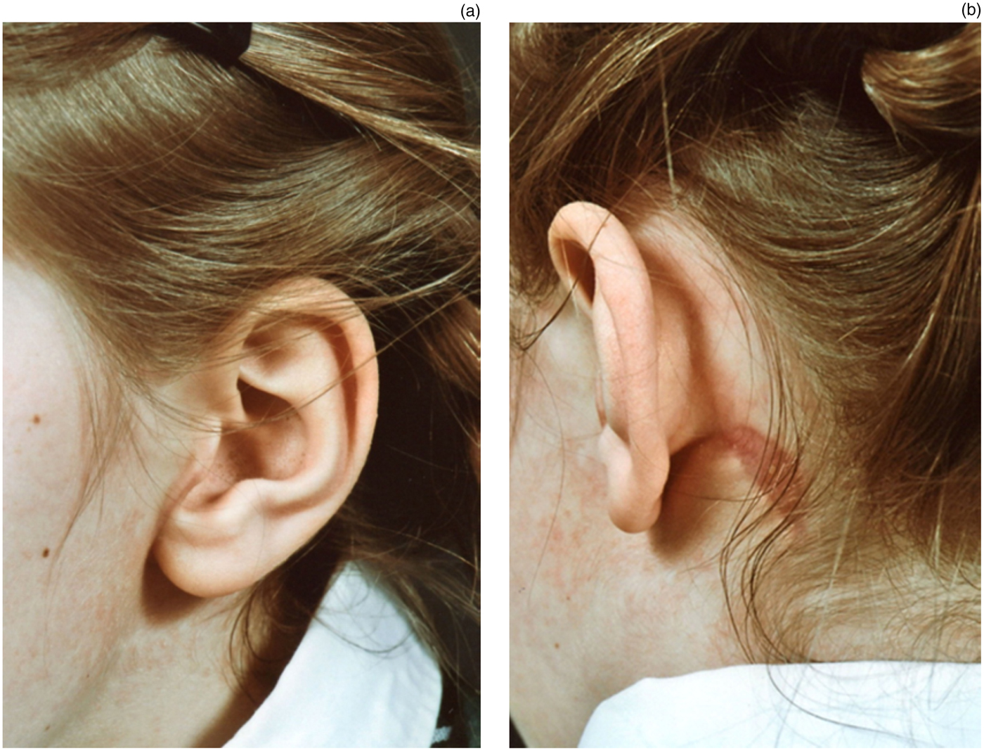
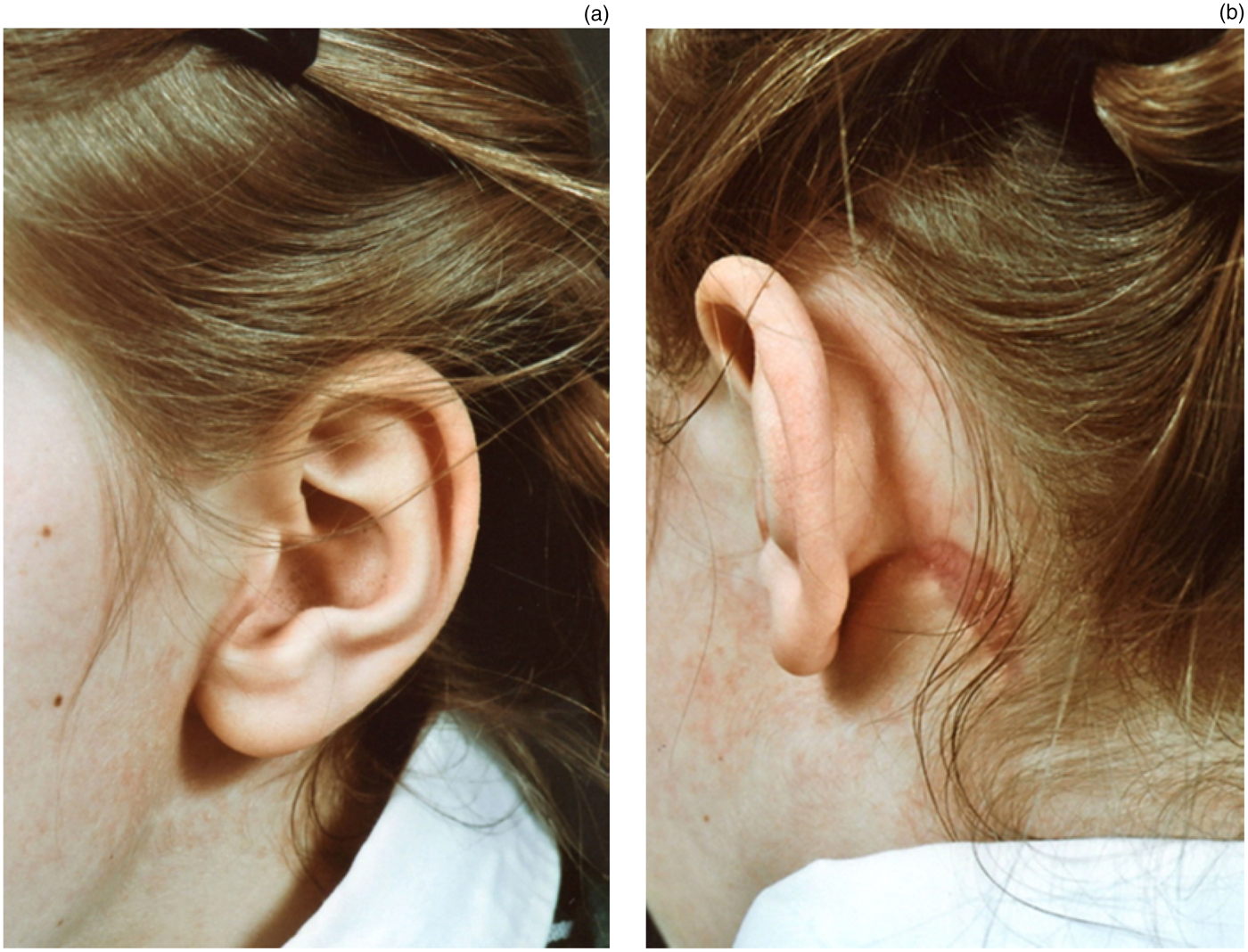
Superficial parotidectomy was subsequently performed using a facelift incision.
Histopathological examination confirmed a diagnosis of pleomorphic adenoma, with clear surgical margins.
At follow up, the patient reported no complications and had a satisfactory cosmetic result (Figure 1). At the time of writing, further follow up was ongoing.
Clinical photograph showing post-operative cosmetic appearance.
Discussion
Salivary gland tumours are rare in the paediatric population;Reference Sultan, Rodriguez-Galindo, Al-Sharabati, Guzzo, Casanova and Ferrari1, Reference Malone and Baker3 thus, there are a limited number of experts who routinely perform surgery on them, and a paucity of literature on the subject.Reference Meiling Xie and Kubba4, Reference Bassu, Parrilla, Rizzo, Almadori, Paludetti and Galli5 There is still ongoing discussion as to the optimal investigation of and surgical options for salivary gland tumours in children, for whom additional considerations apply compared with adults with the same pathology.
It is vital that children with suspected pleomorphic adenoma are seen by an ENT specialist as soon as possible after initial presentation to their general practitioner, to ensure that they are diagnosed quickly and accurately.
A thorough history and examination should always be the initial step in managing these patients.
In terms of imaging, parotid mass US has been proven as the first-line investigation of choice in both adults and children, as it is quick, painless and minimally invasive.Reference Rodriguez, Vargas, Robson, Perez-Atayde, Shamberger and McGill2 It is also able to identify whether or not the mass is part of the parotid gland. However, it has two main limitations. The first is its inability to adequately map the tumour and to clearly establish its extent (for example, whether it involves the deep parotid lobe), as was the case in our patient; this information is important when deciding upon the surgical approach. The other limitation of US is its inability to accurately differentiate between benign and malignant tumours.
Magnetic resonance imaging is therefore vital for tumour mapping.Reference Rodriguez, Vargas, Robson, Perez-Atayde, Shamberger and McGill2, Reference Meiling Xie and Kubba4, Reference Sodhi, Bartlett and Prabhu6 Although this mode of imaging is non-invasive and radiation-free, in a young child it necessitates general anaesthesia, which has its own risks.
Computed tomography has been shown to give inadequate detail and to involve unnecessary irradiation in children, and is therefore not recommended in the imaging of paediatric patients with pleomorphic adenoma.Reference Rodriguez, Vargas, Robson, Perez-Atayde, Shamberger and McGill2, Reference Meiling Xie and Kubba4, Reference Sodhi, Bartlett and Prabhu6
In the adult population, there is no dispute that every patient should undergo fine needle aspiration cytology (FNAC) analysis at the initial stage, in addition to US. The former investigation is a quick and relatively painless procedure which can be performed in the conscious patient and which can supply a histological diagnosis to aid treatment planning. Fine needle aspiration cytology also has very few complications; however, it can give false positive and false negative results, which may increase patient anxiety and occasionally lead to over- or under-treatment.
In children, the use of FNAC is widely disputed. In very young children, it necessitates an additional general anaesthetic, on top of the one needed for surgical excision two weeks later, and it can also cause extra trauma for both the child and their patients while awaiting results. Some experts believe that it is unnecessary to subject young children to FNAC as this investigation will not directly alter the surgical excision treatment plan and therefore has no role. However, others believe that FNAC is worthwhile as it provides information on the histological nature of the mass and therefore enables better preparation prior to surgery. We believe that FNAC should be considered in paediatric patients as a valuable investigation which can give additional, helpful information prior to surgery. However, we advise a degree of scepticism when interpreting the cytology results for paediatric parotid lumps, as rare pathologies may be misinterpreted. The entire clinical picture must therefore be kept in mind. In our patient, both the clinical presentation and the MRI scan suggested benign disease; therefore, we felt that FNAC would not add any information or change the surgical plan, and was thus unnecessary. However, this decision is sometimes difficult and should be left to the individual child's parents and surgeon, following detailed discussion.
Our patient's surgery was performed in a regional head and neck cancer centre, with specialist head and neck pathology and radiology expertise. When required, this centre ensured that any complex salivary gland histology was reviewed by a central salivary gland panel. In our opinion, if such a multi-disciplinary facility is available, in which the required surgery can be performed by a local head and neck surgeon experienced in salivary gland surgery, then the case need not be referred to a specialist paediatric unit.
When planning such surgery in a child, many aspects need to be considered. The size and location of the mass must be taken into account to ensure adequate access, in order to achieve complete excision.Reference Lohuis, Tan, Bonte, Van Den Brekel, Balm and Vermeersch7, Reference Lee, Koh, Kim, Hong, Jeong and Choi8 In children, the recurrence rate for pleomorphic adenoma is 20 per cent, compared with 1 to 9 per cent in adults.Reference Meiling Xie and Kubba4 This means that the planning and execution of surgery need to be done by an experienced surgeon who operates on similar cases on a regular basis.
Preservation of the facial nerve is paramount in children, as in adults. It is also vital to ensure that tumour spillage does not occur as this can lead to recurrence. Tumour spillage takes place when the pseudocapsule or microprojections extending outside the pseudocapsule are cut. In children, the smaller size of facial structures can increase the risk of this happening.Reference Meiling Xie and Kubba4
The other consideration is scarring. In adults, this may take less precedence, but children are more prone to hypertrophic scars. In our case, a superficial parotidectomy was performed via a facelift incision rather than the more conventional modified Blair incision. This improved the patient's cosmetic outcome and gave adequate exposure to the parotid pleomorphic adenoma. We therefore recommend the use of a facelift incision, performed by an experienced surgeon, in paediatric cases of parotid pleomorphic adenoma, following careful patient selection.
• Managing children with parotid pleomorphic adenoma is challenging
• Ultrasonography is the first-line investigation for parotid masses in children and adults
• Fine needle aspiration cytology may be considered on an individual basis for children
• Superficial parotidectomy via facelift incision enables adequate exposure for complete excision, and good cosmesis
Radiotherapy has a very limited role in children with parotid tumours. In recurrent cases, surgery should be considered as the first treatment option. In a very small proportion of cases with affected tumour margins, radiotherapy may be considered; however, the benefits of this must be carefully balanced against the short- and long-term implications for the child.
Conclusion
Management of parotid pleomorphic adenoma in paediatric cases can be very challenging, and requires careful consideration of the individual case in order to select the most appropriate investigation and surgical approach. A multi-disciplinary approach should be adopted for all cases.
Acknowledgements
We thank the patient and her family for their permission and support in the preparation of this paper.