INTRODUCTION
The family Syllidae is one of the most diverse families of polychaetes, with about 74 genera and more than 700 described species (San Martín, Reference San Martín and Ramos2003; Aguado & San Martín, Reference Aguado and San Martín2009; Aguado et al., Reference Aguado, San Martín and Siddall2012). Nygren (Reference Nygren2004) made a general revision of the subfamily Autolytinae. Aguado et al. (Reference Aguado, Nygren and Siddall2007) carried out phylogenetic analysis of the whole family based on molecular data. Aguado & San Martín (Reference Aguado and San Martín2009) performed a phylogenetic analysis based on morphology. All these studies coincide in considering three subfamilies (Exogoninae Langerhans, Reference Langerhans1879; Autolytinae Langerhans, Reference Langerhans1879; and Syllinae Grube, 1850) as monophyletic, while Eusyllinae Malaquin, 1893 was clearly polyphyletic; more recent analysis, based both on morphological and molecular data (Aguado et al., Reference Aguado, San Martín and Siddall2012) showed that some genera are grouped forming a monophyletic group, considering them as Eusyllinae. Additionally, Aguado & San Martín (Reference Aguado and San Martín2009) named another subfamily (Anoplosyllinae Aguado & San Martín, Reference Aguado and San Martín2009) including some genera of ‘eusyllines’. Recently, Aguado & Bleidorn (Reference Aguado and Bleidorn2010) studied the relationships between Anoplosyllinae and the rest of the syllids. All these previous studies noticed the phylogenetic importance of considering the reproductive modes in Syllidae, which together define each subfamily. For instance, all the species in Syllinae reproduce by schizogamic scissiparity (for details about reproductive modes in syllids see Franke, Reference Franke1999; Aguado et al., Reference Aguado, Nygren and Siddall2007, Reference Aguado, San Martín and Siddall2012; Aguado & San Martín, Reference Aguado and San Martín2009).
The genus Megasyllis was erected by San Martín et al. (Reference San Martín, Hutchings and Aguado2008) to include some species of Syllidae previously described in different genera (Syllis, Eusyllis, Typosyllis), but having unique characters, different to any other described genus. These species have a mixture of morphological characters between the subfamily Syllinae and some genera of Eusyllinae, but they all reproduce by schizogamy, the typical reproductive mode of Syllinae. This evidence was strong enough to place them into a new genus in Syllinae.
San Martín et al. (Reference San Martín, Hutchings and Aguado2008) included four species in the genus Megasyllis: M. corruscans (Haswell, Reference Haswell1885), M. heterosetosa (Hartmann-Schröder, Reference Hartmann-Schröder1991), M. inflata (Marenzeller, Reference Marenzeller1879), and M. multiannulata (Aguado et al., Reference Aguado, San Martín and ten Hove2008). These species were characterized by having segments with several annuli, dorsal cirri originating at different levels, pseudoannulated posterior dorsal cirri, opaque bodies with bright colours and a remarkably large body size (>30 mm) (Figure 1A–H). However, there are more species located in different genera which do not match entirely with their generic diagnoses, while they widely coincide with the recent genus Megasyllis except for the body size (about 5 mm). These species were previously identified as incertae sedis in some taxonomic and phylogenetic studies, i.e. the species Pionosyllis marquesensis Monro, Reference Monro1939 and P. procera Hartman, Reference Hartman1965 studied by San Martín & Estapé (Reference San Martín and Estapé1993) and San Martín et al. (Reference San Martín, López and Aguado2009), who suggested that they might belong to Syllinae since they reproduce by schizogamy. Another species with similar characteristics is Odontosyllis glandulosa Augener, Reference Augener1913, which was transferred to Typosyllis by Hartmann-Schröder (Reference Hartmann-Schröder1979). Additionally, there are several species that show notable similarities with Megasyllis, although all of them are relatively small in size. These species are Typosyllis subantennata Hartmann-Schröder, Reference Hartmann-Schröder1984, Typosyllis heterosetosa Hartmann-Schröder, Reference Hartmann-Schröder1991, Typosyllis pseudoheterosetosa Böggemann & Westheide, Reference Böggemann and Westheide2004, and Typosyllis nipponica Imajima, Reference Imajima1966.
Colour photographs of Megasyllis corruscans (A–E) and Megasyllis inflata (?) (F–H); photographed by Denis Riek.

This study provides a revision of the species currently assigned to the genus Megasyllis, together with other conflicting species that share most of the diagnostic characters except for the body size. The conflictive species are recognized herein as new combinations of Megasyllis, and, finally, four new species are described: M. mariandreworum sp. nov. and M. tigrina sp. nov. from Australia, M. chrissyae sp. nov. from The Phillipines, and M. eduardoi sp. nov. from New Zealand.
MATERIALS AND METHODS
The material examined was collected from several collection trips, one to Western Australia (M.T. Aguado and G. San Martín), another to New Zealand (the three authors, E. López, A. Verdes and R. Gallego), as well as material sent by some colleagues and institutions (detailed information is given in the ‘Material Examined’ section of each species). Syllids were sorted using a Nikon SMZ1 stereomicroscope and preserved in 96% ethanol or in 70% ethanol after they were fixed in 10% seawater formalin. The specimens were studied under a Nikon Optiphot microscope with a differential interference contrast system (DIC, Nomarsky). Drawings were made using a camera lucida drawing tube. The width of the specimens, excluding parapodia, was measured at the proventricle level. When available, the specimen length is indicated as follows: small (<5 mm in length), medium (5–10 mm in length) and large (>10 mm in length). The following abbreviations are used for the material examined:
- AM
Australian Museum, Sydney, NSW, Australia;
- BMNH
British Museum of Natural History;
- NMW
Naturhistorisches Museum of Wien, Austria;
- SMF
Senckenberg Museum Frankfurt;
- CASIZ
California Academy of Sciences, Zoology, San Francisco, USA;
- NIWA
National Institute of Water and Atmospheric Research, Auckland;
- MNCN
Museo Nacional de Ciencias Naturales, Madrid, Spain.
RESULTS
The genus Megasyllis is herein reorganized excluding the size as a diagnostic character, since it is not representative for all the species of the group. Megasyllis comprises 14 species, including the four new ones described in this paper. We also provide a key for identification and a re-description of four species, which are new combinations.
SYSTEMATICS
Family syllidae Grube, 1850
Subfamily syllinae Grube, 1850
Genus Megasyllis San Martín, Aguado & Hutchings, Reference San Martín, Hutchings and Aguado2008
Megasyllis San Martín et al., Reference San Martín, Hutchings and Aguado2008: 5–6.
TYPE-SPECIES
Syllis corruscans Haswell, Reference Haswell1885, designated by San Martín et al. (Reference San Martín, Hutchings and Aguado2008).
DIAGNOSIS (EMENDED FROM SAN MARTÍN ET AL., Reference San Martín, Hutchings and Aguado2008)
Body long, robust, dorsally convex, usually of macrofaunal size. Some species larger than 30 mm, and others relatively smaller (about 5–10 mm), all usually brightly coloured. Prostomium with four eyes; usually two lateral swellings; lateral antennae sometimes arising from lateral swellings; median antenna inserted on middle of prostomium. Palps large, broad, fused at their bases. Two pairs of tentacular (peristomial) cirri. Segments bi- or tri-annulated in large specimens, weakly annulated to rough in small ones. Antennae, tentacular and anterior dorsal cirri articulated to weakly articulated; remaining dorsal cirri irregularly wrinkled to smooth, slender, long to moderate in length; sometimes all cirri smooth or articulated. Cirrophores distinct. Large species with longer dorsal cirri arising more dorsally than shorter ones. Pharynx similar in length or shorter than proventricle, provided with a crown of numerous, soft papillae and one single tooth, placed anteriorly, sometimes slightly posterior to the pharyngeal rim; several species with a distinct pigmented band on pharynx. Parapodia with prechaetal and postchaetal lobes. Ventral cirri triangular to conical. Compound chaetae heterogomph, with strongly bidentate falcigers, typically both teeth similar, acute, sometimes proximal tooth larger than distal one. Solitary dorsal and ventral simple capillary chaetae on posterior parapodia; dorsal simple chaetae very thin, distinctly thinner than shafts of compound chaetae. Aciculae straight to slightly acuminate. Reproduction by schizogamic scissiparity by means of sexual stolons, male or female, with four eyes, four short papilliform antennae, and smooth dorsal cirri. Young specimens with all appendages smooth, becoming antennae and anterior cirri progressively articulated.
REMARKS
This genus includes three species originally described within the subfamily Eusyllinae (Pionosyllis marquesensis, P. procera and Odontosyllis glandulosa), and seven other species initially described as Syllinae (Typosyllis nipponica, T. subantennata, T. heterosetosa, T. pseudoheterosetosa, Syllis corruscans, S. inflata and S. multiannulata) as well as four other new species from the Pacific. There are two additional characters which seem to be typical of some species of Megasyllis: presence of a distinct reddish pigmented band in the posterior part of the pharynx (M. glandulosa, M. multiannulata, M. nipponica, M. procera, M. eduardoi sp. nov., and M. tigrina sp. nov.), and a pharyngeal crown with numerous soft papillae, rather than the typical ten of Syllis–Typosyllis.
KEY TO SPECIES OF MEGASYLLIS
1. Large size (more than 30 mm long). Segments distinctly multi-annulated (Figure 1). Longer dorsal cirri arising more dorsally than shorter ones..............2
— Smaller size (less than 30 mm long). Segments indistinctly annulated, or rough. All dorsal cirri almost arising at same level...........................5
2. Some compound chaetae with proximal tooth larger than distal tooth. Blades of most compound chaetae with long, filiform subdistal spines.......................................3
— Compound chaetae with both teeth similar in size. Blades of compound chaetae without long, filiform subdistal spines....................................4
3. Blades of ventral compound chaetae with proximal tooth slightly longer than distal one, more marked on posterior parapodia. Subdistal spines of blades extending to level of proximal tooth.................Megasyllis inflata
— Blades of ventral compound chaetae with proximal tooth distinctly longer than distal one. Subdistal spines of blades extending beyond proximal tooth...............................................Megasyllis heterosetosa
4. Compound chaetae on each parapodium with blades similar in size, relatively short.......Megasyllis multiannulata
— Compound chaetae on each parapodium with long and short blades....................Megasyllis corruscans
5. Posterior compound chaetae without subdistal serrations on shafts...................................6
— Posterior compound chaetae with distinctly subdistal serration on shafts.....................................11
6. Some midbody and posterior chaetae with proximal tooth distinctly longer and larger than distal tooth..............7
— Distal and proximal teeth of blades similar in length and shape..................................9
7. Most anterior segments distinctly pigmented of red-brown with lighter areas; remaining segments with a small band of the same colour. Some midbody and posterior compound chaetae with a bifid distal tooth, acquiring a tridentate appearance. Spines on margin of blades short, except 2–3 distal ones, which are distinctly long.......................Megasyllis chrissyae sp. nov.
— Different colour pattern. Distal tooth of blades entire..................................8
8. Proximal tooth of blades of midbody and posterior compound chaetae distinctly curved, distal tooth much smaller than proximal one; spines on margin short, except some sub-distal ones. Colour pattern unknown (fixed specimens without pigmentation)......................Megasyllis pseudoheterosetosa
— Proximal tooth not curved, distal tooth large; spines on margin moderately long, sub-distal ones slightly long. Distinct colour pattern red and orange...........................Megasyllis mariandreworum sp. nov.
9. Antennae, tentacular and dorsal cirri articulated (except most posterior ones); distinctly pigmented of orange-yellow and a distinct black band of posterior half of pharynx, with numerous epidermal granular inclusions. Shafts of compound chaetae with long and thin subdistal spines on margin..........Megasyllis eduardoi sp. nov.
— Antennae, tentacular and anterior dorsal cirri smooth or irregularly articulated; dorsal cirri from midbody smooth. Different colour pattern. Spines on shafts shorter.........10
10. Prostomium with lateral swellings. Compound chaetae with long and short blades, some provided with long and thin distal spines. Body yellow...............................Megasyllis marquesensis
— Prostomium without lateral swellings. Posterior part of prostomium and peristomium, as well as posterior part of pharynx, with red pigmentation.......Megasyllis glandulosa
11. Segments tri-annulated...............Megasyllis nipponica
— Segments not distinctly annulated.............12
12. Posterior compound chaetae with distinct dorso-ventral gradation in size of blades, bidentate with both teeth similar in size. Segments with two transversal bands of red to brown pigment.........Megasyllis tigrina sp. nov.
— Blades of posterior compound chaetae all short, strongly bidentate; different colour pattern.........13
13. Peristomium covering posterior half of prostomium. Prostomium and most anterior segments dark red, subsequent segments white, and remaining pink to brown (in alcohol), and red spots on the bases of dorsal cirri. Dorsum with rounded granules.....................Megasyllis subantennata
— Peristomium not covering posterior half of prostomium. Red pigments on peristomium and posterior part of pharynx.....................Megasyllis procera
Megasyllis chrissyae sp. nov.
(Figures 2, 3)
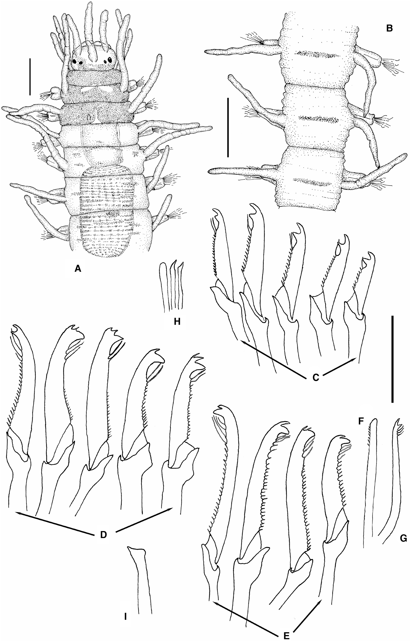
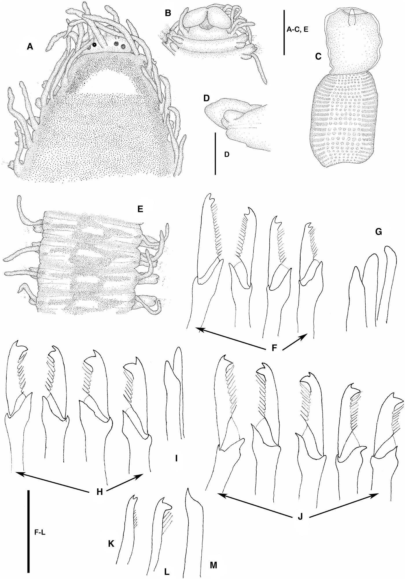
Megasyllis chrissyae sp. nov.: (A) anterior end, dorsal view; (B) midbody segments, dorsal view; (C) compound chaetae, anterior parapodium; (D) compound chaetae, midbody parapodium; (E) compound chaetae, posterior parapodium; (F) dorsal simple chaeta, posterior parapodium; (G) ventral simple chaeta, posterior parapodium; (H) aciculae, anterior parapodium; (I) acicula, posterior parapodium. CASIZ 187469, holotype. Scale bars: A–B, 0.18 mm; C–I, 40 μm.
Colour photographs of Megasyllis chrissyae sp. nov.; (A,B) holotype (CASIZ 187469) and Megasyllis eduardoi sp. nov.; (C) holotype (NIWA 79628).
MATERIAL EXAMINED
Holotype, CASIZ 187469. The Phillipines: Luzón Island, Batangas, Calupan Peninsula, Balayan Bay, Basura dive, 13.76°N 120.92°E, 0–11.5 m depth, Hearst Philippine Biodiversity Expedition 2011. Coll. Christina Piotrowski (Chrissy), 11 May 2011.
DESCRIPTION
Holotype complete, 12 mm long, 0.7 mm wide, with 56 chaetigers, plus an incomplete female stolon with ten chaetigers. Body long, slender, anteriorly and posteriorly broad, distinctly slender on midbody. Marked colour pattern, peristomium and two anterior chaetigers densely pigmented of red-brown, with tow lighter areas on chaetiger 1 and a triangular middle area on chaetiger 2; also pigmented areas on prostomium; from chaetiger 3, segments less pigmentated, with some scattered areas of pigment of same colour, forming a transversal, slender row on midbody (Figures 2A, B, 3A, B), more marked on stolon. Anterior segments smooth (Figure 2A), becoming rough to irregularly annulated from midbody (Figure 2B). Prostomium oval, wider than long, with two pairs of eyes (anterior ones larger) in open trapezoidal arrangement (Figures 2A, 3B). Median antenna inserted on middle of prostomium, about twice longer than combined length of prostomium and palps; lateral antennae shorter than median one, inserted near anterior margin of prostomium (Figure 2A). Palps fused at base with a distinct median groove, ventrally directed, difficult to see dorsally (Figure 2A). Peristomium similar in length to subsequent segments. Dorsal tentacular cirri longer than median antennae, ventral ones shorter than dorsal ones (Figure 2A). Dorsal cirri of chaetiger 1 distinctly longer than antennae, tentacular and subsequent dorsal cirri, anteriorly directed (Figure 2A). Thereafter, cirri alternating in length, all relatively short, slightly longer than body width (Figure 2B). Parapodial lobes relatively short and broad, slighltly bilobed distally (Figure 2A, B). Ventral cirri digitiform, similar in length to parapodial lobes on anterior parapodia, slightly longer than parapodial lobes on posterior parapodia. Anterior parapodia each with about 10–12 compound, heterogomph chaetae, blades strongly bidentate, both teeth similar or proximal one slightly larger than distal, short spines on the edge except 1–2 more distal ones, reaching or even surpassing level of proximal tooth; blades decreasing in length from dorsal (~28 µm) to ventral (~15 µm) (Figure 2C). Number of compound chaetae per parapodium decreasing progressively to posterior part of body to 6–7 on midbody and posterior parapodia; blades of compound chaetae becoming somewhat longer and larger, curved, with thicker shafts and proximal tooth of blades distinctly larger than distal one, and spines on margin moderately long, except 2–3–4 most distal ones, very long (Figure 2D, E); some blades with two minute distal teeth and large proximal tooth (Figure 2D, E). Blades of compound chaetae about 33–12 µm on midbody parapodia, 37–25 µm on posterior parapodia. Posterior parapodia with solitary, dorsal simple chaetae, thin, blunt distally, with short subdistal spines (Figure 2F). Ventral simple chaetae strongly bidentate, distally curved, with subdistal long spines (Figure 2G). Anterior parapodia with three aciculae, one distally blunt and two slender, distally oblique (Figure 2H); from midbody backwards, parapodia with single acicula, acuminate (Figure 2I). Pharynx short, extending through three segments; pharyngeal tooth conical, relatively large, located slightly back from anterior margin (Figure 2A). Proventricle similar in length to pharynx, extending through three segments, with about 20–25 muscle cell-rows (Figures 2A, 3A, B). Pygidium small, with two smooth, unarticulated anal cirri. Regeneration of pygidium and posterior segments ventrally to stolon. Stolon dicerous (Chaetosyllis) (Figure 3A).
REMARKS
Megasyllis chrissyae sp. nov. is characterized by having a distinct colour pattern, different to any other colour pattern of the species within this genus and by the distinct shape of the compound chaetae, with elongated, curved blades, having the proximal tooth distinctly large than proximal one and long distal spines on margin; any species of the genus has also tridentate blades, as some of this species. The most similar species is M. pseudoheterosetosa (Böggemann & Westheide, Reference Böggemann and Westheide2004), from Mahé (Seychelles); the compound chaetae are more or less similar, but that species lacks colour pattern, anterior appendages are more distinctly articulated and the pharynx is longer.
DISTRIBUTION
Only known from the type locality, Luzón Island (The Phillipines).
HABITAT
Coral rubble.
ETYMOLOGY
The species is named after Christina Piotrowski (Chrissy) who collected and kindly gave us the specimen of this new species.
Megasyllis corruscans (Haswell, Reference Haswell1885)
(Figure 1A–E)
Syllis corruscans Haswell, Reference Haswell1885: 734, pl. 50, figures 1–3, pl. 55, fig. 5; 1920: 99, pl. 11, figs 28–31.
Eusyllis brevicirrata Knox & Cameron, Reference Knox and Cameron1971: 25, figures 11–14.
Megasyllis corruscans San Martín et al., Reference San Martín, Hutchings and Aguado2008: 6, figures 2A–E, 3A–C, 4A–E, 5A–F, 6A–F, A–F, 8A–F.
Remarks
A huge species, brightly coloured (Figure 1A–C); a detailed description of this species is in San Martín et al. (Reference San Martín, Hutchings and Aguado2008).
TYPE LOCALITY
Port Jackson, Sydney, NSW, Australia.
DISTRIBUTION
East Australia (NSW) and South Australia; probably also in other Australian areas.
ECOLOGY
All kind of sediments, algae; intertidal to about 18 m.
Megasyllis eduardoi sp. nov.
(Figures 3C, 4, 5)
MATERIAL EXAMINED
New Zealand: holotype and 3 paratypes (NIWA 79628, NIWA 79629). South Island, Akaroa, south dock, 43°48′17.11′′S 172°57′54.71′′E, on sponges and ascidians, 0–1 m depth, 5 February 2012; 3 paratypes (NIWA 79630), North Island, KariKari Peninsula, Matai Bay, 34°49′28′′S 173°24′37′′E, rocky bottom with brown and coralline algae, 3–4 m depth, 30 January 2012; 1 paratype (MNCN 16.01/14721), South Island, Akaroa, north of Matai Bay, 43°47′51.88′′S 172°57′49.51′′E, coralline algae, intertidal, 7 February 2012; 2 paratypes (MNCN 16.01/14722), Akaroa, north of Matai Bay, 43°47′51.88′′S 172°57′49.51′′E, rizoids of Laminarians, 0.5 m depth, 7 February 2012; 1 paratype (MNCN 16.01/14723), Long Bay, 43°50′18.72′′S 173°03′35.07′′E, coralline algae, 0–1 m depth, 9 February 2012.
DESCRIPTION
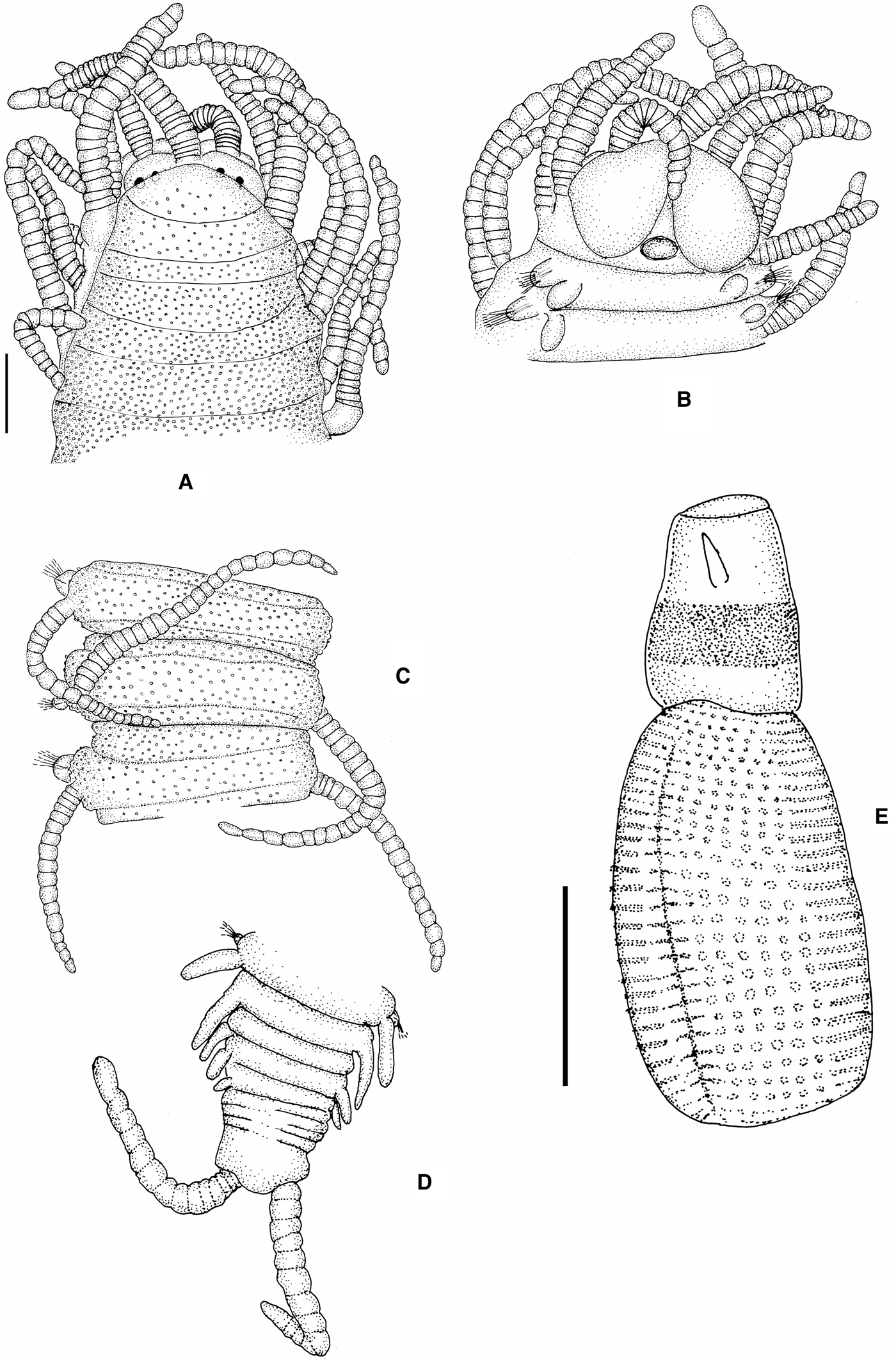
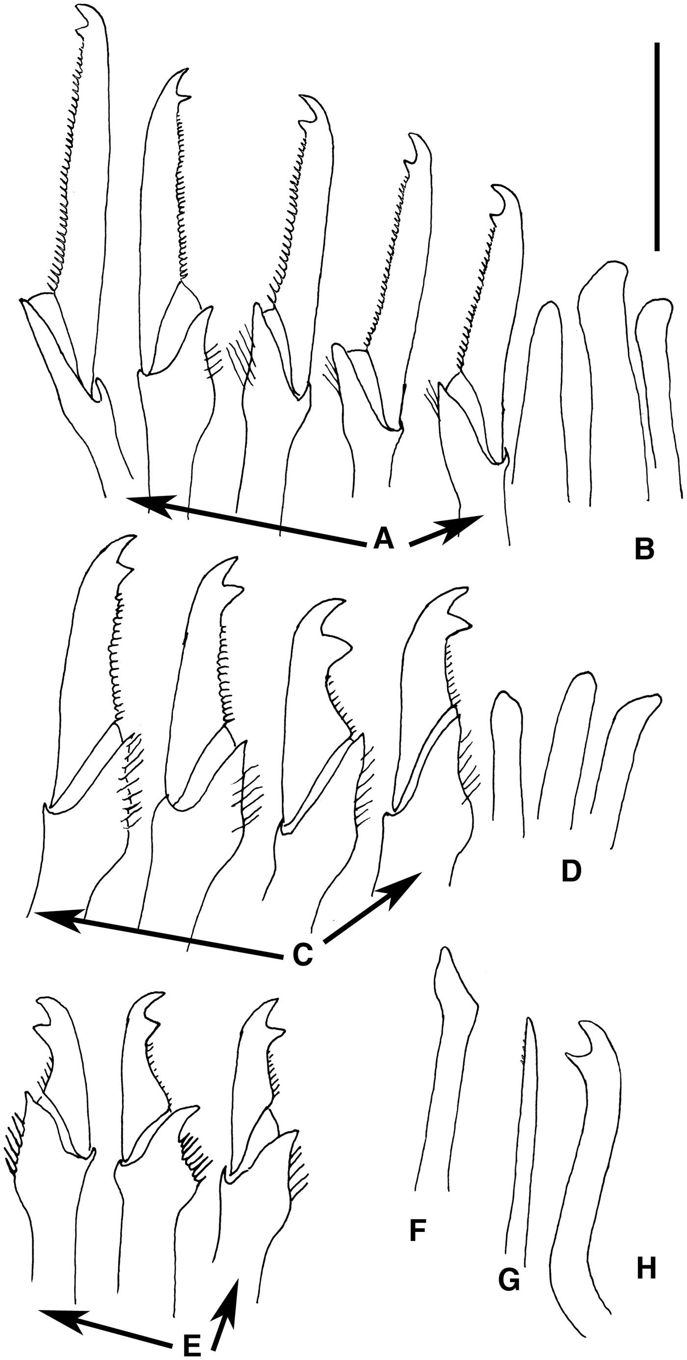
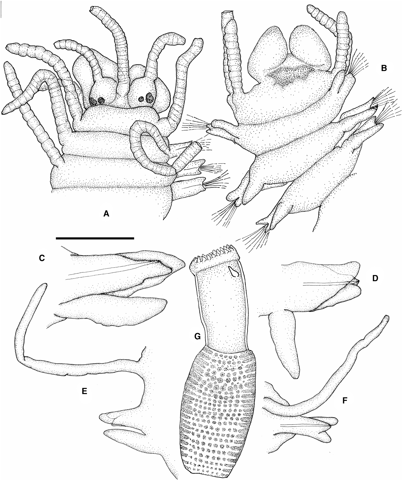
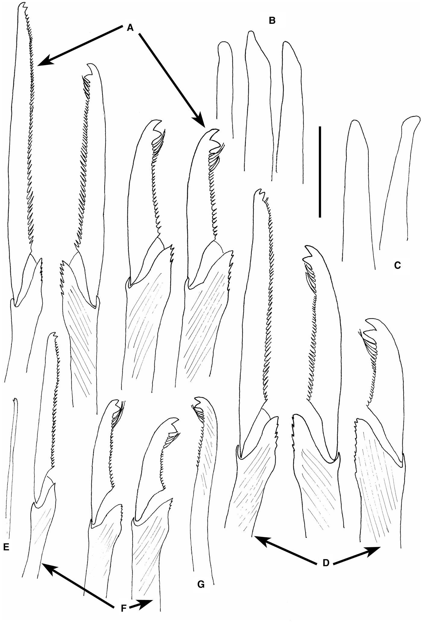
Largest specimen 9.5 mm long, 1 mm wide for 56 chaetigers plus five segments without chaetae and some others in development (Figures 3C, 4D). Body long and slender, anteriorly stout, dorsally convex, bright orange to yellow in alive specimens, with a distinct black transversal band crossing pharynx (Figures 3C, 4E); numerous minute dermal inclusions, appearing verruca on laterals of midbody segments (Figure 4C). Prostomium ovate, broader than long, retracted partially on anterior segments (Figure 4A), provided with two small swellings and four eyes, nearly on line. Median antenna originating between both swellings, in front of eyes, lateral antennae on anterior margin of prostomium; antennae longer than combined length of prostomium and palps together, median antenna longer than lateral ones (Figure 4A), with about 25 articles; lateral antennae with about 15 articles. Palps long, broad, fused basally with a dorsal scar, ventrally folded (Figure 4A, B). Peristomium dorsally reduced; dorsal tentacular cirri distinctly longer than median antenna, with about 30 articles, ventral ones shorter than dorsal ones, similar in length to median antenna, with about 16 articles. Anterior dorsal cirri relatively thick, becoming progressively more elongated to midbody; antennae, tentacular and dorsal cirri of anterior chaetigers and midbody distinctly articulated, only most posterior dorsal cirri weakly articulated or smooth (Figure 4A–D); midbody dorsal cirri alternating long cirri, with about 25 articles, and short, with about 16 articles. Ventral cirri broad, ovate, similar in length to parapodial lobes. Parapodial lobes short, broad. Anterior parapodia each with about 10–13 compound chaetae, diminishing to seven on midbody parapodia and to four on posterior parapodia; shafts of compound chaetae with some thin, long spines distally, more distinct on posterior chaetae; blades of dorsal compound chaetae relatively elongated, bidentate with distal tooth larger in anterior parapodia, and short, straight spines on margin (Figure 5A); blades of medium and ventral compound chaetae shorter, curved, strongly bidentate with both teeth large and divergent, with short basal and median spines on margin (Figure 5C); posterior chaetae similar to those of midbody, but shorter (Figure 5E). Gradation both in shape and length of blades of compound chaetae on anterior and middle parapodia; 40 µm above, 27 µm below on anterior parapodia; 27–30 µm above, 24 µm below on midbody parapodia, all similar, about 17–19 µm on posterior parapodia. Solitary dorsal simple chaetae on posterior parapodia, slender, smooth or with few, minute spines on margin, distally entire (Figure 5G). Solitary ventral simple chaetae on posterior parapodia, stout, sigmoid, distinctly bidentate, with both teeth similar in length and shape, divergent, smooth (Figure 5H). Three aciculae in anterior and midbody parapodia (Figure 5B, D) and two or one on posterior parapodia (Figure 5F), slightly curved distally. Pharynx relatively short, extending through about three to four segments, papillae not seen; pharyngeal tooth long, triangular, placed distinctly in back of anterior rim (Figure 5E). Proventricle barrel shaped, longer than pharynx, through five to six segments, with about 29–33 muscle cell rows.
Megasyllis eduardoi sp. nov.: (A) anterior end, dorsal view; (B) anterior end, ventral view; (C) midbody segments, dorsal view; (D) posterior end, dorsal view. Pharynx and proventricle. Holotype (NIWA 79628). Scale bars: A–D, 0.2 mm; E, 0.1 mm.
Megasyllis eduardoi sp. nov.: (A) compound chaetae, anterior parapodium; (B) aciculae, anterior parapodium; (C) compound chaetae, midbody parapodium; (D) aciculae, midbody parapodium; (E) compound chaetae, posterior parapodium; (F) acicula, posterior parapodium; (G) dorsal simple chaeta, posterior parapodium; (H) ventral simple chaeta, posterior parapodium. Holotype (NIWA 79628). Scale bar: 40 μm.
REMARKS
This species is characterized by its bright orange-yellow uniform colour pattern, with a distinctive broad black band on the middle part of the proventricle. This species has the most distinctly articulated antennae and tentacular cirri. Its compound are similar to those of other species, particularly M. subantennata, M. tigrina, and M. procera, but all these species have different colour pattern and the shafts of the compound chaetae have coarse serration, instead of long, thin spines as happens in M. eduardoi sp. nov.
DISTRIBUTION
New Zealand, South and North Islands (South Pacific).
HABITAT
On low depths, among algae, sponges and ascidians.
ETYMOLOGY
The species is named after our colleague Dr Eduardo López, who participated in the collection and sorting of the specimens.
Megasyllis glandulosa (Augener, Reference Augener1913) comb. nov.
(Figure 6)
Odontosyllis glandulosa Augener, Reference Augener1913: 239, pl. 3, figures 36–37; Day & Hutchings, Reference Day and Hutchings1979: 101.
Typosyllis (Typosyllis) glandulosa Hartmann-Schröder, Reference Hartmann-Schröder1979: 92, figures 82–87; Reference Hartmann-Schröder1980: 50; Reference Hartmann-Schröder1981: 26; Reference Hartmann-Schröder1983: 127.
‘Odontosyllis glandulosa’ Licher, Reference Licher1999 (in part): 297, figure. 114.
MATERIAL EXAMINED
Western Australia: 1 specimen (AM W28369), Geraldton, Love Drummonds, coll. Hartmann-Schröder, October 1975, ZMH P–16940; 1 specimen (AM W36575), north end of beach, Bundegi Reef, Exmouth Gulf, 21°44'S 114°14’E, rocky rubble, sticky sediment and brown algae with epiphytes, 2 m, coll. Stoddart, 4 January 1984; 2 specimens (AM W36850), Port Denison, inside harbour, algae, 1 m, coll. Aguado & San Martín, 22 June Reference Aguado, San Martín and Nishi2008.
DESCRIPTION
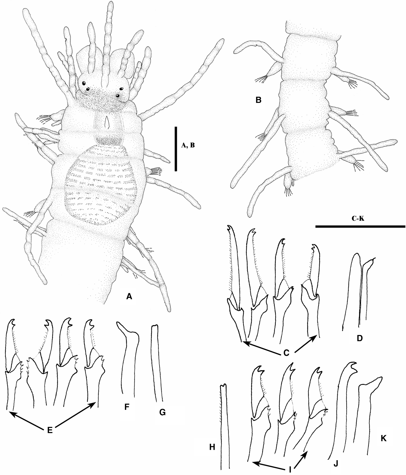
Longest specimen (type) 5.5 mm long, with 35 chaetigers (fide Augener, Reference Augener1913); examined and figured specimen (Figure 6A) 2.6 mm long, 0.18 mm wide, with 30 chaetigers. Body small, fragile, without colour pattern, except for a reddish area on posterior part of prostomium and peristomium (Figure 6A), extending until first chaetiger. Anterior segments smooth (Figure 6A), becoming rough to annulated from midbody (Figure 6B). A reddish transversal band in the inner wall of the pharynx, visible by transparency (Figure 6A). Prostomium wider than long, with two pairs of eyes in open trapezoidal arrangement; eyes similar in size. Median antenna inserted in middle of prostomium, longer than combined length of prostomium and palps, with 7–8 weakly distinct articles; lateral antennae shorter than median one, inserted at anterior margin of prostomium, also with few 6–7 weak articles (Figure 6A). Palps similar in length to prostomium, fused at base with a distinct median groove. Peristomium reddish pigmented, shorter than subsequent segments. Dorsal tentacular cirri longer than median antenna, with nine weak articles, ventral ones about half as long as dorsal ones, weakly articulated (Figure 6A). First dorsal cirri longer than tentacular cirri, weakly articulated, almost smooth (Figure 6A). Dorsal cirri alternating in length towards posterior end, becoming smooth after few anterior segments (Figure 6A, B). Ventral cirri conical, inserted proximally on parapodia, not extending beyond parapodial lobes. Anterior parapodia each with about ten compound, heterogomph chaetae, distal part of shafts smooth or with few small, short spines (Figure 6C); blades bidentate, both teeth similar in size, short spines on edge; decreasing in length from dorsal (~38 µm) to ventral (~25 µm). Number of compound chaetae per parapodium decreasing progressively to posterior part of body; midbody parapodia with five compound chaetae, posterior parapodia with two to three chaetae; blades of compound chaetae in midbody (Figure 6E) quite similar, about 20–24 μm;, posterior compound chaetae with shorter blades (Figure 6I), 22–20 μm, more strongly bidentate, with thicker shafts and few, small subdistal spines. Posterior parapodia with solitary, dorsal simple chaetae bifid, thin, with distal spines (Figure 6G, H). Ventral simple chaetae solitary bidentate, ~ two times thicker than dorsal one, distally curved, strongly bidentate (Figure 6J). Anterior parapodia with two aciculae, one distally rounded and other slender, distally oblique with thin arista (Figure 6D); from midbody backwards, parapodia with single, distally oblique acicula (Figure 6F, K). Pharynx small, extending through two segments; pharyngeal tooth conical, relatively long, located in the middle of pharynx (Figure 6A). Proventricle extending through three segments, with about 18–22, not well marked, muscle cell-rows (Figure 6A).
Megasyllis glandulosa (Augener, Reference Augener1913) comb. nov.: (A) anterior end, dorsal view; (B) midbody, dorsal view; (C) compound chaetae, anterior parapodium; (D) aciculae, anterior parapodium; (E) compound chaetae, midbody; (F) acicula, midbody; (G) dorsal simple chaeta, midbody; (H) dorsal simple chaeta, posterior parapodium; (I) compound chaetae, posterior parapodium; (J) ventral simple chaeta, posterior parapodium; (K) acicula, posterior parapodium; AM W36575. Scale bars: A–B, 0.1 mm; C–K, 40 μm.
TYPE LOCALITY
Sharks Bay, Western Australia.
DISTRIBUTION
Western Australia.
HABITAT
Algae and sediments, intertidal and subtidal on shallow depths.
Megasyllis heterosetosa (Hartmann-Schröder, Reference Hartmann-Schröder1991)
Typosyllis (Typosyllis) heterosetosa Hartmann-Schröder, Reference Hartmann-Schröder1991: 30, figures 30–35.
‘Typosyllis’ heterosetosa (sic) Licher, Reference Licher1999: 298, figure 115.
Megasyllis heterosetosa San Martín et al., Reference San Martín, Hutchings and Aguado2008: 8, figure 9A–F, 10A–E.
REMARKS
A detailed description of this species is in San Martín et al. (Reference San Martín, Hutchings and Aguado2008).
TYPE LOCALITY
Heron Island, Queensland, Australia.
DISTRIBUTION
East Australian coasts (Queensland, New South Wales) and Japan.
HABITAT
Coralline algae and muddy sand, seagrasses; intertidal to subtidal on shallow depths.
Megasyllis inflata (Marenzeller, Reference Marenzeller1879)
(Figure 1G, H)
Syllis inflata Marenzeller, Reference Marenzeller1879: 131, pl. 3, figure 5; Izuka, Reference Izuka1912: 190, pl. 20, figures 9, 10; Okuda, Reference Okuda1938: 91; Uschakov & Wu, Reference Uschakov and Wu1962: 58.
Syllis kinbergiana Haswell, Reference Haswell1885: 739, pl. 51, figures 1–3.
Syllis corruscans (?) Augener, Reference Augener1913: 208. Non Haswell, Reference Haswell1885: 734.
Syllis (Typosyllis) kinbergiana Haswell, Reference Haswell1920: 98, pl. 11, figures 23–27, pl. 12, figures 1, 2.
Typosyllis inflata Imajima & Hartman, Reference Imajima and Hartman1964: 136.
Eusyllis inflata Imajima, Reference Imajima1966: 99, figure 32a–o.
Eusyllis brevicirrata Hartmann-Schröder, Reference Hartmann-Schröder1985: 67, figures 11–13; Reference Hartmann-Schröder1986: 41. Non Knox & Cameron, Reference Knox and Cameron1971: 25.
Dentatisyllis inflata Ding, Licher & Westheide, Reference Ding, Licher and Westheide1998: 39.
Megasyllis inflata San Martín et al., Reference San Martín, Hutchings and Aguado2008: 12: figures 11A–F, 12A–C, 13A–E, 14A–F, 15A–F.
REMARKS
The specimens in Figure 1G, H likely represent this species, but we are not sure. A detailed description of this species is in San Martín et al. (Reference San Martín, Hutchings and Aguado2008).
TYPE LOCALITY
Eno-sima Island, Japan.
DISTRIBUTION
Japan and Australia (South Australia, Victoria, Western Australia, New South Wales, Queensland).
HABITAT
Under rocks, among seagrasses, algae, clay and silty clay, from intertidal to subtidal up to 9 m deep.
Megasyllis mariandreworum sp. nov.
(Figure 7)
MATERIAL EXAMINED
Holotype (AM W35406): Australia, New South Wales: Botany Bay, 400 m north of Towra Point, 33°59′50′S 151°10′21′E, sand, 2.1 m, Capa, Attwood, coll. Murray & Stuckey, 7 March 2009.
DESCRIPTION
Holotype complete, 8.5 mm long, 0.7 mm wide, with 70 chaetigers. Body relatively long, with a marked colour pattern, orange anterior segment, with a semicircular white area on most anterior ones (Figure 7A); transversal orange bands with a rectangular median area in midbody (Figure 7E), disappearing on posterior segments. Anterior segments smooth (Figure 7A), becoming rough to annulated from midbody (Figure 7E). Prostomium oval, wider than long, with two pairs of eyes similar in size, in open trapezoidal arrangement (Figure 7A). Median antenna inserted in middle of prostomium, slightly longer than combined length of prostomium and palps; lateral antennae shorter than median one, inserted near anterior margin of prostomium (Figure 7A). Palps equal or longer than prostomium, fused at base with a distinct median groove, ventrally directed (Figure 7B). Dorsal tentacular cirri longer than median antenna, ventral tentacular cirri about half as long as dorsal ones (Figure 7A). Anterior dorsal cirri somewhat longer than dorsal tentacular cirri (Figure 7A). Thereafter, dorsal cirri alternating in length, all relatively short; shorter than body width (Figure 7A, E). Parapodial lobes with distinct prechaetal and postchaetal lobes (Figure 7D). Ventral cirri triangular to conical, inserted proximally on parapodia, extending beyond parapodial lobes (Figure 7D). Anterior parapodia each with about ten compound, heterogomph chaetae; blades bidentate, both teeth similar or proximal one slightly larger than distal, short spines on cutting edge except for more distal ones that are longer; blade length decreasing from dorsal (~20 µm) to ventral (~15 µm) (Figure 7F). Number of compound chaetae per parapodium decreasing progressively to posterior part of body; 4–5 on posterior parapodia; from midbody backwards, blades of compound chaetae becoming somewhat larger, with thicker shafts and proximal tooth distinctly larger than distal one; spines on margin cutting edge moderately long, most distal ones longer (Figure 7H, J). Blades of compound chaetae similar in length throughout. Posterior parapodia with solitary, dorsal simple chaetae bidentate, thin, with subdistal spines and proximal tooth slightly larger than distal one (Figure 7K). Ventral simple chaetae bidentate, distinctly thicker than dorsal one, distally curved, strongly bidentate, with subdistal part having long spines; proximal tooth much larger than distal one (Figure 7L). Anterior parapodia with three aciculae, two distally rounded and other slender, distally oblique (Figure 7G); from midbody backwards, parapodia with two aciculae, one straight and one somewhat oblique (Figure 7I) and later single, distally oblique acicula (Figure 7M). Pharynx short, extending through three segments; pharyngeal tooth conical, relatively large, located on anterior margin (Figure 7C). Proventricle similar in length to pharynx, extending through three segments, with about 30 muscle cell-rows (Figure 7C). Pygidium small, with two rough, unarticulated anal cirri.
Megasyllis mariandreworum sp. nov.: (A) anterior end, dorsal view; (B) anterior end, ventral view; (C) pharynx and proventricle; (D) parapodial lobe, midbody, dorsal view; (E) midbody, dorsal view; (F) compound chaetae, anterior parapodium; (G) aciculae, anterior parapodium; (H) compound chaetae midbody; (I) aciculae, midbody; (J) compound chaetae, posterior parapodium; (K) dorsal simple chaeta; (L) ventral simple chaeta; (M) acicula, posterior parapodium. AM W35406. Scale bars: A–C, E, 0.18 mm; D, 0.5 mm; F–L, 20 μm.
REMARKS
Megasyllis mariandreworum is mainly characterized by having a distinct colour pattern, different to any other colour patterns of the species within this genus and by the distinct shape of the compound chaetae, with blades having the proximal tooth distinctly large than proximal one and long distal spines on the cutting edge.
ETYMOLOGY
The species is named after María Zamora and Andrew McCarthy who kindly helped the second author during her stay in New York.
DISTRIBUTION
Australia (New South Wales).
HABITAT
Sand on shallow depths.
Megasyllis marquesensis (Monro, Reference Monro1939) comb. nov.
(Figures 8, 9)
Pionosyllis marquesensis Monro, Reference Monro1939: 387, text-figure 299.
MATERIAL EXAMINED
5 syntypes, (BMNH 1932.7.8.17–27), Marquesas Islands, coll. Crossland.
DESCRIPTION
Largest complete specimen 34 mm long, 1.2 mm wide for 145 chaetigers. Body long and slender, anteriorly stout, dorsally convex, without colour markings (preserved specimens); however, according to the notes of Crossland (who collected the specimens) ‘Colour yellow deeper posteriorly’. Prostomium ovate, broader than long, provided with two conspicuous swellings and four eyes; placed nearly on a line, lateral eyes slightly larger than median ones. Median antenna originating between swellings, lateral antennae on anterior margin of prostomium; lateral antennae relatively short, longer than prostomium and palps together; median antenna longer than lateral ones (Figure 8A). Palps long, broad, fused basally with a dorsal scar, ventrally folded (Figure 8A, B). Peristomium well defined; dorsal tentacular cirri somewhat longer than median antenna; ventral tentacular cirri shorter than dorsal ones, similar in length to median antenna. Anterior dorsal cirri relatively thick, as long as body width; antennae, tentacular cirri and dorsal cirri of anterior chaetigers weakly articulated; subsequent dorsal cirri long, slender and smooth (Figure 8E, F), alternating long and short cirri; long cirri about 1/3 longer than body width; short cirri as long as body width. Ventral cirri triangular to ovate as long as parapodial lobes (Figure 8C, D). Parapodial lobes elongate, with an entire, triangular prechaetal lobe and an entire postchaetal lobe on anterior parapodia (Figure 8C), with bifid lobes on remaining parapodia (Figure 8D–F). Anterior parapodia each with about 16 compound chaetae, diminishing to 13 on midbody parapodia and to six on posterior parapodia; shafts of compound chaetae with some spines distally; blades of dorsal compound chaetae long, bidentate with both teeth relatively short, similar in size, having short, straight spines on cutting edge (Figure 9A, D, F); blades of medium and ventral compound chaetae shorter, slightly curved, bidentate with both teeth large and divergent; short basal and median spines on cutting edge; a few distal spines long, upward dressed, reaching and even surpassing level of proximal tooth. Gradation both in shape and length of blades of compound chaetae; blades of dorsal and ventral compound chaetae on anterior parapodia 76 and 37 µm long, respectively; blades of dorsal and ventral compound chaetae on midbody parapodia 65 and 35 µm long, respectively; blades of dorsal and ventral compound chaetae on posterior parapodia 44 and 24 µm long, respectively. Solitary dorsal simple chaetae on posterior parapodia, thin, slender, smooth, bifid at tip (Figure 9E). Solitary ventral simple chaetae on posterior parapodia, stout, sigmoid, bidentate, with both teeth similar in length and shape, divergent, subdistal spines on cutting edge short, except few distal ones, long, upward dressed and surpassing level of proximal tooth (Figure 9G). Three aciculae in anterior and midbody parapodia (Figure 8B) and two on posterior parapodia, one straight, slightly oblique distally, other distally blunt, slightly curved distally (Figure 9C). Pharynx relatively short, extending through about six segments, anterior rim with about 22 soft papillae; pharyngeal tooth triangular, placed slightly in back of anterior rim (Figure 8G). Proventricle barrel shaped, somewhat longer than pharynx, with about 28 muscle cell rows.
Megasyllis marquesensis (Monro, Reference Monro1939) comb. nov.: (A) anterior end, dorsal view; (B) anterior end, ventral view; (C) parapodial lobes, posterior view, anterior segment; (D) parapodial lobes, anterior view, midbody; (E) posterior parapodium, anterior view; (F) posterior parapodium, posterior view; (G) pharynx and proventricle. BMNH 1932–7–8–17–27. Scale bars: A–B, 0.4 mm; C–F, 68 μm; G, 1 mm.
DISTRIBUTION
Only known from the type-locality. Marquesas Islands (Central Pacific).
HABITAT
Among weed lining pool on the shore shelf and under stones.
Megasyllis multiannulata (Aguado, San Martín & Nishi, Reference Aguado, San Martín and Nishi2008) comb. nov.
Syllis multiannulata Aguado et al., Reference Aguado, San Martín and ten Hove2008: 536, figures 8–11.
Megasyllis marquesensis (Monro, Reference Monro1939) comb. nov.: (A) compound chaetae, anterior parapodium; (B) aciculae, anterior parapodium; (C) aciculae, posterior parapodium; (D) compound chaetae, midbody parapodium; (E) dorsal simple chaeta; (F) compound chaetae, posterior parapodium; (G) ventral simple chaeta. BMNH 1932–7–8–17–27. Scale bar: 40 μm.
Megasyllis subantennata (Hartmann-Schröder, Reference Hartmann-Schröder1984) comb. nov.: (A) anterior end, dorsal view; (B) midbody segments, dorsal view; (C) pygidium; (D) parapodial lobe, ventral view; (E) compound chaetae, anterior parapodium; (F) aciculae, anterior parapodium; (G) compound chaetae, midbody; (H) aciculae, midbody; (I) compound chaetae, posterior parapodium; (J) dorsal simple chaeta, posterior parapodium; (K) ventral simple chaeta, posterior parapodium; (L) acicula, posterior parapodium. AM W36502. Scale bars: A–C, 0.375 mm; D, 92 µm; E–L, 40 μm.
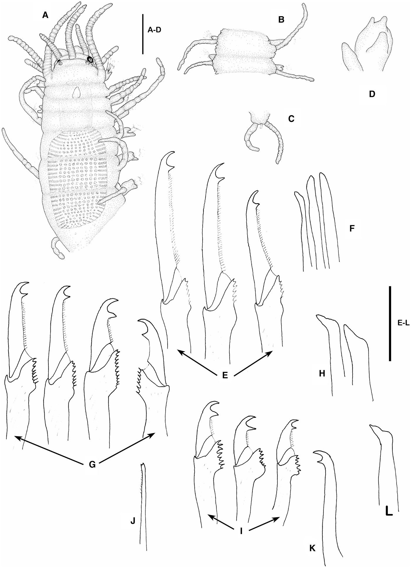
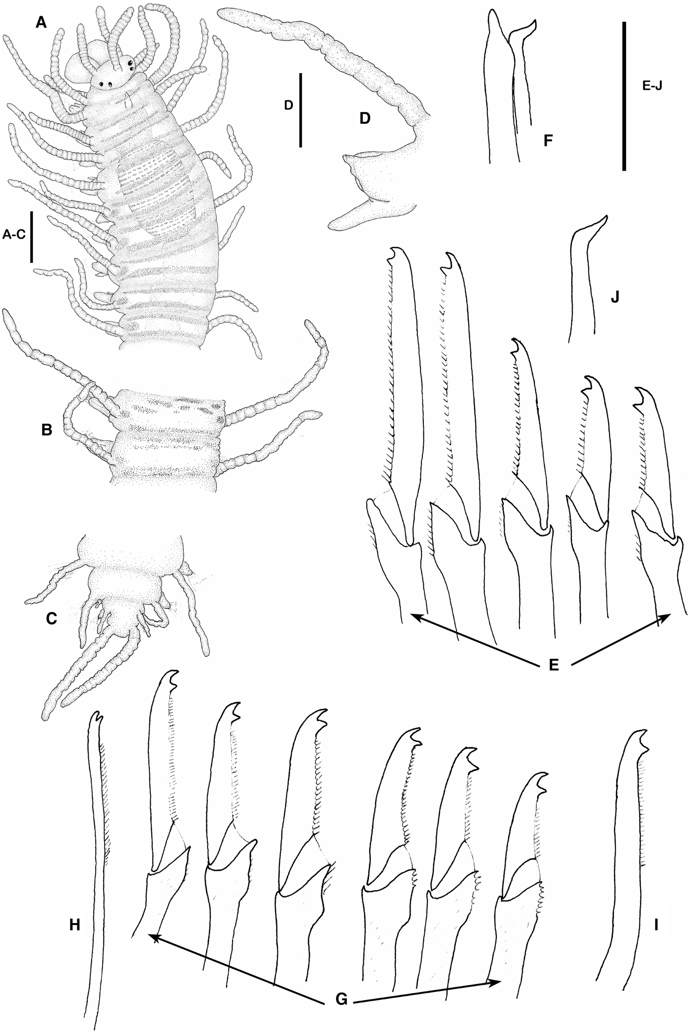
Megasyllis tigrina sp. nov.: (A) anterior end, dorsal view; (B) midbody segments, dorsal view; (C) posterior end, dorsal view; (D) midbody parapodium, anterior view; (E) compound chaetae, anterior parapodium; (F) aciculae, anterior parapodium; (G) compound chaetae, posterior parapodium; (H) dorsal simple chaeta, posterior parapodium; (I) ventral simple chaeta, posterior parapodium; (J) acicula, posterior parapodium. Holotype, AM W36503. Scale bars: A, C–D, 0.18 mm; E–J, 40 μm.
MATERIAL EXAMINED
Japan: 3 specimens (MNCN 16.01/14726) Shiraiso coast, Manazuru Peninsula, Sagami Bay, 35°09′14′′N 139°08′49′′E, intertidal, rocky shore, seagrass and algae, coll. Nishi, May 2002; 2 specimens (MNCN 16.01/14724) Mangokuura, Ishinomaki, Miyagi, Tohoku, 39°02′N 141°51′E, intertidal, coarse sand, coll. Nishi, June 2002; 1 specimen (MNCN 16.01/14725), Shiraiso coast, Manazuru Peninsula, Sagami Bay, 35°09′14′′N 139°08′49′′E, intertidal to subtidal rocky shore, seagrass and algae, coll. Nishi, September 2004.
REMARKS
Some of these additionald specimens (not types) showed some variability in the shape of the shafts and the length of the dorsal cirri. In some specimens, the distal end of the posterior shafts was curved and the dorsal cirri are longer than those originally described. Additionally, we found some specimens with simple dorsal and ventral chaetae. Aguado et al. (Reference Aguado, San Martín and ten Hove2008) did not describe these chaetae since the type series lacked these features. These simple chaetae are the same as those of M. nipponica. Some specimens showed the surface with small spherical granules visible by transparency.
TYPE LOCALITY
Shiraiso coast, Manazuru, Sagami Bay, Japan.
DISTRIBUTION
North-West Pacific Ocean (Japan).
HABITAT
Seagrasses and algae, rocky shore, intertidal to subtidal.
Megasyllis nipponica (Imajima, Reference Imajima1966) comb. nov.
Typosyllis nipponica Imajima, Reference Imajima1966: 266–268, figure 57; Reference Imajima2003: 165–166; Licher Reference Licher1999: 121–124.
REMARKS
Although we have not examined specimens, the characters shown in the previous descriptions agree with the diagnosis of Megasyllis and so we include this species in this genus.
TYPE LOCALITY
Funadomari, Rebun Island, Japan.
DISTRIBUTION
Japan.
HABITAT
Intertidal.
Megasyllis subantennata (Hartmann-Schröder, Reference Hartmann-Schröder1984) comb. nov.
(Figure 10)
Typosyllis (Typosyllis) subantennata Hartmann-Schröder, Reference Hartmann-Schröder1984: 17, figures 20–22; 1986: 39.
‘Odontosyllis glandulosa’ Licher, Reference Licher1999 (in part): 297.
MATERIAL EXAMINED
Western Australia: 1 specimen (AM W36502), Red Bluff, Kalbarri, 37°42′S 114°04′E, on seagrass in shallow-water sand and on rocky shore, coll. Springthorpe, 10 January 1984; 15 specimens (AM W36849), Blue Holes, Kalbarri, algae, 1 m, coll. Aguado & San Martín, 19 June Reference Aguado, San Martín and Nishi2008; 1 specimen (AM W36848), Rat Island, Abrolhos Islands, algae, 1 m, coll. Aguado & San Martín, 21 June Reference Aguado, San Martín and Nishi2008; 1 specimen (AM W36627) Inshore limestone ref., Ned’s Camp, Cape Range National Park, 21° 59′S 113° 55′E, very fine sediment and sand from patches in reef, 1 m, coll. Stoddart, June 1984.
DESCRIPTION
Largest complete specimen about 12 mm long, 0.8 mm wide, with about 104 chaetigers. Body relatively long, stout, strongly convex anteriorly, becoming distinctly slender from proventricular segments backwards, with some rounded, small dermal inclusions on dorsum. Dorsum of segments more or less annulated after proventricular segments (Figure 10B), smooth on anterior segments (Figure 10A). Live specimens with dark prostomium and most anterior segments; subsequent segments white, and remaining segments pink to brown, with red spots on bases of dorsal cirri. Prostomium wider than long, covered partially by peristomium, with two pairs of eyes in open trapezoidal arrangement, anterior ones slightly larger than posterior ones (Figure 10A). Median antenna inserted on middle of prostomium, longer than combined length of prostomium and palps, with about 23, more or less distinct, short articles; lateral antennae shorter than median one, inserted in front of anterior margin of prostomium, with about 15 articles (Figure 10A). Palps similar in length to prostomium, fused at base with a distinct median groove. Peristomium shorter than subsequent segments, covering posterior part of prostomium (Figure 10A). Dorsal tentacular cirri longer than lateral antennae, with about 14 articles, ventral tentacular cirri about half as long as dorsal ones, weakly articulated (Figure 10A). First dorsal cirri longer than tentacular dorsal cirri, directed anteriorly more or less well defined, with about 22 articles (Figure 10A). Thereafter, cirri alternating in length, becoming smooth after few anterior segments (Figure 10A, B). Parapodia with prechaetal and postchaetal lobes, plus an accessory, digitiform, ventral lobe, dorsal to ventral cirri (Figure 10D). Ventral cirri conical, inserted proximally on parapodia, not extending beyond parapodial lobes (Figure 10D). Anterior parapodia each with about 15 compound, heterogomph chaetae, distal part of shafts provided with serrations (Figure 10E), blades bidentate, both teeth similar, acute, well separated to each other, with short spines on cutting edge, decreasing in length from dorsal (~80 µm) to ventral (~60 µm). Number of compound chaetae per parapodium decreasing progressively to posterior part of body; midbody parapodia with 12 compound chaetae, posterior parapodia with ten chaetae; shafts of compound chaetae increasing in thickness as more ventral and more posterior, with stronger serrations (Figure 10E, G, I); most ventral blades shorter and larger; blades of compound chaetae in midbody, about 50 µm dorsally to 40 µm ventrally; posterior compound chaetae with shorter blades (Figure 10I), 32–26 µm in length, more strongly bidentate. Posterior parapodia with solitary, dorsal simple chaetae bidentate, very thin, with distal spines (Figure 10J). Ventral simple chaeta, much thicker than dorsal one, distally curved, strongly bidentate, distal tooth larger (Figure 10K). Anterior parapodia with three to four aciculae, two to three distally blunt and other slender, distally oblique (Figure 9F); two aciculae distally oblique from midbody (Figure 10H), posterior parapodia with single, distally oblique acicula (Figure 10L). Pharynx short, extending through three segments; pharyngeal tooth conical, relatively large, located near anterior margin (Figure 10A). Proventricle extending through three segments, with about 26–33, not well marked, muscle cell-rows (Figure 10A).
DISTRIBUTION
South and Western Australia.
HABITAT
Algae and sand, intertidal to subtidal in shallow depths.
Megasyllis procera (Hartman, Reference Hartman1965) comb. nov.
?Pionosyllis malmgreni McIntosh, Reference McIntosh1869: 414–415, pl. 15, figure 21, pl. 16, figure 10; Ehlers, Reference Ehlers1913: 475; Day Reference Day1967: 264, figure 12.8i–k.
Pionosyllis procera Hartman, Reference Hartman1965: 79–80, figure 10 c, d; San Martín & Estapé Reference San Martín and Estapé1993: 227–231, figures 1, 2; López & San Martín, Reference López and San Martín1994: 134.
Pionosyllis micropharyngea Hartmann-Schröder, Reference Hartmann-Schröder1974: 126, pl. 10, figures 89–92.
REMARKS
San Martín & Estapé (Reference San Martín and Estapé1993) provided a redescription of P. procera and described the sexual stolons. Subsequently, López & San Martín (Reference López and San Martín1994) suggested that it could be a synonymous of P. malmgreni. We have tried to examine the type specimens of P. malmgreni species but it was not possible, since they have been probably lost. However, it is easy to perceive after a detailed examination of the original description and drawings of McIntosh (Reference McIntosh1869) that Pionosyllis malmgreni belongs to Megasyllis as herein defined. Moreover, McIntosh described a sexual stolon in process of budding, similar to that described by San Martín & Estapé for P. procera.
TYPE LOCALITY
New England Continental Slope; between Atlantis Canyon and Block Canyon.
DISTRIBUTION
Southern and south-western Africa, Cuba, Atlantic coast of USA, Cape Verde Islands.
HABITAT
Under littoral stones, fine sand, algae, inside coralline rocks, from intertidal to 200 m depth.
Megasyllis pseudoheterosetosa (Böggemann & Westheide, Reference Böggemann and Westheide2004) comb. nov.
Typosyllis pseudoheterosetosa Böggemann & Westheide, Reference Böggemann and Westheide2004: 409, figures 2a–e, 3a–n.
MATERIAL EXAMINED
Holotype (SMF 11309), Anse Forbans, Mahé (Seychelles), radial zone from coral rubble, 18 March 2000.
REMARKS
The holotype has the pharyngeal tooth slightly back than that was drawn originally and the segments are dorsally rough.
TYPE LOCALITY
Mahé (Seychelles), Indian Ocean.
DISTRIBUTION
Only known from the type locality.
MATERIAL EXAMINED
Holotype (AM W36503) and 3 paratypes (AM W36504). Holotype and one paratype, Australia, New South Wales: Bowen Island, Jervis Bay, 35°07.49′S 150°45.77′E, small, white sponge with pink lobes, on a seagrass bed, 7 m, coll. Serov & Wilson, 8 December 1993; 2 paratypes (AM W36505), south-west of Bowen Island, Jervis Bay, 35° 7.49′S 150° 47.77′E, rock on sandy bottom covered with bryozoa and polychaetes, 7 m, coll. Serov & Wilson, 8 December 1993.
DESCRIPTION
Holotype complete 8 mm long, 0.4 mm, with 56 chaetigers; developing one sexual stolon of 14 segments, without notochaetae, 2.5 mm long. Body relatively long, with a marked colour pattern; two transversal rows of black or red pigment on dorsum of each segment and another oval reddish pigmented area on bases of dorsal cirri (Figure 11A); transversal rows divided in several smaller ones in midbody (Figure 11B), disappearing on posterior segments (Figure 11C). Anterior segments smooth (Figure 11A), becoming rough to annulated from midbody (Figure 11B). A reddish transversal area in the inner wall of the pharynx, visible by transparency. Prostomium oval, wider than long, with two pairs of similar eyes in open trapezoidal arrangement (Figure 11A). Median antenna inserted on middle of prostomium, slightly longer than combined length of prostomium and palps, with about 15, more or less well-defined, articles; lateral antennae shorter than median one, inserted near anterior margin of prostomium, with about 12–13 articles (Figure 11A). Palps similar or longer than prostomium, fused at base with a distinct median groove (Figure 11A). Peristomium shorter than subsequent segments. Dorsal tentacular cirri longer than median antenna, with about 18, more or less well defined articles, ventral tentacular cirri about half as long as dorsal ones (Figure 11A). First dorsal cirri longer than tentacular dorsal cirri, with about 23 articles (Figure 11A). Thereafter, cirri alternating in length, and becoming less articulated (Figure 11B, D), to smooth posteriorly (Figure 11C). Parapodial lobes with prechaetal and postchaetal lobes; prechaetal lobe more triangular and larger than the other one (Figure 11D). Ventral cirri conical, inserted proximally on parapodia, extending beyond parapodial lobes (Figure 11D). Anterior parapodia each with about 14 compound, heterogomph chaetae, distal part of shafts provided with spines (Figure 11E), blades bidentate, both teeth similar in size, short spines on cutting edge, decreasing in length from dorsal (~80 µm) to ventral (~40 µm). Number of compound chaetae per parapodium decreasing progressively to posterior part of body to ten on posterior parapodia; blades of compound chaetae in posterior parapodia somewhat shorter and larger, with thicker shafts (Figure 11G), 52 μm (long dorsal) blades to 35 μm (ventral blades). Posterior parapodia with solitary, dorsal simple chaetae bidentate, very thin, with subdistal spines (Figure 11H). Ventral simple chaetae solitary, distinctly thicker than dorsal one, distally curved, strongly bidentate, with subdistal short spines (Figure 11I). Anterior parapodia with two aciculae, one distally blunt and other slender, distally oblique (Figure 11F); from midbody backwards, parapodia with single, distally oblique acicula (Figure 11J). Pharynx small, extending through three segments; conical, relatively large tooth on anterior margin (Figure 11A). Proventricle extending through three segments, with about 33 muscle cell-rows (Figure 11A). Pygidium small, with two rough, unarticulated anal cirri (Figure 11D).
REMARKS
Megasyllis tigrina is characterized by having a distinct colour pattern, formed by two reddish transversal stripes on dorsum of each anterior segment, different to any other pattern of the species within this genus. The compound chaetae are very similar to those of M. subantennata, but the teeth of the blades are not so separated, and the shafts have much smaller distal serrations.
DISTRIBUTION
New South Wales, Australia.
HABITAT
In sponges, sandy bottoms with bryozoans, subtidal in shallow depths.
ETYMOLOGY
The name refers to the transversal stripes, similar to the pattern of a tiger.
DISCUSSION
The genus Megasyllis, has a wide distribution through the Indo-Pacific region from Japan (three species: M. inflata, M. multiannulata, M. nipponica), The Phillipines (one species, M. chrissyae); Marquesas Islands (M. marquesensis); Seychelles (M. pseudoheterosetosa); Australia (seven species, M. corruscans, M. glandulosa, M. heterosetosa, M. inflata, M. marianandreworum, M. subantennata, M. tigrina); and New Zealand (one species, M. eduardoi). All of them have a more or less restricted distribution; the only species reported from more than one area is M. inflata, reported in Japan and Australia. Probably, there are still numerous species to be discovered in the area.
By contrast, there is a single species reported outside the Indo-Pacific region, M. procera, and this species is widely distributed (southern and south-western Africa, Cuba, the Atlantic coast of USA, Cape Verde Islands, and also in Brazil, Karla Paresque, personal communication).
The group could be originated in the central Pacific, perhaps in Australia, where more species are reported, and more widely are distributed, and were diverging in different areas. One species could have come to the eastern Pacific before the closure of Panamá Isthmus, about 3.2–3.5 millions years ago (Coates et al., Reference Coates, Jackson, Collins, Cronin, Dowsett, Bybell, Jung and Obando1992; Glynn & Maté, Reference Glynn and Maté1996; San Martín et al., Reference San Martín, López, Redondo, Capa, Cladera, Laborda and Castroviejo1997) and cross to the Atlantic Ocean, where it spread out. So, there are two areas of distribution of Megasyllis, one with numerous species (Pacific) and another with a single known one (Atlantic). Up to now, no species of this group is known in the east Pacific Ocean. However, this is only a hypothesis, impossible to be checked, since the effort of sampling has been unequal in all areas, and new reports and species could be discovered in the future.
ACKNOWLEDGEMENTS
We are grateful to Kate Attwood, Anna Murray, Pat Hutchings, (AM), A. Muir and M. Lowe (BMNH) for the kind loan of specimens. Thanks to E. Nishi (Mazanuru Marine Laboratory, Japan) for specimens of M. multiannulata. Christina Piotrowski, California Academy of Sciences, provided us with some syllids from The Phillipines; among them appear a new species of this genus. We greatly appreciate the help of Jane Fromont (Western Australian Museum) during our collecting field trip (M.T. Aguado and G. San Martín). Many thanks to Aida Verdes, Eduardo López and Ramón Gallego for collecting and sorting the material during the New Zealand field trip. Dr Geoff Read (NIWA, Wellington) offered us useful advice about collection sites in New Zealand. We are also very grateful to Denis Riek for sharing his beautiful colour pictures (Figure 1) and Pat Hutchings for her useful advices.
FINANCIAL SUPPORT
This paper is a contribution of the project ‘Caracterización Taxonómica y Sistemática de la familia Syllidae (polychaeta) basada en datos moleculares y morfológicos. El problema de las especies cosmopolitas y Biodiversidad en el Pacífico’, funded by the ‘Ministerio de Ciencia e Innovación’ of the Spanish Government, Project No. CGL2009–12292 BOS.