Introduction
The term otosclerosis was coined by Adam Politzer in 1893.Reference Politzer1 Apparently, the fissula ante fenestram is unique to humans.Reference Anson, Bast, Shambaug and Glasscock2 Anson and Bast related the histologically unstable cartilage in the fissular region to the development of otosclerosis.Reference Anson, Bast, Shambaug and Glasscock2 The pathological tissue that is the active focus of otosclerosis consists of either newly formed cartilage or bone.Reference Glasscock and Gulya3 Malleoincudal dislocation may occur, particularly when there are otosclerotic foci above the oval window. Secondary torsional stresses on the stapes pulling the incus down are believed to be responsible for malleoincudal dislocation.Reference Swartz and Loevener4
This obliquity and downward displacement of the stapes towards the promontory increases the distance between the stapes and the horizontal portion of the facial nerve. The obliquity of the stapes in otosclerotic ears also reduces the space between the stapes and the inferior border of the oval window niche when compared to non-otosclerotic ears, where the stapes lies perpendicular to the oval window.
After stapes surgery, the incus can revert back to its original position as a result of biological remodelling and the subsequent pull by the superior incudal ligament. This can lead to long-term failures in stapes surgery.
This study aimed to establish objectively the obliquity of the stapes towards the promontory using intra-operative measurements in otosclerosis patients, and to devise methods to overcome the delayed complications of stapes surgery.
Objectives
These were: to establish the obliquity of the stapes observed in otosclerosis patients during stapes surgery, to analyse some of the late complications and failures in stapes surgery, and to understand how appreciation of this obliquity can aid in the development of techniques to avoid these issues.
Materials and methods
A prospective observational study was conducted in our centre (following clearance from the institutional ethics committee) from January 2013 to December 2014. The study comprised 20 patients, divided into 2 groups: a study group consisting of 10 patients with otosclerosis and a control group comprising 10 (non-otosclerosis) patients with sudden sensorineural hearing loss (SNHL). All the sudden SNHL patients underwent tympanometry with reflex study. Only those with either ipsilateral or contralateral reflex positive results were selected to exclude any otosclerosis in this group.
All patients (in both the study and control groups) were evaluated intra-operatively after obtaining informed consent. Measurements (described below) were taken in the 10 otosclerosis patients during stapes surgery under local anaesthesia using 90-degree measuring picks. These were compared with measurements for the 10 sudden SNHL patients who underwent intratympanic steroid therapy by exploratory tympanotomy.
Intra-operative measurements
In the otosclerosis and non-otosclerosis patients, two distances were measured intra-operatively using 90-degree measuring picks of various sizes, ranging from 0.1 to 2.5 mm (Figure 1).
Ninety-degree picks were used to record intra-operative measurements (a); the picks were of various sizes, ranging from 0.1 to 2.5 mm (b).

The first distance measured was from the upper margin of the posterior stapes crura to the facial prominence of the horizontal portion of the facial nerve. The second distance measured was from the upper margin of the posterior stapes crura to the inferior border of the oval window niche. Care was taken to avoid facial nerve damage with the picks during measurements.
The measurements were conducted by a single researcher to avoid inter-observer variation. The facial prominence (prominentia canalis facialis) is the bony bulge of the distal part of the tympanic facial nerve, in the tympanic cavity, superior to the stapes. This is one of the main landmarks in stapedotomy or chronic otitis media surgery. The rate of facial canal dehiscence is high in this area.Reference Ulug5Figure 2 shows the distance measured from the facial prominence to the upper margin of the stapes crura in otosclerosis and non-otosclerosis patients.
Schematic picture showing the increase in length from facial nerve to upper margin of stapes crura due to obliquity of stapes, named the ‘Pisa sign’, in otosclerosis. Bold lines indicate distance measurement between facial nerve and upper margin of stapes crura (short bold line in non-otosclerosis, long bold line in otosclerosis). Arrows indicate the incus being pulled down and torsional effect on the incus due to otosclerosis as indicated by the incus drawn in a dotted line.
Results
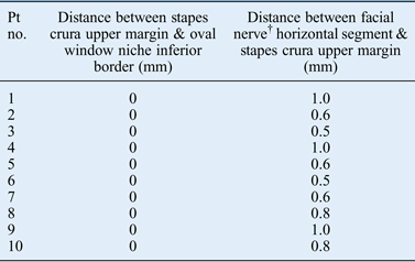
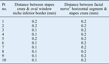
The mean distance (± standard deviation) from the upper margin of the posterior stapes crura to the facial prominence in otosclerotic ears was 0.74 mm (± 0.20 mm). In non-otosclerotic ears, the same distance was 0.20 mm (± 0.0 mm). The mean distance from the upper margin of the posterior stapes crura to the inferior border of the oval window niche in otosclerosis patients was not recordable (i.e. 0 mm) as the stapes crura was in contact with the oval window niche as a result of the obliquity and downward displacement of the stapes. In non-otosclerotic ears, the distance measured was 0.13 mm (± 0.04 mm).
Statistical analysis was conducted with SPSS® software (version 17.0) using the paired t-test. The differences were found to be statistically significant (p < 0.0001) (Tables I–III).
Intra-operative measurements in otosclerosis patients*
* Measured using 90-degree picks. †Prominentia canalis facialis. Pt no. = patient number
Intra-operative measurements in non-otosclerosis patients*
* Measured using 90-degree picks. †Prominentia canalis facialis. Pt no. = patient number
Paired samples t-test results
SD = standard deviation; SEM = standard error of the mean; CI = confidence interval; df = degrees of freedom
Figures 3 and 4 are intra-operative views showing distance measurements in otosclerosis and non-otosclerosis patients using 90-degree measuring picks. Figures 5 and 6 are graphs showing the intra-operative measurements recorded in otosclerosis and non-otosclerosis patients.
(a) Intra-operative view showing 1 mm distance between stapes crura and facial prominence in otosclerosis. (b) Intra-operative view showing no gap between stapes crura and inferior border of oval window niche due to leaning of stapes towards promontory in otosclerosis.
(a) Intra-operative view showing 0.2 mm distance between facial prominence and stapes crura during intratympanic treatment for sudden sensorineural hearing loss (SNHL). (b) Intra-operative view showing 0.1 mm distance between stapes crura and inferior border of oval window niche during intratympanic treatment for sudden SNHL.
Middle-ear distance between facial nerve and stapes crura measured using picks in otosclerosis and non-otosclerosis patients.
Middle-ear distance between stapes crura and inferior border of oval window niche in otosclerosis and non-otosclerosis patients.
Discussion
Otosclerosis has been described as pleomorphic bone dyscrasia. Fixation of the stapes begins as a calcification of the annular ligament joining the oval window otosclerotic lesion with the stapedial footplate. The stapes then becomes fixed by the lesion itself. The process then appears to flow across the ligament onto the footplate of the stapes, totally obliterating any remnants of the original annular ligament. Resorption of the otic capsule bone and replacement with a hypercellular woven bone occurs, which undergoes further remodelling resulting in sclerotic mosaic architecture.Reference Shea, Shea, Glasscock and Gulya6
The most common area of stapes fixation is the anterior crus in the region of the embryonic fissula antefenestram.Reference Ruedi7–Reference Shambaugh10 Because of the metamorphosis of the lining cartilage, the fissula is thought to be an area of histological instability.Reference Anson, Cauldwell and Bast11–Reference Anson, Cauldwell and Bast13 The progression and extension of the abnormal remodelling process are dependent on an underlying genetic defect (COL1A1 gene) in collagen metabolism that results in the generation of an unstable extracellular matrix with a high propensity for remodelling.Reference Glasscock and Gulya3
Biological remodelling is the norm in the oval window area in otosclerosis, resulting in a torsional effect on the ossicular chain via the incudostapedial joint due to the obliquity of the stapes towards the promontory.
Torsional effect
The downward displacement of the stapes due to otosclerosis also affects the incus via the incudostapedial joint and it gets pulled down. Malleoincudal dislocation may occur as a secondary torsional sequela. A secondary incus subluxation as a torsional sequela may occur, and the appearance may be identical to that which occurs as a result of trauma.Reference Swartz and Loevener4 This feature may account for persisting conductive hearing loss following a successful stapes operation. After stapes surgery, the incus can revert back to its original position as a result of biological remodelling and the subsequent pull by the superior incudal ligament.
These secondary torsional stresses can lead to late complications in stapes surgery; to date, these have not been objectively evaluated and documented. In studies by Maru et al. and Zhu et al., the mean distance measured between head of the stapes and the facial nerve using a stereomicroscope was 1.58 mm and 1.18 mm (± 0.42 mm) respectively.Reference Zhu, Sun, Zhang, Sun and Jiang14, Reference Maru, Cheita, Mogoanta and Prejoianu15
As the oval window, stapes head and facial nerve are not in the same horizontal plane, we measured the distance between the upper margin of the posterior stapes crura and the facial prominence of the horizontal portion of the facial nerve by a simpler method, using 90-degree measuring picks. The mean distance between the upper margin of the posterior stapes crura and the horizontal portion of the facial nerve in otosclerotic ears was 0.74 mm (± 0.21 mm). However, the distance measured between the upper margin of the stapes crura and the horizontal segment of the facial nerve in non-otosclerosis patients was 0.20 mm (± 0.00 mm). There was no gap between the upper margin of the posterior crura and the inferior border of the oval window niche in otosclerosis patients because of the obliquity of the stapes. However, the distance measured between the upper margin of the posterior crura and the inferior border of the oval window niche in non-otosclerosis patients was 0.13 mm (± 0.05 mm).
This study confirms that the distance between the stapes and the facial nerve is greater in otosclerosis patients than in non-otosclerosis patients. Because of the obliquity of the stapes in otosclerosis, there is no gap between the stapes and the inferior border of the oval window niche. The differences between the groups were found to be statistically significant (paired t-test, p < 0.0001).
Implications for stapes surgery
Historically, surgical procedures for the correction of otosclerosis have been modified, abandoned and subsequently re-established.
Lateralised piston syndrome
A review, by Lagleyre et al., of 1289 consecutive stapes operations revealed 119 revisions (9 per cent), from which 22 cases (18.5 per cent) could be classified as lateralised piston syndrome which presented as delayed conductive hearing loss in 95.5 per cent of cases.Reference Lagleyre, Calmels, Escude, Deguine and Fraysse16 Pre-operative computed tomography scans showed a lateralised piston out of the fenestra in 81 per cent of cases and in contact with the tympanic membrane in 54.5 per cent of cases. Revisions revealed lateral displacement of the piston out of the entire oval window in 86 per cent of cases, with closure of the fenestra in all cases and incus necrosis in 77 per cent of cases.Reference Lagleyre, Calmels, Escude, Deguine and Fraysse16
In a study by Ying et al., on patterns of failure for a heat-activated crimping prosthesis in stapedotomy, there was an 11 per cent rate of possible lateral displacement of the prosthesis.Reference Ying, Hillman and Chen17
Lesinki, in 2003, reviewed his series of 279 cases of revision stapes surgery performed with a laser (as cited by Myers and CarrauReference Myers and Carrau18).The most common finding was a displaced or malfunctioning prosthesis (81 per cent), followed by erosion of the incus (30 per cent). Correction of prosthetic slippage and problems with the incus was statistically more successful than reopening of a bony window closure.
A wire/connective-tissue prosthesis used with stapedectomy can migrate towards the superior edge of the oval window. This has been found to induce conductive hearing loss or even SNHL, and vertigo.Reference Pauw, Pollak and Fisch19
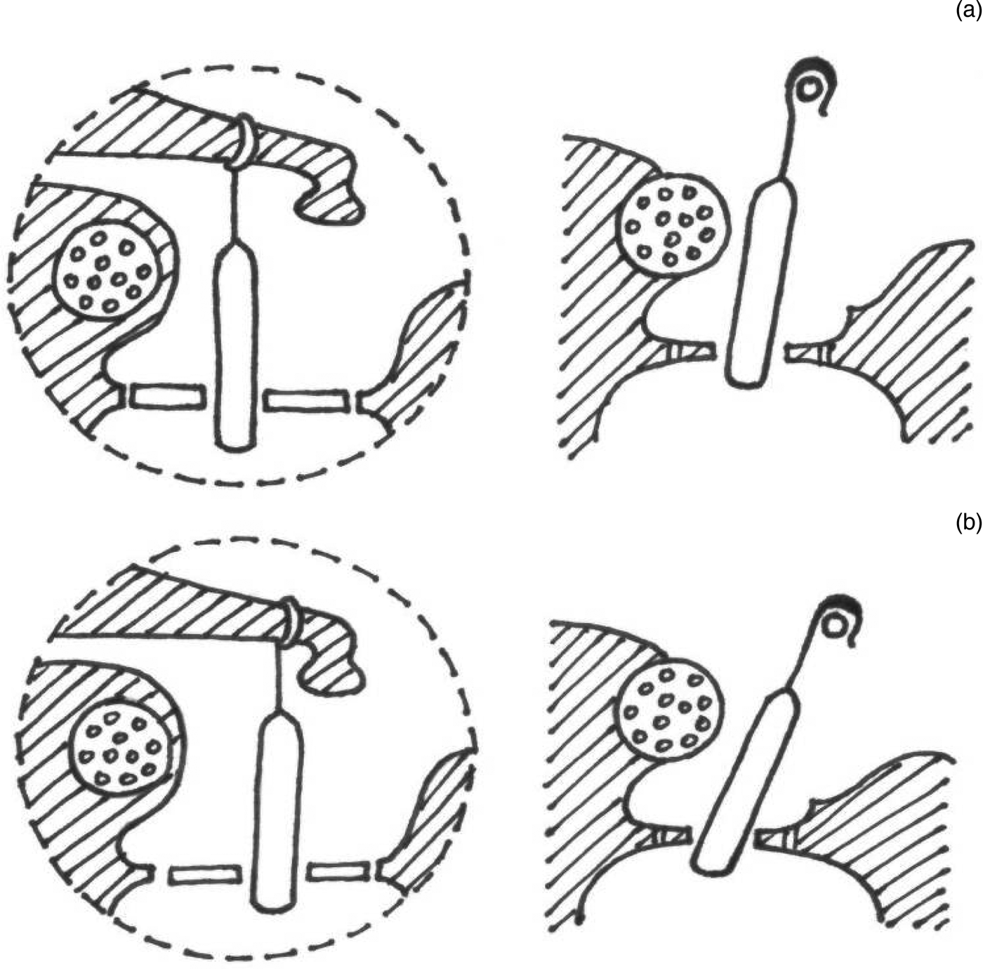
Lateralised piston syndrome and displacement of the prosthesis may be due to biological remodelling of the incus, as the torsional effect of otosclerotic foci is absent following stapes surgery. The senior author's personal unpublished data support this: in cases of revision stapes surgery with delayed conductive hearing loss, about 60 per cent of cases had lateral displacement of the prosthesis. For this reason, it is better to crimp the prosthesis lower down the incus to avoid lateralisation, and to prevent the prosthesis coming into contact with the facial nerve. The slinging of the chorda tympani nerve above and over the crimped piston, placing a soft tissue shim anterior and superior to the junction of the piston stem and fenestra mimicking the force of the original otosclerotic foci, also prevents the piston from coming into contact with the horizontal segment of the facial nerve during the post-operative period (Figures 7–9).
(a) Piston crimped high with less distance between piston and facial nerve. (b) Piston crimped low with more space from facial nerve, which prevents piston from coming into contact with facial canal.
Anchoring of the piston by chorda tympani nerve to prevent ‘lateralised piston syndrome’ of prosthesis.
Lower crimping of the Kurz soft clip piston and soft tissue (vein graft) placed as a shim superior and anterior to piston, thus preventing superior and lateral displacement out of vestibule and preventing piston coming into contact with facial canal.
Space for instrumentation
Increased superior distance between the facial nerve and stapes because of the obliquity of the stapes allows space for perforators, drills and lasers for otosclerosis surgery. This also helps to cut and fracture the stapes crura downwards by force towards the promontory. This step prevents destabilising of the footplate: too much displacement of the stapes during fracturing of the crura is avoided by contact of the stapes with the inferior margin of the oval window niche (Figures 10–12).
Carbon dioxide laser stapedotomy intra-operative view, showing leaning of stapes providing space for laser spot.
(a) Space for instrumentation due to obliquity of stapes (‘Pisa sign’). (b) Space for instrumentation as a result of Pisa sign (leaning of stapes towards promontory, increasing the distance between facial nerve and stapes head).
Stapes crura fractured by force using Heermann's hook downwards towards promontory, preventing destabilising of footplate.
Loose wire syndrome and precise crimping of prosthesis
Inaccurate crimping of the prosthesis may lead to loose wire syndrome. McGee, in 1981 (as cited in NuriozgirginReference Nuriozgirgin20), reported on 43 patients with 1 or more symptoms from the triad that is typical for loose wire syndrome (i.e. hearing loss, poor speech discrimination and distortion of sound). At revision surgery, a loose attachment of the prosthesis at the long process of the incus was found. This suggests that fear of incus erosion or necrosis due to over-crimping is unwarranted. Lesinki, in 2002 (as cited in NuriozgirginReference Nuriozgirgin20), evaluated the findings of 260 patients who underwent revision stapes surgery. Varying degrees of incus erosion were found in 91 per cent of patients.
Erosion of the incus is caused by the continuous vibration of biological bone against the displaced and fixed prosthesis. Nuriozgirgin reported that erosion of the long process of the incus and dislocation of the prosthesis were the most frequent reasons for revision operations.Reference Nuriozgirgin20 Ying et al. found that reopening of the nitinol hook off the incus was another common associated finding.Reference Ying, Hillman and Chen17 Most incus erosion occurs gradually from vibration of the incus against a fixed and displaced prosthesis, and not from over-crimping. Early surgical revision of patients with conductive hearing loss following stapedectomy should help to prevent complete incus erosion, making the conductive repair simpler.Reference Carrasco and Pillsbury21
We have found that the obliquity of the stapes in otosclerosis, leading to a torsional effect on the ossicular chain, can lead to loose wire syndrome. When remodelling of the incus occurs, the incus tries to go back to its original position, leading to displacement of the prosthesis. This could also lead to reopening of the nitinol hook, as mentioned by Ying et al.Reference Ying, Hillman and Chen17 Hence, precise and tight crimping of the prosthesis at the lower end of the incus is important to avoid the torsional effect on the piston and loose wire syndrome.
Dehiscent facial nerve
Nuriozgirgin emphasises that in stapes surgery special attention should be given to anatomical variations related to the stapes and the oval window during the operation.Reference Nuriozgirgin20 A deeply situated narrow oval window and facial nerve anatomical variations are problems frequently encountered during primary operations.Reference Nuriozgirgin20
Tange and de Bruijn investigated the incidence of dehiscence in the horizontal facial canal in otosclerosis patients.Reference Tange and de Bruijn22 Facial nerve dehiscence was observed in 14 (3.27 per cent) of the 427 patients who underwent stapedotomy. Facial nerve dehiscence localised to the horizontal segment of the facial nerve is rather common in adults, and has been described in 52–74 per cent of cases in temporal bone studies.Reference Tos23
Obliquity of the stapes and consequent space for instrumentation helps in cases of dehiscent facial nerve. Crimping the piston low on the incus avoids contact of the piston with the facial nerve (Figures 13 and 14).
• The oblique nature of stapes with downward displacement was confirmed to be a unique feature and a new clinical sign of otosclerosis
• Mean distance between facial nerve and upper margin of stapes crura in otosclerosis patients was 0.74 mm, compared with 0.2 mm in non-otosclerosis patients
• Downward stapes displacement produces torsional force via the incudostapedial joint pulling the incus down, thus stretching the superior incudal ligament
• After stapedotomy, the force caused by downward displacement of stapes is absent and the incus moves up via biological remodelling
• Movement of incus can pull the stapes piston out of the fenestra, which can lead to prosthesis displacement
• Precise and tight crimping of the piston low in the incus is suggested as a solution
Dehiscent facial nerve in stapes surgery. The increased distance as a result of obliquity of stapes provides space for instrumentation.
(a) Dehiscent facial nerve and piston crimped high comes into contact with the facial nerve. (b) Dehiscent facial nerve and piston are crimped low to avoid piston coming into contact with facial nerve.
The method of intra-operative measurement used in this study to confirm the obliquity of the stapes in otosclerosis is simple and reliable when compared to the telemanipulator systems used in other studies.Reference Maier, Strauss, Bauer, Grasser, Hata and Lueth24
Conclusion
Increased distance between the stapes and horizontal portion of the facial nerve due to the obliquity of the stapes towards the promontory is caused by otosclerotic foci. This obliquity of the stapes is a result of the torsional effect of otosclerosis on the stapes, which also has an effect on the rest of the ossicular chain. It correlates with late complications and failures in stapes surgery, and helps us to devise ways to avoid these problems. The senior author suggests that the term ‘Pisa sign’ may be considered to describe this obliquity of the stapes, as knowledge of the leaning tower of Pisa is well known.
Acknowledgement
The authors wish to sincerely thank the MCV Memorial ENT Trust Hospital (Pollachi, India) for providing the support and material for this study.