Introduction
Telomeres are short noncoding DNA repeats located at the end of chromosomes that maintain the integrity of the genetic code throughout consecutive replication cycles. Telomere length provides an estimate of the number of replication cycles that have occurred and has been proposed to be a measure of cellular senescence and disease susceptibility.Reference Factor-Litvak and Susser1 Indeed, shorter telomeres have been linked to numerous diseases associated with aging including cardiovascular disease,Reference Samani, Boultby and Butler2 diabetes, and various forms of cancer.Reference Martínez and Blasco3 Given these associations, telomere attrition has also been explored as a potential mechanism by which stress contributes to disease development in later life.Reference Epel, Blackburn and Lin4 Recent studies using biomarkers of stress exposure have described negative correlations between stress and telomere length,Reference Woody, Hamilton and Livitz5 such that individuals with higher levels of stress exposure have shorter telomeres. However, because of the cross-sectional designs of these studies, it can be difficult to establish whether physiological stress precedes telomere attrition, was predicted by it, or if they were simply both due to another common exposure or process. It may be the case that relatively greater physiological stress exposure during life stages where chronic illness is rarer (e.g., early adulthood) may adversely influence telomere length years later, potentially conferring a greater propensity for premature aging and subsequent illness.
Stress vulnerability can be assessed in a number of different ways, but salivary cortisol levels and greater relative right frontal alpha asymmetry at rest are two particularly useful biomarkers of this construct. Each measure is presumed to tap different physiological systems and accordingly can provide unique insights into stress regulation and vulnerability arising from disparate biological pathways. Salivary cortisol is a well-validated biomarker of hypothalamic–pituitary–adrenal (HPA) axis activity,Reference Ockenfels, Porter and Smyth6–Reference Khoury, Gonzalez and Levitan8 and we generated a measure of afternoon baseline cortisol averaged across three samples. Afternoon cortisol levels have been hypothesized to serve as a more stable measure of cortisol concentration that is less susceptible to the influence of circadian rhythm than morning cortisol measures.Reference Burke, Davis and Otte9 Greater relative right frontal electroencephalogram (EEG) alpha asymmetry at rest has been linked to reduced emotional regulation in response to challenge and greater stress, anxiety, and depressive symptoms.Reference Coan and Allen10,Reference Reznik and Allen11 It has been proposed that the balance of electrical activity between the left and right prefrontal cortex reflects the relative activity of the approach and withdrawal systems, respectively. These systems are thought to govern higher order behavioral responses to the environment, such that approach behaviors engage with pleasant stimuli while withdrawal behaviors avoid aversive stimuli. Greater right asymmetry therefore is indicative of a greater tendency toward aversion and a reduced receptivity to pleasant environmental cues.Reference Jesulola, Sharpley and Bitsika12 Here, we recorded resting brain electrical activity (EEG) from the left and right frontal brain regions and computed a measure of relative frontal EEG asymmetry. The present study assessed these two distinct physiological measures of stress vulnerability in early adulthood (when participants were 22–26 years of age).
Recent research has suggested that a dose–response effect exists between stress and shorter average telomere length.Reference Price, Kao and Burgers13,Reference Drury, Theall and Gleason14 While the majority of these studies measure telomere length during childhood, Entringer et al. noted an association between maternal stress during pregnancy and shorter telomere length in young adult offspring.Reference Entringer, Epel and Kumsta15 However, a recent study comparing telomere length between preterm and term infants reported that those born preterm actually had longer telomeres than their term counterparts during infancy.Reference Vasu, Turner and George16 An association between pain exposure related to neonatal intensive care and a reduction in telomere length among preterm infants has also been observed,Reference Provenzi, Giorda and Fumagalli17 suggesting that prematurity may instead confer a susceptibility to postnatal stress, reflected in telomere length reduction, rather than influencing telomere length directly.Reference Provenzi, Scotto di Minico and Giorda18 However, absent within the literature is an investigation of telomere length in adult survivors of preterm birth, and whether individual differences in stress vulnerability conferred by the stresses associated with this can impact telomere length and attrition. Understanding how the early-life adversities associated with preterm birth interacts with later stress vulnerability to affect telomere length could provide important insights into observed associations among the early-life environment, telomere attrition, and disease susceptibility.
Extremely low birth weight survivors (ELBW; <1000 g) are exposed to severe early-life adversity which can lead to differences in their ability to manage physiological stress compared to their normal birth weight (NBW; >2500 g) counterparts.Reference Mathewson, Pyhälä and Hovi19–Reference Kajantie, Phillips and Andersson21 Such differences in stress appraisal and regulation may contribute to the increased susceptibility to chronic physicalReference Irving, Belton and Elton22 and mental illness.Reference Van Lieshout, Boyle and Saigal23,Reference Goffinet24 Indeed, the prenatal risk factors,Reference Goffinet24 early postnatal experiences (i.e., Neonatal intensive care unit intervention),Reference Mooney-Leber and Brummelte25 and long-term health outcomes associated with ELBW birth suggest that in addition to being a compromised birth outcome, ELBW birth may be a marker of early-life stress exposure and perhaps increased stress vulnerability. The physiological stress conferred by ELBW birth can have persistent deleterious effects on stress regulationReference Harris and Seckl26 via its influence on HPA axis developmentReference Murgatroyd and Spengler27 and function,Reference Entringer, Kumsta and Hellhammer28 with implications for the development of somatic illness.Reference Van Lieshout, Boyle and Favotto29 Furthermore, it has been hypothesized that the health problems encountered by adult ELBW survivors are not necessarily a result of exposure to severe early-life stress, but may instead represent a broader pattern of accelerated biological aging conferred by that stress exposure.Reference Mathewson, Van Lieshout and Saigal30 Emerging research has sought to identify genomic indicators of cellular senescence that may precede the development of age-related disease, and telomere length measurement represents an exciting candidate predictor.
Given this background, we predicted that ELBW and NBW adults would exhibit a negative association between two measures of stress vulnerability (salivary cortisol and EEG-based frontal alpha asymmetry) in early adulthood (22–26 years) and telomere length measured in adulthood (30–35 years). We predicted that participants with greater stress vulnerability (i.e., relatively higher baseline afternoon salivary cortisol and greater relative right frontal alpha asymmetry at rest) would have shorter telomere lengths on average. However, because of their exposure to early-life adversity and the influence it may have exerted on their stress regulatory systems, we also hypothesized that ELBW adults would be differentially susceptible to stress exposure than NBW controls. Accordingly, we predicted the strength of associations between our stress vulnerability markers and telomere length would be stronger for ELBW than NBW adults.
Methods and materials
Study population and overview
The sample for this study comprised participants recruited to a longitudinally followed cohort study of 179 ELBW survivors (recruited at birth between January 1, 1977, and December 31, 1981) and 145 NBW control participants matched on age, sex, and familial socioeconomic status (who were recruited at age 8). This study is the oldest of its kind and was created to investigate the consequences and outcomes of such severe (and historically fatal) premature birth. At the 22–26-year assessment, 142 ELBW survivors and 133 NBW control participants remained enrolled in the study. At the 30–35-year assessment, 99 ELBW survivors and 89 NBW controls remained enrolled. A subset of 69 participants (34 ELBWs, 35 NBWs) provided buccal samples for telomere measurement. This subset did not significantly differ from participants who did not elect to undergo telomere measurement. Of these, 38 participants (16 ELBWs, 22 NBWs) also had complete baseline afternoon salivary cortisol and resting EEG measures at age 22–26 and were therefore eligible for inclusion in the analyses below. A total of 38 participants had telomere and frontal asymmetry data, and 38 participants had telomere and salivary cortisol data, although only 35 participants had data on all three measures. Only participants who were right handed and free of neurosensory impairment and major mental illness completed the EEG and cortisol measures at age 22–26. These criteria were set in an attempt to reduce variability in EEG data in order to test a particular disparate hypothesis regarding frontal alpha asymmetry in ELBW adults (published separately).Reference Schmidt, Miskovic and Boyle31 A small number of participants completed both of these measures but were not available at the 30–35-year visit. Finally, funding for telomere assays was not secured until after the 30–35-year visit was underway and so it was not possible to secure samples for all eligible individuals.
Baseline afternoon salivary cortisol measurement (22–26 years)
Three salivary cortisol samples were gathered approximately 30 min apart in the early afternoon (average time: 1 pm), representing a baseline afternoon measure of circulating cortisol. Each participant was asked to refrain from eating or drinking 1 h prior to coming to the laboratory and to report on any atypical recent life events, as these factors are known to influence cortisol levels. Each participant was asked to provide three individual saliva samples during the laboratory visit. Passive saliva was collected by having each participant expectorate into a 50 µl cryogenic tube. Approximately, 50 µl of saliva were collected at each sample.
All saliva samples were transported on ice and stored at −20°C prior to assaying. Saliva was centrifuged at 3000×g for 15 min, and only the supernatant was assayed in the Neuroendocriniology Laboratory at Brock University (St. Catharines, Ontario). All enzyme immunoassays were carried out on NUNC Maxisorb plates. Cortisol antibodies (R4866) and corresponding horseradish peroxidase conjugate were obtained from C. Munro of the Clinical Endocrinology Laboratory, University of California, Davis. Steroid standards were obtained from Steraloids, Inc. (Newport, Rhode Island). Each sample was assayed in duplicate and averages were used. Interplate variation (Coefficient of variation) was 6.45%, while intraplate variation was 6.51%. Because all three salivary cortisol samples were correlated (p’s < .05), they were aggregated and an average measure of baseline salivary cortisol (expressed in ng/ml) was computed. The natural log (ln) of this composite measure was then computed to reduce skewness.
Resting EEG measurement (22–26 years)
Regional EEG was measured during a 2-min resting baseline (1 min eyes open, 1 min eyes closed). EEG measurements of this length are known to provide a reliable estimate of resting EEG asymmetry.Reference Schmidt, Miskovic and Boyle32,Reference Somsen, Van’t Klooster and Van Der Molen33 EEG was collected using a lycra stretchable cap. The cap electrodes are positioned according to the 10/20 system of the International Federation.Reference Jasper34 The experimenter used the blunt end of a Q-tip in combination with an abrasive gel (Omni-prep) and gently abraded each electrode surface. Each electrode site was then filled with a small amount of electrolyte gel which served as a conductor. Electrode impedances below 10 K ohms per site and within 500 Ω between homologous sites were considered acceptable. EEG was recorded from the left and right anterior and posterior regions of the scalp (i.e., mid-frontal: F3, F4; central: C3, C4). EEG activity was collected in posterior regions in order to ensure asymmetry differences were specific to the frontal region. All active EEG sites were referenced to Cz during acquisition. The five channels were amplified by individual SA Instrumentation Bioamplifiers, with filter settings for all channels set at 1 Hz (high pass) and 100 Hz (low pass). The data from all five channels were digitized online at a sampling rate of 512 Hz.
The EEG data were visually scanned for artifact due to movement (e.g., eye blinks, body movements) and edited out using software developed by James Long Company (EEG Analysis Program, Caroga Lake, NY). If artifact was present in one channel, then data in all channels were excluded. All artifact-free EEG data were analyzed using a discrete Fourier transform (DFT), with a Hanning window of 1 s width and 50% overlap. Regional EEG power (in uV2) was derived in the alpha (8–13 Hz) frequency band separately for the eyes open (EO) and eyes closed (EC) conditions. It is well documented that this EEG frequency band is linked to individual differences in stress vulnerability.Reference Coan and Allen10,Reference Davidson35 Because EEG power in the EO and EC conditions was highly related for each of the sites (r’s > .80, p’s < .01), a composite measure of resting EEG alpha power was computed separately for each EEG site by averaging power in the EO and EC conditions. This aggregate measure is known to produce a more reliable estimate of EEG power and asymmetry than separate EO and EC conditions.Reference Tomarken, Davidson and Wheeler36 A separate EEG asymmetry measure was then computed for each region (e.g., F4 alpha power minus F3 alpha power). Because EEG power is inversely related to activity, negative values on the frontal asymmetry metric reflect greater relative right EEG activity. The two study groups did not differ in the amount of EEG artifact data removed or in the amount of artifact-free EEG data used in the analyses. Two participants with frontal asymmetry values exceeding 3 SD were excluded from the analysis below.
Telomere length (30–35 years)
DNA samples were extracted from buccal cells for telomere length measurement. Because of their noninvasiveness and ease of collection, buccal cells are commonly used in clinical researchReference Drury, Theall and Gleason14,Reference Kroenke, Epel and Adler37 and are correlated with measurements derived from other cell types.Reference Thomas, O’ Callaghan and Fenech38–Reference Gramatges and Bertuch40 Samples were analyzed in the Centre for Environmental Epigenetics and Development at the University of Toronto – Scarborough Campus (Toronto, Ontario). DNA was isolated from buccal swabs using the QIamp DNA investigator kit (Qiagen) according to the manufacturer’s instructions. Absolute telomere length measurement was acquired using a modified quantitative polymerase chain reaction (qPCR) assay described elsewhere,Reference O’Callaghan and Fenech41 performed on a StepOne Plus realtime PCR system (ABI).
Participants provided a cheek swab sample during the 30–35-year visit. Cheek swabs were placed in 0.1 M buffer solution to produce a cell suspension prior to DNA isolation. To reduce pipetting error, we utilized a normalization plate in which all DNA samples are standardized to a concentration of 5 ng/μl. qPCR was completed in triplicate for all samples. Unlike previous qPCR telomere assays, this method utilizes an oligomer standard of 14 TTAGGG repeats to generate an absolute measure of length in kilobase pairs (kbp). Telomere-fwd primer (TTAGGG) cycling conditions were as follows: 10 min at 95 °C, followed by 40 cycles of 95 °C for 15 s, 60 °C for 1 min, followed finally by a melt curve. The kilobase pairs (kb)/reaction value acquired via qPCR was then utilized to calculate total telomere length in kbp per human diploid genome. Finally, this number was divided by 92 (referring to the total number of telomeres on 23 pairs of chromosomes in human cells) to provide telomere length in kbp.
Covariates
Cortisol sample collection time
Although sample collection was highly regimented and cortisol samples measured at midday are thought to be less susceptible to temporal differences in sample collection, cortisol is circadian rhythm susceptible.Reference Daly and Evans42 As a result, we adjusted all models in which cortisol was a predictor by including time of day as a covariate.
Sex
As some evidence suggests that telomere length may differ between males and females,Reference Benetos, Okuda and Lajemi43 this variable was controlled for in our analyses.
Body mass index
Because body mass index can influence cortisol concentrationsReference Incollingo Rodriguez, Epel and White44 and premature survivors may exhibit greater BMI,Reference Crane, Yellin and Ong45 we adjusted for BMI at 22–26 years of age.
Postnatal steroid exposure
A subset of ELBW survivors received postnatal corticosteroid therapy. As the clinical rationale for its application nor its subsequent effect upon HPA axis development is well understood, we adjusted for any exposure to it in our models.
Familial socioeconomic status at age 8
As familial socioeconomic status (SES) is a risk factor for premature birth and may itself be an environmental stressor that could potentially influence telomere attrition,Reference Winchester, Sullivan and Roberts46 we controlled for SES at participant age 8 in our analyses. Familial SES at age 8 was measured using the Hollingshead 2-factor index (occupational prestige, educational level of head of household) of social position, with 1 = highest SES quintile, 5 = lowest quintile.Reference Hollingshead47
Statistical analyses
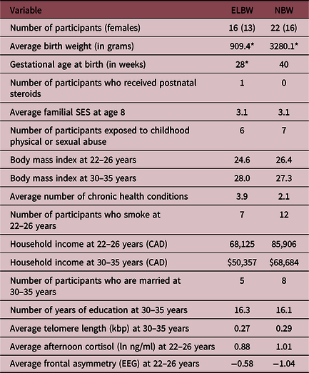
The characteristics of ELBW and NBW participants were compared using student’s t-tests (for continuous variables) and χ2 tests (for categorical variables) and are summarized in Table 1. We used linear regression models to evaluate the association of baseline afternoon salivary cortisol or resting frontal EEG alpha asymmetry (as predictors in separate models) upon telomere length stratified by each birth weight group. We also re-ran these models with a stress vulnerability measure × birth weight group interaction term to formally test a moderation effect of early-life stress exposure (ELBW status) upon the observed association between stress measure (either cortisol or frontal EEG asymmetry) and telomere length. Statistical analyses were conducted using IBM SPSS Statistics 22 (IBM SPSS Statistics, IBM Corporation).
Birth and sociodemographic characteristics of ELBW survivors and NBW control participants

ELBW, extremely low birth weight; NBW, normal birth weight.
Chronic health problems: a self-report measured defined as current non-psychiatric health problems lasting ≥6 months which was drawn from a 16-item list of common disorders including: visual problems, learning disabilities, asthma, heart disease, and diabetes.
Frontal asymmetry (EEG): positive values represent left asymmetry, negative values right asymmetry.
*Denotes statistically significant difference between ELBW and NBW groups at corresponding follow-up period (p = 0.05).
Results
Table 1 contains the characteristics of study participants. There were expected differences in birth weight, gestational age, and postnatal steroid exposure. Although there were no significant differences in the ratio of male/female participants between groups, there were significantly more female participants than males within each group, supporting our inclusion of participant sex as a covariate in adjusted analysis. After pooling ELBW and NBW participants into a single group, sex failed to moderate associations between cortisol and frontal asymmetry in the prediction of telomere length, and there were no statistically significant associations in either the whole group or birth weight grouped models when they were stratified by sex. There were no significant differences in exposure to childhood abuse or prevalence of smoking between groups; two common factors thought to influence telomere length. Likewise, although NBW controls had nominally greater household income at both timepoints, this difference was not statistically significant. There were no significant differences in telomere length, baseline afternoon salivary cortisol, or resting frontal EEG alpha asymmetry between birth weight groups. Importantly, there were no demographic differences, or differences in baseline afternoon salivary cortisol concentration/resting frontal EEG alpha asymmetry between participants who completed telomere measurement compared to those who did not, suggesting that there were no other significant predictors of attrition that might have influenced the observed results. Likewise, there was no evidence of attrition effects within each birth weight group. Although approximately 4% of the originally recruited study were non-Caucasian, after attrition, all participants in the presented analyses are Caucasian. Finally, among participants included in this study, there was a significant association between baseline afternoon salivary cortisol and resting frontal EEG alpha asymmetry (β = −0.46, p = 0.005), suggesting that increases in baseline afternoon salivary cortisol were associated with increases in resting right frontal EEG alpha asymmetry across all participants. As well, this modest relation also suggested these measures were possibly tapping similar phenomenon but were physiologically distinct measures of stress vulnerability. Exploratory analyses pooling all participants from ELBW and NBW groups revealed that after adjustment for all covariates, as well as birth weight group, neither cortisol (β = −0.21 p = 0.288) nor frontal asymmetry (β = −0.31, p = 0.133) at 22–26 years of age significantly predicted telomere length at 30–36 years of age.
Baseline afternoon salivary cortisol and telomere length
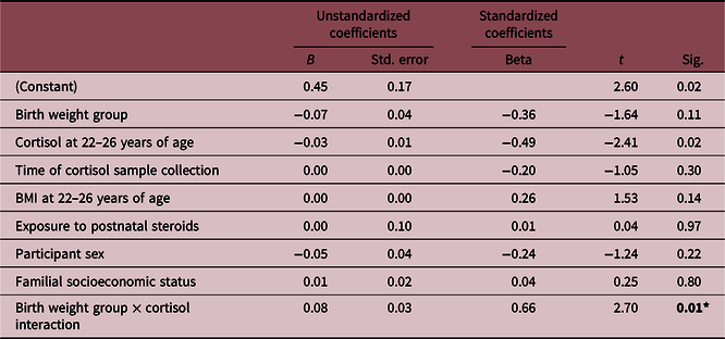
Before and after model adjustment for cortisol samples collection time, sex, BMI, postnatal steroid exposure, and familial SES, a statistically significant interaction was observed between baseline afternoon salivary cortisol concentration at 22–26 years of age and birth weight predicting telomere length at 30–35 years of age (β = 0.66, p = 0.012) (Table 2). These results were then stratified by birth weight group using one-way analysis of covariance (adjusting for all covariates). A negative association was noted between baseline afternoon salivary cortisol concentration and telomere length only among NBW control participants (β = −0.66, p = 0.019), such that as salivary cortisol values increased, telomere length decreased. No such association was observed among ELBW survivors (β = 0.45, p = 0.253).
Baseline afternoon salivary cortisol and telomere length in ELBW and NBW adults

ELBW, extremely low birth weight; NBW, normal birth weight.
*Denotes statistically significant effect (p < 0.05).
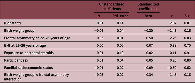
Frontal EEG alpha asymmetry at rest and telomere length in ELBW and NBW adults

ELBW, extremely low birth weight; NBW, normal birth weight.
*Denotes statistically significant effect (p < 0.05).
Resting frontal EEG alpha asymmetry and telomere length
Analyses of resting frontal EEG alpha asymmetry were performed using the same covariates listed previously, save cortisol sample collection time. After adjustment, the interaction term of this model was not significant (β = −0.34, p = 0.158) (Table 3). However, as we were also interested in evaluating the strength and direction of association between this stress predictor and telomere length within each birth weight group independently, we next stratified our analyses. Consistent with our cortisol findings, NBW controls exhibited a statistically significant association between greater right frontal asymmetry at rest (suggestive of increased stress vulnerability) at 22–26 years of age and shorter telomere length at 30–35 years of age (β = 0.48, p = 0.042). However, no association was found between frontal EEG asymmetry at rest and telomere length among ELBW survivors (β = −0.19, p = 0.651).
Discussion
Among NBW participants, two independent physiological markers of stress vulnerability, baseline afternoon salivary cortisol concentration, and resting right frontal EEG alpha asymmetry measured in early adulthood (22–26 years of age) predicted telomere length measured nearly a decade later (30–35 years of age). Although preliminary and limited by sample size, our results support previous research linking stress biomarkers with shortened telomere lengthReference Epel, Blackburn and Lin4,Reference Zalli, Carvalho and Lin48 and potentially extend these earlier findings by providing data on the long-term consequences of stress. The use of two reliable physiological measures of stress vulnerability also suggests that a link may exist between exposure to chronic stress and health and aging.
Pooling data from both birth weight groups failed to show associations between either stress vulnerability marker or telomere length, and contrary to our a priori hypothesis, associations were not observed between our stress vulnerability measures and telomere length among ELBW survivors. While these findings may be due to the limited statistical power associated with our small sample size, it also raises questions of whether survivors of prematurity may begin with longer telomeres to being with,Reference Vasu, Turner and George16 have telomeres that are less susceptible to shortening, or that ELBW individuals manifest unique pathways linking stress and telomere attrition.
While it is highly unlikely that ELBW survivors are exposed to less stress than their NBW peers, in keeping with the match/mismatch hypothesis,Reference Nederhof and Schmidt49,Reference Santarelli, Lesuis and Wang50 ELBW survivors could exhibit a unique stress response phenotype as a consequence of their exposure to early-life adversity. Certainly, they have observable differences in stress handling, highlighted by their propensity toward risk aversion (with lower rates of risk-taking, alcohol, and substance use), while still manifesting higher rates of anxiety and depression.Reference Van Lieshout, Boyle and Saigal23 In NBW samples, elevated circulating cortisol may reduce telomerase activity,Reference Choi, Fauce and Effros51 and severe early-life stress may reduce glucocorticoid receptor expression,Reference Daskalakis, Kloet and Yehuda52 potentially ‘uncoupling’ cortisol-mediated stress responses from telomerase activity. Alternatively, it may be that despite increased stress exposure, ELBW survivors have more specific phenotypic adaptations that increase telomere repair mechanisms such as telomere reverse transcriptase (hTERT) activity. Finally, our findings may be accounted for by ELBW infants being born with (or maintaining) longer telomeres in the months following birth, a difference which is not yet overcome by the fourth decade of life.Reference Vasu, Turner and George16 As telomere data were only available at a single timepoint, we do not know the average length of the participants’ telomeres at birth or telomere length contemporaneously with our stress measures at 22–26 years of age.
Although this study provides novel evidence linking stress vulnerability measures to telomere shortening in adulthood, as well as examining telomere length in ELBW adults for the first time, a number of important limitations warrant discussion. First, our small sample size limited our statistical power to detect interactions between the stress vulnerability measures and telomere length and reduces the precision with which we might estimate the magnitude of the effect. It would be of value for more contemporary cohorts with larger numbers of participants to attempt to replicate these exploratory findings in the future. Moreover, significantly more females than males were included in this study, which could further limit the generalizability of our findings. The subsample of 22–26-year-old participants who completed EEG and cortisol measures further constrained our sampling frame and could reduce the external validity of our findings. Therefore, replication of these findings with larger longitudinal studies is needed to more conclusively confirm the associations observed. Future studies may also consider exploring alternative measures of stress vulnerability (e.g., oxidative stress, circadian rhythm disruption) to generate a more comprehensive assessment of the links between physiological stress and telomere length. Given that we did not have historical biological samples for cohort members, we were unable to make telomere measurements contemporaneously with our stress vulnerability measures, and so it is difficult to determine whether stress led to accelerated rates of telomere shortening in the subsequent decade, or whether they already had shorter telomeres. Subsequent follow-up visits with this cohort will seek to compare physiological stress measures and telomere length at the same timepoint. Moreover, as this study utilized buccal cell samples to measure telomere length, it is possible that measurement in lymphocytes or other cell lineages may generate different values. However, there is evidence to suggest that good correlation exists between telomere length in different cell lines,Reference Gramatges and Bertuch40,Reference Finnicum, Dolan and Willemsen53 suggesting that the observed associations reported in this paper would be well conserved if telomere length measurements were performed using blood samples. For example, although we controlled for socioeconomic status at age 8 in an attempt to control for the prevailing social and environmental conditions of the participants’ childhood and adolescence, we did not control for SES at age 30–35. Finally, as this study was observational in nature, we cannot definitively establish causal associations between stress during childhood/adolescence/early adulthood and adult telomere length. Basic science research utilizing animal models could provide additional opportunities to establish whether these pathways are causal.
These preliminary data support the existence of an association between individual differences in stress vulnerability during early adulthood and reduced telomere length a decade later among individuals born at NBW, but not preterm. Limitations notwithstanding, these preliminary findings also raise new questions about the development of the physiological stress responses and their sequelae in preterm survivors, as well as their consequences for aging and cellular senescence in later life. It is important that these are addressed so that we can optimize the health of all adults.
Acknowledgments
None.
Financial Support
This research was supported by a Canadian Institutes of Health Research grant (CIHR, TMH-103145, awarded to Dr. Louis Schmidt).
Conflicts of Interest
The authors have no conflicts of interest to disclose.
Ethical standards
The authors assert that all procedures contributing to this work comply with the ethical standards of the Tri-Council Policy Statement (TCPS), Ethical conduct for Research Involving Humans, and with the Helsinki Declaration 1975, as revised in 2008, and have been approved by the Hamilton Integrated Research Ethics Board.