I. INTRODUCTION
Fabrication of nanostructured materials over an array of chemical compositions, shapes, and size with novel properties constitutes a progressively evolving branch of nanotechnology.Reference Nalwa1–Reference Gupta, Goel, Mehrotra and Kandpal3 It is the phenomenon of bottom-up directed growth of building blocks with nanoscale features to organize themselves into functional structures, as driven by the energetics of the system. More specifically, development of π-conjugated organic polymeric systems in one-dimensional (1D) forms, such as nanowires, nanofibers, nanorods, nanotubes, etc., has become one of the most potential and innovative area of research in the recent years.Reference Gupta, Goel, Mehrotra and Kandpal3–Reference Liu, Li, Xiao, Gan, Jiu, Li, Jiang, Zhu, Yu, Xiang and Chen5 These structures display excellent configurations for producing molecular wires/tubes, which act as fundamental units in nanodevices. Their unique size-dependent properties bestow them superior and indispensable characteristic properties that facilitate potential applications in nanotechnological fields like optics, electronics, optoelectronics, electrochemical, biological devices, etc.Reference Nalwa6, Reference Kaushik, Solanki, Ansari, Ahmad and Malhotra7 However, to manipulate molecules into desired structures, simple and novel chemical methodologies are required.
Till date, synthesis and characterization of many nanostructured organic polymers have been reported through different techniques so as to develop nanosystems of desired functionalities.Reference Goel, Gupta, Singh, Mehrotra and Kandpal8–Reference Maynor, Filocamo, Grinstaff and Liu10 An intriguing root to construct nanosystems has also been to deposit polymeric systems on suitable substrates in a spatially controlled fashion to improve their properties at nanodimensions. In this regard, various strategies like sol–gel,Reference Kaushik, Solanki, Ansari, Ahmad and Malhotra7 pulsed laser deposition,Reference Rebollar, Gaspard, Oujja, Villavieja, Corrales, Bosch, Georgiou and Castillejo11 Langmuir–Blodgett,Reference Lü, Rua, Ji, Ren, Cong and Zhao12 self-assembly,Reference Stamm and Sydorenko13 soft lithography,Reference Liu and Lee14 thermal evaporation,Reference Yong-Jun, Jian-Bao and Jin-Hui15 etc. have been developed in an overdrive mode to make smart materials exercising the knowledge of the characteristic properties of the nanodimensioned polymeric systems. These major advances have propelled the chemists into the unique position of gaining fundamental insights into the dynamics and reactivity of nanostructured systems and furthermore to exploit this knowledge for the bottom-up approach to nanofabrication, which promises to revolutionize future device miniaturization and electronics. Due to these reasons, convenient and cost-effective methods are being explored that could be directly and easily patterned on solid substrates. These substrates act as templates and assist in the fabrication of nanodimensioned structures of various polymers,Reference Madou16 with the aim to extend the applications of nanomaterials. In this way, the concrete problems, such as assembly and recycling of the synthesized nanostructures obtained through template and template-free routes, can also be overcome while manufacturing practical nanodevices.
Of the many organic systems, polypyrrole (PPY) has been an extensively investigated conducting polymer due to its outstanding material properties.Reference Yan, Zhang, Shen, Chen, Shi and Zhang17, Reference Goel, Mazumdar and Gupta18 PPY has attracted considerable interest of both the academic and industrial researchers, displaying wide range of applications in multidisciplinary fields, such as batteries, supercapacitors, microwave shielding, sensors, electrochromic devices, corrosion protection, etc.Reference Yan, Zhang, Shen, Chen, Shi and Zhang17, Reference Goel, Mazumdar and Gupta18 Besides the various chemicalReference Goel, Gupta, Singh, Mehrotra and Kandpal19, Reference He, Yang and Li20 and electrochemicalReference Akinyeye, Michira, Sekota, Al-Ahmed, Baker and Iwuoha21, Reference Jerome, Labaye and Jerome22 techniques applied for the synthesis of PPY nanostructures, few reports are also available that discuss the fabrication of PPY nanostructures on suitable substrates.Reference Wang, Mo, Ge, Tian, Wang and Wang23 Nevertheless, there is a persistent quest to develop better and less sophisticated fabrication routes to obtain novel PPY nanostructures for desired applications in nanotechnology. Direct fabrication of PPY nanostructures with controlled morphology, orientation, and surface architectures is still a significant challenge. Furthermore, gaining better control over the structure of matter has been a primary project of a scientific research. Improved strategies and combinations for obtaining PPY nanostructures with increased regularity in its polymeric backbone are important for enhancing a more defined 1D structure. Methods are being investigated that could control the size and shape of obtained nanostructures since the properties of assembled materials are strongly dependent on their intermolecular interactions and thus on their dimensional structures.Reference Goel, Gupta, Singh, Mehrotra and Kandpal8 This will further help in establishing a direct relationship between the “morphology” and the desired “material properties” of PPY. Such possibilities may as well authorize PPY as a highly potential candidate for specific technological purposes like static or magnetic shield coating, sensors and molecular devices, or modified electrodes.
In the present study, a simple and direct chemical route of fabricating 1D nanostructures of PPY on glass substrates is discussed using an in situ vapor phase chemical oxidative polymerization method. Using a similar strategy, Goel et al. have earlier reported the fabrication of 1D nanostructures of anthracene,Reference Gupta, Goel, Mehrotra and Kandpal3 polyaniline,Reference Gupta, Goel, Singh, Mehrotra and Kandpal24 polyindene, and polyindole on glass substrates.Reference Goel, Mazumdar and Gupta25 Analogous to the approach, herein, the effect of dopants has been explored to study the morphological characteristics of fabricated PPY nanostructures.
II. EXPERIMENTAL PROCEDURE
A. Materials used
All the chemicals were purchased from Sigma-Aldrich. Pyrrole (98%) was distilled under reduced pressure, before the initiation of polymerization reaction. Other reagents, such as the dopants like hydrochloric acid (HCl), ferric chloride (FeCl3), p-toluene sulfonic acid (p-TSA), camphor sulfonic acid (CSA), and polystyrene sulfonic acid (PSSA) and the solvents like methanol, acetone, and chloroform, were used as received. Carborundum powder having a grain size of 125 μm was used for grounding purpose.
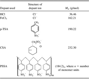
The chemical structures and molecular weights of different dopants involved in the fabrication process are given in Table I.
Chemical structures and molecular weights of the dopants used in the fabrication of PPY nanofibers.

B. Methodology
1. Preparation of substrates
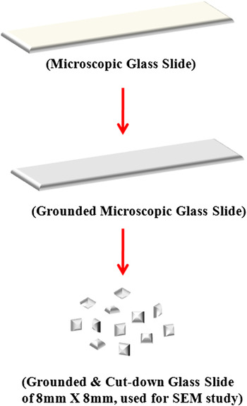
The microscopic glass slides ground on one surface (for 10 min) using carborundum powder were used as substrates for the fabrication. Grounding was done for firm adhering of reactant molecules to the substrate surface. Additionally, some of these ground glass slides were also cut down into the dimensions of 8 mm × 8 mm and were used for scanning electron microscopy (SEM). Figure 1 shows the diagrammatic representation of preparation of the microscopic glass slides for the fabrication process. Further, the slides were thoroughly washed with deionized water and different solvents like methanol, acetone, and chloroform. Finally, the ground slides were dried under vacuum before being used as substrates.
Diagrammatic representation of preparation of microscopic glass slides as substrates for fabrication process.

2. Fabrication process
Nanostructures of PPY doped with different dopants were fabricated on the prepared glass slides using an in situ vapor phase chemical oxidative polymerization method. The experimental setup involved in the fabrication process is reported in the previous works based on a similar study focused on polyindene and polyindole nanostructures.Reference Goel, Mazumdar and Gupta25
The reaction was conducted in a covered glass chamber, which was preheated in a furnace at a constant temperature of ∼180 °C for half an hour. For a particular set of fabrication process, pyrrole (∼3 mL) and the dopant (1.4 mmol) were mixed evenly in a watch glass. The watch glass was then placed at the bottom of the closed glass chamber. Few (two to three) ground microscopic slides were also set aside into the closed chamber. The small (8 × 8 mm microscopic ground slides) substrates were adjusted in a grooved aluminum plate and sited near the watch glass carrying the “pyrrole and dopant” mixture. The reaction chamber was placed in the furnace, maintained at ∼180 °C. The substrates were exposed to the reaction vapor for ∼15 min, after which all the substrates were removed from the reaction chamber, allowed to cool and finally kept in a desiccator.
C. Characterization
Light microscopy of all the fabricated PPYs was performed using Olympus BX 60 microscope having eyepiece magnifications of 20x and 50x. SEM was conducted using LEO 435VP variable pressure scanning electron microscope. The sample preparation for the SEM analysis was done by pasting the fabricated slides of PPYs on sampling troughs of aluminum stalks using carbon tapes, followed by covering them with a thin layer (10–20 Å) of sputtered gold.
III. RESULTS AND DISCUSSION
A. Light microscopy
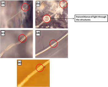
Primarily, the growing pattern of the PPY structures fabricated on the surfaces of glass slides was investigated using an optical microscope. The microscopic images of PPY structures fabricated on the glass surface in presence of different dopants are shown in Fig. 2. Figures 2(a)–2(e) represent the optical microscopic images for PPY–HCl, PPY–FeCl3, PPY–p-TSA, PPY–CSA, and PPY–PSSA, respectively. Formation of smooth tubular/fibrillar structures exhibiting straight fluorescent channels is observed in all the PPY micrographs.
Optical light micrographs of PPY doped with different doping agents (a) HCl, (b) FeCl3, (c) p-TSA, (d) CSA, and (e) PSSA.

It is examined that all the PPYs exhibit exclusively 1D nanostructures and are independent of the nature of dopant used in the polymerization process. Nearly, all the structures are straight, well elongated, and possess a uniform diameter individually. However, the dimensions (diameter) of these PPY structures are found to vary from dopant to dopant, subsisting in the respective PPY molecular matrixes. Differences in the diameters of the doped PPY structures can be attributed to the variations in the size, i.e., the bulkiness of the dopant ions incorporated into the respective PPY molecular chains during the polymerization process. These variations cause discrepancies in the thickness, which lead to the development of straight fibers to thick tubule-like PPY structures during the fabrication process. In other words, although the choice of dopant is not critical to the fabrication of 1D PPY structures, but it does affect their diameters.
Additionally, it is observed that temperature conditions and the duration of polymerization process have a profound influence in defining the shape and size of the fabricated structures. In the present study, the in situ vapor phase chemical oxidative polymerization process was carried out at 180 °C for 15 min only. Repeated experimental attempts have illustrated that by regulating the temperature and duration of the polymerization process, thick tubular to fine fibrous nano-/microdimensional forms of PPY can be obtained. Thus, the quality of polymeric systems depends on the arrangement of atoms present in their molecular chains, which in turn depends upon the fabrication conditions prevailing during the in situ polymerization reaction.
B. Scanning electron microscopy
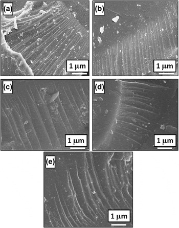
To closely investigate the morphology and estimate the size of the PPY structures so obtained, the SEM micrographs were collected. Figures 3(a)–3(e) show the SEM images of PPY–HCl, PPY–FeCl3, PPY–p-TSA, PPY–CSA, and PPY–PSSA nanostructures grown on glass substrate by in situ vapor phase chemical oxidative polymerization method, respectively.
SEM images of PPY nanostructures fabricated on glass substrate with different doping agents (a) HCl, (b) FeCl3, (c) p-TSA, (d) CSA, and (e) PSSA.

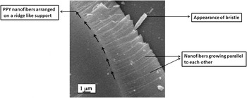
The images [Figs. 3(a)–3(e)] show the appearance of 1D nanofibers growing mostly parallel to the substrate—the etched glass surface. Formation of such arrays in a well-aligned and ordered manner is observed in all cases. These PPY fibers have an average diameter of 70 nm and lengths in micrometers. The nanofibers have a smooth surface and appear to be arranged in a comb-like fashion, emerging out of a common ridge-like support. A clear view of a typical SEM image of the fabricated PPY–dopant nanofibers showing such a brush-/comb-like growing pattern is shown in Fig. 4. Similar brush- or comb-like growing pattern of PPY nanostructures has been earlier reported by some researchers using nanoporous template membranes.Reference Yan, Zhang, Shen, Chen, Shi and Zhang17, Reference Demoustier-Champagne, Duchet and Legras26 Warren et al.Reference Warren, Walker, Anderson, Rhodes and Buckley27 have also reported hexagonal/octagonal PPY particles on glass slides having diameter ∼100 nm by vapor deposition. Very few reports are available for fabrication of well-aligned nanostructures of PPY without the support of template membranes.Reference Wang, Mo, Ge, Tian, Wang and Wang23 Fibrous morphology of PPY nanostructures has only been observed during electrochemical deposition on graphite electrode.Reference Wang, Mo, Ge, Tian, Wang and Wang23
A typical SEM image of PPY-doped nanofibers, depicting a brush-/comb-like growth pattern.

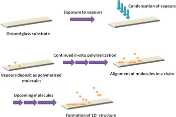
The key reason for the growth of ID PPY nanofibers on the glass substrate can be attributed to the gradual and consistent pace of the in situ vapor phase chemical oxidative polymerization reaction.Reference Goel, Mazumdar and Gupta25Scheme 1. depicts the growth mechanism of 1D PPY nanostructures fabricated on the glass substrate. The fabrication process undergoes in three steps in the following manner. First, pyrrole reacts with the dopant molecules, and concurrently, their vapors (polymerized product) are exposed to the glass substrate in the closed reaction chamber. In the second step, the vapors condense on the surface of glass substrate, and finally, a series of such condensed vapors adhere onto the substrate and form polymeric chains by their intermolecular interactions. It is the slow and constantly occurring polymerization reaction that plays a vital role in governing the growth of the fabricated structures. Since, in the vapor phase, the molecular concentration of polymer is low, consequently, only few nucleation sites are created on the surface of glass substrates. The driving force for 1D growth is determined by the small number of incoming atoms that could adhere at entire surface of the substrate and then migrate to the growing tip of the nanofibers.Reference Goel, Mazumdar and Gupta25 The regular and low speed reaction during the fabrication process is benefited in forming integrated and ordered structures on the glass surface. In other words, the gradual evaporation → condensation → adherement of polymeric molecules enables the molecules to adjust the orientation and organize the molecular chain in an orderly fashion. Therefore, better aligned and ordered arrangements of polymeric molecular chains are developed.
Growth mechanism of 1D PPY nanostructures on glass substrate.

The work highlights the use of commonly available “glass” as an important substrate for technical applications and its significant role in the development of nanodevices. The undertaken in situ polymerization reaction emerges as a simple and cost-effective method in contrast to the tedious and expensive routes to fabricate PPY nanostructures, which involve sophisticated techniques and/or instrumentation. Principally, the essence of the fabrication method is the self-alignment of the molecules that adhere with one another and propagate the polymeric chain in 1D, without the aid of any template membrane or use of any directing molecules like surfactants.
The glass substrates can directly be used for analysis of the infrared (IR) spectroscopic study of the fabricated structures. As such, these can act as substitutes for the conventional IR substrates for collecting information regarding the molecular properties of the fabricated structures. It is well known that there is an enormous advantage to collect information about the structural molecular properties of nanostructured systems through IR spectroscopy.Reference Goel, Gupta, Singh, Mehrotra and Kandpal8, Reference Goel, Mazumdar and Gupta25, Reference Goel28 Generally, to access the spectral details of a system in the mid-infrared range, a material, which is insoluble in water and IR transparent to the spectral range, is required for the sampling purpose.Reference Goel28 These requirements are met only with specialized expensive salt windows, e.g., calcium fluoride, barium fluoride, or potassium bromide. However, reports are available in literature that confer that the ordinary glass may in principle be used as a substrate for IR measurements in this region.Reference Shaw, Eysel, Liu and Mantsch29 The results are much accurate and compete with those achieved using conventional mid-infrared substrates. Besides, the investigations have been applied as an alternative for the clinical and/or diagnostic fields.Reference Shaw, Eysel, Liu and Mantsch29
The method can be further applied for developing convenient and economical nanodevices and/or functional glass for sensing, optoelectronic, and photocatalysis. Characterization of the structural chemical properties of the fabricated structures before and after a suitable chemical modification reaction can accomplish such functions. For instance, the author has reported application of anthracene-based nanostructures fabricated on glass substrate for sensing purpose by analyzing the spectral characteristics after exposure to NH3 vapors.Reference Gupta, Goel, Mehrotra and Kandpal3 Anthracene-based nanostructures fabricated on glass substrate by the reaction of anthracene 9-carboxylic acid with calcium oxide through chemical vapor deposition technique were characterized using Fourier transform infrared spectroscopy, nuclear magnetic resonance spectroscopy, and gas chromatography–mass spectroscopy.Reference Gupta, Goel, Mehrotra and Kandpal3 IR studies of the nanostructures revealed the presence of C = O groups at 1725 cm−1, characteristic to the six- to seven-membered ring ketones. However, the structural properties monitored after exposure to NH3 vapors, indicated strong covalent modification in the spectral profile of the nanostructures. Exposure to the vapors led amidation of the carbonyl groups, which was clearly indicated by the presence of IR band at 1680 cm−1. In addition, strong N–H stretch band was observed at 3300 cm−1 in contrast to the O–H stretch band at 3414 cm−1before derivatization. The glass substrates may also provide greater surface area and facilitate the interaction of chemical molecules with the nanostructured systems, thereby become an alternative material with improved sensitivity and fast response behavior. Likewise, entrapment of solar energy rays by the fabricated nanostructures can provide enhanced absorbed energy and may be exploited for solar energy and other similar systems.
IV. MECHANISM
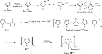
The proposed mechanism pathway for the formation of different doped PPY nanostructures is shown in Scheme 2. The mechanism for the formation of PPY follows that a neutral pyrrole molecule undergoes oxidation in the presence of the dopants and generates its radical cation through electron transfer process.Reference Goel, Mazumdar and Gupta18 The pyrrole radical cation combines with consecutive radical cation, followed by rearrangement, and results in a dimer of pyrrole. The dimer is oxidized to form a new radical cation, which reacts either with a radical cation monomer or a radical cation dimer to form a trimer or tetramer, respectively. The process is repeated several times until a positively charged molecular chain of PPY is formed.Reference Goel, Mazumdar and Gupta18 Consequently, the respective negatively charged dopant ions attach at the positive sites of the PPY backbone as dopants and form series of counter anions integrated into the PPY matrix.Reference Goel, Mazumdar and Gupta18 Thus, the different dopants facilitate both the oxidation and the doping process.
Proposed mechanism for the formation of PPY doped with different dopant ions.

The different dopants of varying structures and ionic size shall exercise a significant influence on the characteristic features of the fabricated nanostructures and their potential application in the nanotechnological areas. Among the different conducting polymers, PPY holds an outstanding position and is known for its intrinsic conducting properties. The unique optical and electrical properties exhibited by the nanostructured polymeric systems like PPY are definitely a consequence of their low dimensionality and quantum confinement effect that enables them to display outstanding applications in areas of nano-optics and nanoelectronics. Introduction of different types of dopants in the nanostructured PPY is expected to further improve its optical and electrical properties and enhance applications in different fields. Additionally, conductivity significantly varies as a function of dopant occurring in the molecular matrix of a polymeric system.Reference Liu, Li and Zhu2 The size of the doped ion also has a significant effect on the structure of the molecular chains of the conducting polymer and its conjugation length. The double charged doped ions have much more effect on the molecular structure than the single charged doped ions. The high charge distorts the polymeric chains and disturbs its conjugate. Such studies require careful investigations before being applied to the nanotechnological devices.
V. CONCLUSIONS
In summary, a simple and convenient in situ vapor phase chemical oxidative polymerization method has been explored to fabricate 1D nanostructures of PPY on the surface of glass substrates in presence of different dopants. The study reports brush-like growing pattern for the PPY nanofibers, which is not very commonly reported in the literature without the aid of template membranes and/or sophisticated techniques or surfactants. The fabrication method affords well-defined smooth surfaced PPY nanofibers, which may find applications in developing practical and economical nanodevices and/or functional glass for sensing, optoelectronic, photocatalysis, and solar energy systems.
The chemical structure and size of dopant ion shall exercise significant influence on the optical, electrical, and conductive properties of the fabricated PPY nanofibers and their potential application in the nanotechnological areas. The work contributes to the existing knowledge of chemical routes available for the fabrication of 1D nanostructures of PPY on desired substrates. It merits further detailed investigation and outlines technological applications of fabricated PPY nanofibers in the nanodevices where consistency and quality solely depend upon the structural and morphological characteristics of the polymer.
ACKNOWLEDGMENT
The author is grateful to Dr. Ranjana Mehrotra (National Physical Laboratory, New Delhi) for providing the research facilities.