Implications
A comprehensive description of all genes expressed in mammalian oocytes important for affecting ‘oocyte competence’ is still pending. The present work reports identification of some unknown transcripts identified in a buffalo oocyte-specific subtracted cDNA library, which were found to be expressed either exclusively or preferentially in oocytes up to several folds higher than other tissues examined. The high abundance pattern of these transcripts selectively in oocytes suggests that some of these could have a link with oocyte development and thereby have potential as markers of fertility. Data generated in this study will aid future investigation into poor reproductive rate and compromised success rate of assisted reproductive technologies in buffalo.
Introduction
Maturation of mammalian oocytes presents a protracted developmental process whereby an undifferentiated female germ cell prepares itself for undertaking the process of fertilization and guides the newly formed zygote through early developmental stages until the embryo's own genome becomes eligible to undertake its own transcription (Yamashita et al., Reference Yamashita, Mita, Yoshida and Kondo2000; Zuccotti et al., Reference Zuccotti, Merico, Cecconi, Redi and Garagna2011). Transcriptionally, the period of oocyte maturation displays a typical cycle of mRNA synthesis and degradation starting with a very high turnover rate, which goes silent toward the end (Tomek et al., Reference Tomek, Torner and Kanitz2002). A very fundamental yet practically relevant question that still remains unresolved is ‘what makes an oocyte competent’? The ability to select the most competent oocytes out of a pool of oocytes will contribute to the success rate of many assisted reproduction technologies (ARTs) procedures awaiting large-scale implementation for genetic improvement of livestock, as well as for identifying new drug targets for infertility management (Armstrong, Reference Armstrong2001).
Recent published literature suggests that discovery of novel transcripts in oocytes is still an open research agenda (Klatsky et al., Reference Klatsky, Wessel and Carson2010). Continued identification of new oocyte-specific genes and their functional annotation is providing additional insight into the molecular mechanism involved in folliculogenesis, oogenesis, fertilization and early embryogenesis (Bettegowda et al., Reference Bettegowda, Yao, Sen, Li, Lee, Kobayashi, Patel, Coussens, Ireland and Smith2007; Wells et al., Reference Wells, Alfarawati and Fragouli2008; Luo et al., Reference Luo, Kleemann, Ashraf, Shaw and Murphy2010). Several techniques such as differential-display reverse transcription-PCR, microarray hybridization, serial analysis of gene expression and cDNA library construction have been used to reinforce the inventory of oocyte-expressed genes. Each of these techniques, however, suffers from the disadvantage of biased detection of more abundant transcripts and often misses rarely or lowly expressed transcripts. Suppressive subtractive hybridization (SSH) provides an attractive solution to identify rare transcripts, which may encode regulatory proteins and have crucial cellular role (Zeng and Schultz, Reference Zeng and Schultz2003). The advantage of this method stems from its ability to normalize the mRNA population so that abundant mRNAs are reduced, whereas rare transcripts are enriched (Robert et al., Reference Robert, Barnes, Hue and Sirard2000).
Buffalo (Bubalus bubalis) is an economically important dairy animal species in many Asian, African and Mediterranean countries because of its immense potential in agriculture, milk and meat production (Nanda and Nakao, Reference Nanda and Nakao2003). Poor reproductive efficiency of buffalo, however, is a major hindrance toward implementing planned breeding improvement programs in this species. Late maturity, poor estrus expression, prolonged calving intervals, seasonal reproductive patterns and low conception rates directly affect the propagation of good germplasm in buffalo (Nandi et al., Reference Nandi, Raghu, Ravindranatha and Chauhan2002; Drost, Reference Drost2007). Although several ARTs have been demonstrated in this species, the overall success rates of these techniques remain low, thus constraining their widespread adoption (Singh et al., Reference Singh, Chauhan, Singla, Gautam, Verma, Manik, Singh, Sodhi and Mukesh2009). Delineating the exact cellular mechanism of oocyte function and identification of new genes in oocytes of this species would not only help to understand the buffalo oocyte biology better, but would also provide markers to study the oocyte competence phenomena in any species. The present study was undertaken to identify developmentally important genes expressed in buffalo oocyte during the maturation process using a complementary approach of SSH and customized cDNA macroarray analysis.
Material and methods
Buffalo oocytes and somatic tissues
Buffalo ovaries were collected from the abattoir and cumulus oocyte complexes (COCs) were aspirated from all visible follicles irrespective of the follicular diameter. COCs were classified as A, B and C grades based on cumulus cell and ooplasm characteristics as described by Kumar et al. (Reference Kumar, Verma, Roy, Rajput, Ojha, Anand, Yadav, Arora, De, Goswami and Datta2011). About 200 COCs under each grade were cultured at 38.5°C under 5% CO2 in TCM-199 HEPES-modified (Sigma, St. Louise, MO, USA) medium supplemented with 10% fetal bovine serum, 0.01% sodium pyruvate, 0.005% glutamine, 0.005% streptomycin, along with 5 μg/ml follicle-stimulating hormone(FSH-p) and 10 μg/ml leutinizing hormone. Batches of COCs were retrieved at 0, 8, 16 and 24 h of in vitro maturation culture and denuded completely by vortexing for 3 to 5 min. After washing, all oocytes were pooled (2400 oocyte) and stored at −80°C in 500 μl Trizol (Invitrogen, Carlsbad, CA, USA). Isolated cumulus cells were collected separately after washing three times with 1× phosphate-buffered saline and preserved in Trizol at −80°C. Buffalo somatic tissues (heart, kidney, intestine, liver, lung, muscle and spleen) were also collected at the abattoir in RNA later (Ambion, Drive Foster, CA, USA) and stored at −80°C till RNA isolation.
RNA extraction
Total RNA was isolated from somatic tissues (lung, kidney, liver, heart, intestine, spleen and cumulus cells) and the oocyte pool using Trizol protocol as per the manufacturer's instructions. The concentration and purity of the isolated RNA was determined using a NanoDrop ND-1000 spectrophotometer and integrity of the total RNA was assessed using a Bio-Analyzer 2100 (Agilent, New Delhi, India). RNA samples with RNA integrity number values of 7 and above were processed further for library preparation. Equal amounts of total RNA from each somatic tissue were pooled and adjusted to a final concentration of 0.5 mg/ml (used as ‘driver’ in the SSH protocol). RNA isolated from oocytes was precipitated using 1 μg glycogen to obtain a final concentration of 0.5 mg/ml and used as a ‘tester’ RNA. Total RNA (2 μg) isolated from oocytes and somatic tissues were incubated with DNase-I (Fermentas, Burlington, ON, Canada) to remove contaminating genomic DNA and purified by organic extraction (Chomczynski and Sacchi, Reference Chomczynski and Sacchi1987). After DNase-I treatment, the genomic DNA contamination was negated by buffalo genomic DNA-specific primer amplification with the 10 ng of RNA from each extracted samples.
Construction of cDNA library
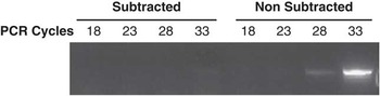
Equal quantity (1 μg) of the total RNA isolated from the oocytes (tester RNA) and somatic tissues (driver RNA) were processed simultaneously for cDNA synthesis using the Super SMART PCR cDNA synthesis kit (Clontech, Mountain View, CA, USA) following manufacturer's instructions. Synthesized cDNA was amplified by long-distance PCR (LD-PCR) to enrich full-length cDNA population. It was followed by RsaI digestion of tester and driver double-stranded (ds)-cDNA to prepare blunt-end cDNA for SSH experiments. In the next step, SSH was performed to subtract the oocyte cDNA (tester) with excess of cDNA from somatic tissues (driver) using the PCR-Select cDNA subtraction Kit (Clontech) as per the manufacturer's instructions. After SSH, the resultant primary and nested PCR products were amplified for 27 cycles. Diluted PCR products (10×) were further used for secondary amplification for 10 cycles to obtain the subtracted cDNA, representative of oocyte transcripts only. The subtraction efficiency was analyzed by PCR amplification of the subtracted and non-subtracted cDNAs using the constitutively expressed gene GAPDH using buffalo-specific primers (forward 5′CCAGAATATCACCCCTGCTT3′ and reverse 5′CCCAGCATCGAAGGTAGAAG3′). The PCR amplification was performed for 18, 23, 28 and 33 number of cycles with thermal conditions of 95°C for 2 min followed by ‘n’ number of cycles at 95°C for 15 s, 56°C for 30 s, 72°C for 30 s and final incubation at 72°C for 5 min. Five microliters of PCR product was collected at 18, 23, 28 and 33 cycles and analyzed on 2% agarose gel (Figure 1). The secondary PCR products obtained after the SSH experiment was purified using Qiagen column, cloned into pGEMT easy vector (Promega, Madison, WI, USA) and transformed into XL-Blue Escherichia coli competent cells (Invitrogen) to establish an oocyte-specific subtracted cDNA library. Transformed colonies were randomly picked up and screened for the recombinant clones by colony PCR using a vector-specific M13 primer. PCR was performed in 50 μl at 95°C for 3 min followed by 30 cycles of 95°C for 30 s, 50°C for 15 s and 72°C for 1 min 30 s, and finally incubated at 72°C for 10 min. Cloning efficiency was analyzed by running 10 μl of PCR product on 1.5% agarose gel electrophoresis. Clones with a minimum inset size of 200 bp were processed for sequencing.
Amplification results of GAPDH from subtracted and non-subtracted cDNA at respective PCR cycle numbers.

Sequence analysis of the cloned expressed sequence tags (ESTs)
Putative identity of the sequences was determined using DNA (megablast, blast-n, t-blastx) and protein (blastx) sequence search tools against several databases (Nucleotide, ESTs, RefSeq RNA, PDB, Swiss-Prot, Genome) for different mammalian species (human, bovine, ovine, murine) available at National Center for Biotechnology Information (NCBI; http://www.ncbi.nlm.nih.gov) using e-value 10−2 to 10−5. On the basis of the results, sequences were classified as known, uncharacterized, unknown and novel sequences. Known sequences were defined as cases where a high degree of the homology was found with already identified genes available in mammalian databases. Sequences that did not match with known genes but possessed homology with EST databases were grouped as uncharacterized, and those that did not show any significant similarity with nucleotide and the EST database but showed some similarity with unannotated genomic DNA in bovine (considered homologous species to buffalo) were classified as unknown. Novel ESTs were designated as the cases where no match could be established using different genomic and EST databases available. Sequences included in known, uncharacterized and unknown categories were further analyzed against Bos taurus genome database available at NCBI to search the 5′untranslated region (UTR), 3′UTR, poly-A tail and other structural elements. Potential open reading frames (ORFs) in the identified sequences were searched for all the unknown and novel sequences using Geneious Pro (Drummond et al., Reference Drummond, Ashton, Cheung, Heled, Kearse, Moir, Stones-Havas, Thierer and Wilson2007) and sequences having more than 200 bp ORF length were considered as putative coding RNA. Analyzed sequences were submitted to the EST database at NCBI GenBank.
Nonradioactive labeling of cDNA
Both oocyte- and tissue-specific nonradioactive Digoxigenin (DIG)-labeled probes were synthesized using the super smart cDNA synthesis kit (Clontech) with minor modifications. In brief, 1 μg each of oocyte and somatic tissue RNA were used as templates to prepare full-length cDNA. The purified cDNAs were used as templates to generate the DIG-labeled ds-cDNA probe from oocytes and somatic tissues using LD-PCR amplification. Total 100 μl PCR reaction mixture was prepared containing 120 ng of ss-cDNA, 10 μl of 10× PCR buffer (Clontech), 10 μl of 10× PCR DIG probe synthesis mix (Roche, Mississauga, ON, Canada), 10 μl of 10× deoxyribonucleotide triphosphate (dNTP) stock solution (Roche), 2 μl of 12 μM 5′ PCR primer-IIA (Clontech) and 2 μl of 50× advantage cDNA pol mix (Clontech). Identical PCR reactions were carried out with cDNA from both oocytes and somatic tissues. The PCR mixture was subjected to a total of 30 cycles using thermal cycles of 95°C for 1 min, followed by 10 cycles at 95°C for 20 s, 65°C for 30 s, 68°C for 3 min and remaining 11 to 30 cycles were performed at 95°C for 20 s, 65°C for 30 s, 68°C for 3 min plus additional 40 s for each successive cycle and final incubation at 68°C for 7 min. Labeled ds-cDNA were purified using size exclusion column chromatography (Clontech) to remove the unincorporated DIG-labeled dNTPs.
cDNA macroarray hybridization
A custom cDNA macroarray was prepared for all the identified and nonredundant clones. For this, the inserts were amplified using M13 primer (as described above) and purified using QIAquick PCR purification kit (Qiagen, Valencia, CA, USA). Each purified PCR products (10 ng) were blotted in duplicate onto Hybond N+ membrane (GE healthcare, Gurgaon, Haryana, India) using vacuum slot blot as per the manufacturer's instructions. The blotted DNA was cross-linked to the membrane using UV linking under 70 000 μJ/cm2 followed by baking at 80°C for 2 h. Housekeeping genes, GAPDH and ACTB, were also blotted on the same membrane as positive controls. Custom cDNA macroarrays were hybridized with DIG-labeled cDNA probes generated from either oocytes or somatic tissue RNA. Prehybridization (to reduce the background) and hybridization of the labeled probe were performed using the standardized protocol (Sambrook et al., Reference Sambrook, Fritsch and Maniatis1989) and were immunodetected using the DIG labeling detection system (Roche) according to the manufacturer's instructions. Images obtained after hybridization of the probe generated from oocyte and somatic tissues were recorded on X-ray film and data were compared between oocytes and somatic tissue by quantification of the signal using Image-J software.
Reverse transcription (RT)-PCR
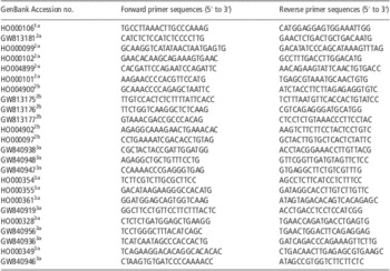
RT-PCR was performed to validate the results obtained from macroarray hybridization-based differential screening. In total, 24 clones with more than 10-fold overexpression in oocytes as compared with somatic tissues were selected to design sequence-specific primers (Table 1). For cDNA synthesis, 500 ng of total RNA each from oocytes and pooled somatic tissues were used to synthesize single-strand cDNA using SMART scribe reverse transcriptase (Clontech). After purification, 10 ng of oocyte cDNA and 100 ng of somatic tissues cDNA were used as templates in RT-PCR analysis of selected transcripts. The expression status of the identified novel gene (BOE283N) was studied independently in different somatic (cumulus, heart, lung, kidney, spleen, intestine) and reproductive tissues (oocyte, testis, COC) using equal quantities of cDNA (100 ng). PCR reaction was performed on each cDNA sample under the following conditions: 95°C for 2 min; 30 cycles at 95°C for 30 s, 58°C for 15 s and 72°C for 20 s; and then 72°C for 5 min.
Primers used for RT-PCR analysis of 24 genes selected for confirming their oocyte-specific expression

RT-PCR = reverse transcription-PCR; EST = expressed sequence tags; ORF = open reading frame.
aESTs having more than 200 bp ORF; bESTs without ORF.
1Novel EST.
2Unknown EST.
3Uncharacterized EST.
Results
Validation of the SSH library
An average of 4.2 μg of total RNA was recovered from a pool of 2400 oocytes (1.75 ng/oocyte). Total RNA (1 μg) isolated from oocyte and pool of somatic tissues was subjected to ds-cDNA synthesis and amplification following LD-PCR. SSH was performed with good-quality amplified cDNA and subtraction efficiency was evaluated by detecting the housekeeping gene (GAPDH) in both subtracted and non-subtracted cDNA pools. The PCR product of GAPDH appeared to be detectable after amplification of 33 PCR cycles with subtracted cDNA compared with 28 cycles without subtraction. This indicated a reduction of GAPDH transcript by more than 30-fold after subtraction (Figure 1). After cloning and transformation, more than 2000 clones from the subtracted library were amplified by PCR using M13 primers against the vectors. This procedure revealed that 1875 clones from the bovine library were positive insert-containing clones. The insert size of the clones ranged from 0.1 to 2.0 kb, and the average fragment insert size was ∼500 bp, which is in agreement with that statistically predicted by RsaI digestion.
Sequence analysis of SSH cDNA clones
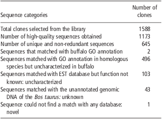
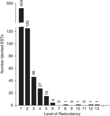
Table 2 presents a summary of sequence results from our cDNA library. A total of 1173 high-quality sequences were obtained, which were found to represent a maximum of 645 unique sequences based on clustering analysis results. Approximately 85% of the genes were present once or twice in the library, confirming effective normalization of the subtraction procedure used (Figure 2). The level of redundancy in the library was found to be ∼45%. Further, putative identity of the 645 unique sequences were determined using BLAST search against NCBI databases and classified them in known, uncharacterized, unknown and novel categories. BLAST analysis revealed that 77.21% (498) of the unique sequences with significant similarity with known genes, 15.97% (103) matched with functionally unannotated ESTs of bovine, ovine and murine origin, and hence were classified as uncharacterized sequences. About 6.5% (43) of sequences did not match with any nucleotide or EST databases but showed similarities with unannotated sequences from the bovine genome and thus were classified as unknown sequences. One sequence was found to be novel, as it did not match with any reported sequence available in GenBank.
Summary of the sequences generated from oocyte-specific cDNA library
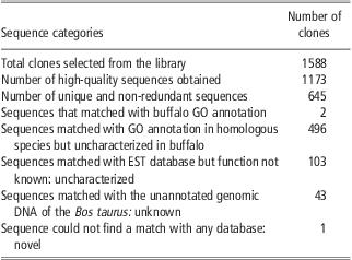
GO = gene ontology; EST = expressed sequence tags.
Frequency distribution of assembled expressed sequence tags (EST) sequences obtained in cDNA library representing the normalization effect of suppressive subtractive hybridization procedure and level of redundancy in cDNA library. The X-axis represents the number of times the same sequence is represented in the library.

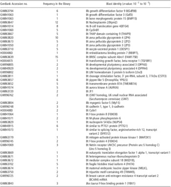
On the basis of the information available from GenBank database, we observed that for the cDNA corresponding to known genes (498) in our subtracted library 69% of the cDNA contained UTRs at 5′ or 3′ end. A majority of these clones had a 3′ UTR region (51%) and a smaller number had a 5′ UTR region (18%). Confirmation of the absence of housekeeping genes (GAPDH and ACTB) and detection of several oocyte- and embryo-specific genes like zona pellucida genes (zp1-4); gdf3 and 9, bmp15, jy1, btg4, npm2, nalp14, osp1 (Table 3) in subtracted oocyte cDNA pool authenticated the quality of our SSH library.
Identified oocyte-specific genes in our library that are involved in different stages of oocyte and preimplantation embryo development in bovine, human, ovine and murine

Functional annotation of ESTs
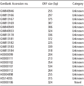
Functional annotation of identified known genes was carried out using the gene ontology (GO) program as per panther classification system (http://www.pantherdb.org). Identified genes were classified into three categories of cellular function, biological process and cellular component (Figure 3). It was observed that in cellular function category, the most abundant sequences were associated with binding (35.8%) and catalytic activity (29.3%). For biological process, the most frequent process identified was the cellular and metabolic process (46%) followed by cell communication (9.0%) and cell cycle (8.1%). The most frequent cellular components identified were intracellular (56.2%) in which a majority of the sequences were associated with cytoskeleton. Eighteen unknown and the novel ESTs in the present library were confirmed as having a potential ORFs with a minimum length of 200 bp (Drummond et al., Reference Drummond, Ashton, Cheung, Heled, Kearse, Moir, Stones-Havas, Thierer and Wilson2007; Table 4).
Functional gene ontology of identified annotated sequences represented in the library categorized as ‘Cellular functions’ ‘Cellular components’ and ‘Biological process’.

List of the EST (18 unknown and 1 novel) with >200 bp ORF predicted
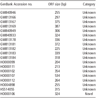
EST = expressed sequence tags; ORF = open reading frame.
Screening of ESTs for oocyte specificity by cDNA macroarray
The results of differential screening of the ESTs in oocyte v. somatic tissues are presented in Figure 4. Most of the 645 spotted PCR products of the selected clones were observed to yield hybridization signals with the probe made from the pool of oocytes RNA (Figure 4a). Signals obtained from equivalent spots hybridized with probe made from other somatic tissue cocktail are presented in Figure 4b. In either case, the signal intensity of each spot was normalized against that of GAPDH and was compared between oocytes and somatic tissues. Differential signal intensity of a majority of spots hybridized with oocyte v. tissue probe suggested that about 21% of these are exclusively oocyte-expressed genes, whereas the remaining genes were expressed preferentially in oocytes. Only a few spots (16) were found positive for both the tissue and oocyte blots and hence could be designated as false positives. The group interpreted as exclusively oocyte-expressed genes represented already known oocyte-specific genes such as GDF3, GDF9, BMP15, JY1, NPM2, NALP14, OSP1, zp2, zp3 and zp4. A summary of the preferentially expressed known ESTs are provided in Table 5.
Hybridization signals from selected clones using probes made from the pool of oocytes (a) and the pool of somatic tissues (b). The housekeeping gene GAPDH is in the white circle and ACTB in the white square. False positives are represented by black dotted circles and the genes highly abundant in oocyte are in black circles.

Summary of the known ESTs showing more than 5-fold preferential expression in oocyte v. somatic tissue

EST = expressed sequence tags; Bb = Bubalus bubalis; Bt = Bos taurus; Hs = Homo sapiens.
RT-PCR validation of oocyte-specific ESTs
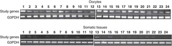
Subsequent to differential screening of identified ESTs by cDNA macroarray hybridization, the next level of validation was undertaken to confirm their oocyte-specific expression using a list of 24 representative genes, which revealed at least 10-fold higher expression in oocytes than other tissues. These 24 genes included 12 uncharacterized, 11 unknown and the isolated novel gene. The comparative expression profile of these ESTs in oocytes v. somatic tissues confirmed high specificity of these ESTs in oocytes (Figure 5). As a strategy to completely negate the possibility of false-positive interpretation about their oocyte-specific expression, we used 10-fold excess cDNA from somatic tissues than that from oocytes while setting up the RT-PCR assay. Results revealed that at least eight ESTs (five uncharacterized and three unknown) were expressed exclusively in oocytes and the remaining ones were barely detectable in a pool of somatic tissues. Interestingly, the novel gene (BOE283N) was found to be expressed only in oocytes and testis and no other somatic tissues tested (Figure 6).
Differential expression status of 24 unknown genes in oocytes v. somatic tissues. Lane 1: identified novel gene, Lanes 2 to 12: uncharacterized genes and Lanes 13 to 24: unknown genes.

Expression pattern of the novel expressed sequence tags (BOE283N) in different somatic and reproductive tissues. O = oocytes; Cu = cumulus; COC = cumulus oocyte complex; Te = testis; He = heart; Lu = lung; Ki = kidney; In = intestine; and Sp = spleen.

Discussion
Over the last decade, vast insight has been gained about molecular events involved in oocyte function in several mammalian species, by identifying genes preferentially expressed in oocytes (Bettegowda et al., Reference Bettegowda, Yao, Sen, Li, Lee, Kobayashi, Patel, Coussens, Ireland and Smith2007; Gaska et al., Reference Gaska, Pellestor, Assou, Loup, Anahory, Dechaud, De Vos and Hamamah2007; Baillet et al., Reference Baillet, Pépin, Cabau, Poumerol, Pailhoux and Cotinot2008; Parrish et al., Reference Parrish, Siletz, Xu, Woodruff and Shea2011). In spite of their economic importance in dairy production, however, buffalo remains a relatively unexplored species particularly in terms of explaining their reproduction disadvantages. The genomic and EST database of this species also remains grossly incomplete. The present study targeted isolation and identification of oocyte-expressed transcripts in buffalo, which might help in understanding the typical molecular mechanism of oocyte maturation and to discover new oocyte-specific genes, which will enrich knowledge about the reasons for reproduction constraints in this species.
Different strategies have been used in the past to isolate differentially expressed tissue-specific genes under given biological situations. In recent times, several high-throughput sequencing methods have evolved, which allow reading millions of nucleic acid sequences in short time, thereby expanding the scope of gene discovery beyond what was possible with standard dye terminator methods (Schuster Reference Schuster2008; Tucker et al., Reference Tucker, Marra and Friedman2009). However, SSH as a method was chosen in the current study as it does not require a priori sequence knowledge and can be used with a low quantity of RNA obtained from less abundant tissues such as oocytes and embryos. To increase the possibility of oocyte-specific gene discovery, cDNA library was generated by using tester mRNA isolated from a carefully selected pool of oocytes representing all morphological types and physiological status, and it was subtracted by the driver mRNA prepared from a pool of somatic tissues representing diverse cell types. Testis was purposely not included among the driver population so that ESTs specific to germ cells of both sexes are represented in our library. In the analyzed sequence repository, we observed that the chance of obtaining a new independent clone was more than 50% even after sequencing of 800 clones. This confirmed the efficiency of cDNA subtraction strategy used in the present study, which was better than reported earlier (Hennebold et al., Reference Hennebold, Tanaka, Saito, Hanson and Adashi2000). The custom macroarray approach used in the present study represents a quick and simple method of high-throughput screening of tissue-specific cDNA libraries. It was observed that the intensity of the hybridization signals of individual ESTs was positively correlated with its level of redundancy reflecting the fidelity of subtracted cDNA library construction (Sahu and Shaw, Reference Sahu and Shaw2009). Large-scale sequence analysis data lead us to suggest that the subtraction method followed in generating the SSH library was potent enough to exclude common genes in oocytes and other somatic tissues. False-positive cases in the library were only about 2% (16 out of 645).
In this study, we identified the 496 known, 103 uncharacterized, 43 unknown and 1 novel EST in our library, which are not previously reported to be expressed in buffalo oocytes and submitted these to the EST database (Table 2). On the basis of the GO annotation (of the identified known ESTs), it was determined that ∼94% of transcripts detected in our library were involved in crucial molecular events associated with oocyte and preimplantation embryonic development in different mammalian species such as bovine (Dalbie's-Tran and Mermillod Reference Dalbiès-Tran and Mermillod2003; Fair et al., Reference Fair, Carter, Park, Evans and Lonergan2007; Ghanem et al., Reference Ghanem, Hölker, Rings, Jennen, Tholen, Sirard, Torner, Kanitz, Schellander and Tesfaye2007; Salilew-Wondim et al., Reference Salilew-Wondim, Rings, Holker, Gilles, Jennen, Tholen, Havlicek, Besenfelder, Sukhorukov, Zimmermann, Endter, Sirard, Schellander and Tesfaye2007), ovine (Baillet et al., Reference Baillet, Pépin, Cabau, Poumerol, Pailhoux and Cotinot2008), human (Assou et al., Reference Assou, Anahory, Pantesco, Le Carrour, Pellestor, Klein, Reyftmann, Dechaud, De Vos and Hamamah2006) and mice (Su et al., Reference Su, Sugiura, Woo, Wigglesworth, Kamdar, Affourtit and Eppig2007). Identified genes were also classified as oocyte-specific genes involved in folliculogenesis, oogenesis, fertilization, zygotic genome activation and preimplantation embryo development (Table 3). Sequence analysis of identified unknown and novel ESTs revealed that at least 18 of these were matching with the intergenic part of the bovine chromosomes and presented a poly-A tail with potential ORF, which qualified them as the protein coding transcripts. The remaining unknown ESTs with poly-A tail of more than 200 bp size but having no apparent coding clue can be speculated as potential oocyte-expressed long noncoding RNA, which have recently been suggested as the key regulators of embryogenesis (Pauli et al., Reference Pauli, Rinn and Schier2011). Detailed sequence analysis of the identified novel EST revealed 324 bp continuous ORF encoding a 108-amino acid polypeptide that was found to have similarity with mammalian transmembrane proteins and thus could qualify to be a potentially important oocyte functional gene. This will, however, require further functional validation. To our knowledge, this is the first report on molecular description of buffalo oocytes at the mRNA level. This would help to understand the apparently unresolved problems with buffalo reproduction shortcomings and compromised ART results in this species (Singh et al., Reference Singh, Chauhan, Singla, Gautam, Verma, Manik, Singh, Sodhi and Mukesh2009).
Conclusion
The present study reports isolation of 44 new oocyte transcripts identified from an SSH cDNA library prepared from buffalo oocytes taken from diverse follicular milieu. Functional identity of the associated genes/ESTs is yet to be established in any mammalian species. It was interesting to observe that some of these genes are expressed exclusively in oocytes and some were detected preferentially in oocytes to the extent of more than 80-fold over a variety of somatic tissues tested. The current cDNA library was validated with more than one approach to confirm its high oocyte specificity. However, further characterization of identified transcripts by full-length cDNA amplification and functional annotation is required. The next order of validating their functional relevance would be to gather experimental evidence for loss of function using knockout or gene-silencing models. There is a possibility that some of these identified genes might emerge as important candidate genes involved in oogenesis and early follicle development, which could subsequently lead to the discovery of new potential oocyte competence markers. To our knowledge, this is the first report providing detailed information on the transcriptional control of buffalo oocytes, a species with acknowledged reproductive problems.
Acknowledgment
This work was supported under World bank NAIP project C4056 to the corresponding author.
Supplementary material
For supplementary material referred to in this article, please visit http://dx.doi.org/doi:10.1017/S1751731112001620