Introduction
Alzheimer's disease (AD) is a neurodegenerative disease characterized by memory disorder, visuospatial disorder, attention deficit and executive dysfunction as its core symptoms (Delbeuck et al., Reference Delbeuck, Van Der Linden and Collette2003; Baudic et al., Reference Baudic, Barba, Thibaudet, Smagghe, Remy and Traykov2006). In patients with early AD or mild cognitive impairment (MCI), atrophy of the entorhinal cortex and hippocampus is a characteristic finding of brain magnetic resonance imaging (MRI) studies, and these observations serve as valuable biological markers for predicting prognosis (Ferreira et al., Reference Ferreira, Diniz, Forlenza, Busatto and Zanetti2009; Schroeter et al., Reference Schroeter, Stein, Maslowski and Neumann2009). A previous study has reported that hippocampal atrophy of AD patients progresses with the course of aging and the dementia stage (Raji et al., Reference Raji, Lopez, Kuller, Carmichael and Becker2009). The association between memory disorder (episodic, recent and delayed recall memory) and atrophy of the hippocampus, entorhinal cortex and medial temporal cortex has often been reported in neuroimaging study (Tulving and Markowitsch, Reference Tulving and Markowitsch1998). On the other hand, some controversial concepts concerning the abnormal functional connectivity between the hippocampus and the neocortical associative areas during the initial stage have been reported (Delbeuck et al., Reference Delbeuck, Van Der Linden and Collette2003; Bai et al., Reference Bai2009). In some studies and reviews that elucidated the pathogenesis of episodic memory disorders with functional MRI (fMRI), the authors stated that those disconnections might cause both afferent and efferent corticocortical neurocircuit disruption, or disconnection syndrome (Delbeuck et al., Reference Delbeuck, Van Der Linden and Collette2003; Bai et al., Reference Bai2009).
Among the cognitive impairments observed during early AD and amnestic MCI (A-MCI), executive dysfunctions are recognized as a frequently occurring memory disorder (Baudic et al., Reference Baudic, Barba, Thibaudet, Smagghe, Remy and Traykov2006; Traykov et al., Reference Traykov2007; Hanyu et al, Reference Hanyu, Sato, Takasaki, Akai and Iwamoto2009). Some previous studies have implicated an association with damage to plural cortical areas, rather than a localized area, as being involved in the pathogenesis of executive dysfunction; moreover, the disconnection of neural networks between the anterior and posterior cerebral areas has been hypothesized to be involved in the initial stages of AD (Collette et al., Reference Collette, van der Linden, Delrue and Salmon2002; Delbeuck et al., Reference Delbeuck, Van Der Linden and Collette2003). A positron emission tomography (PET) study has shown that the neural networks between the hippocampus and prefrontal cortex areas influenced the executive functions, including the working memory, in healthy controls (Takahashi et al., Reference Takahashi2007).
A significant association between memory disorder and hippocampal atrophy has been previously reported (Tulving and Markowitsch, Reference Tulving and Markowitsch1998). However, the effect of hippocampal atrophy on executive dysfunction as a non-memory disorder in patients with early AD and A-MCI has not been previously described. Recently, Hirata et al. (Reference Hirata2005) developed a statistical imaging analysis system, known as voxel-based specific regional analysis for Alzheimer's disease (VSRAD), using Statistical Parametric Mapping 2002 (SPM2) (Wellcome Department of Imaging Neuroscience, London) to quantify the hippocampal volume using an unbiased automated method. The region of interest (ROI) was fixed within bilateral hippocampal formations, including the entorhinal cortex and parahippocampal gyrus, and the severity of the gray matter atrophy was indicated by the Z-scores; the standardized deviation value was determined by comparing the data with that for age-matched healthy volunteers. Such Z-scores could also discriminate patients with early-stage AD from healthy volunteers with an accuracy of 87.8%. Some previous clinical studies used a Z-score to investigate risk factors, including lifestyle-related disease, of hippocampal atrophy in patients with AD or MCI and to prove the clinical significance of such factors (Hirata et al., Reference Hirata2005; Anan et al., Reference Anan2010). In the present cross-sectional study, we investigated how the hippocampal volume in patients with early AD and A-MCI influenced executive function among non-memory cognition based on observations made using the VSRAD system.
Methods
Subjects
One-hundred-and-fifty-seven consecutive patients with AD and A-MCI who had been referred to the Jikei University Kashiwa Hospital outpatient clinic were enrolled in this study. All the patients were diagnosed as having probable AD or A-MCI according to the National Institute of Neurology and Communicative Disorder and Stroke/Alzheimer Disease and Related Disorder Association (NINCDS/ADRDA) criteria (McKhann et al., Reference McKhann, Drachman, Folstein, Katzman, Price and Stadlan1984) or the diagnostic criteria for amnestic MCI (Petersen et al., Reference Petersen2001) after an examination of the patients' past medical history, an evaluation of physical or neurological examination results, routine blood tests, and magnetic resonance imaging (MRI) findings by a geriatric psychiatrist. Our A-MCI included both amnestic MCI-single domain and multiple domain type (Petersen et al., Reference Petersen2001). To recruit patients with early-stage AD or A-MCI and homogeneous memory impairments, we selected 120 patients (70 AD subjects: 71.4% female, 77.2 ± 6.6 years; 50 MCI subjects: 38% female, 74.3 ± 5.0 years) whose Mini-mental State Examination (MMSE) scores were ≥ 15 points (Folstein et al., Reference Folstein, Folstein and McHugh1975; Baudic et al., Reference Baudic, Barba, Thibaudet, Smagghe, Remy and Traykov2006), global Clinical Dementia Rating (CDR) scores were 0.5 or 1.0 (Hughes et al., Reference Hughes, Berg, Danziger, Coben and Martin1982), and Behave-AD (Reisberg et al., Reference Reisberg, Borenstein, Salob, Ferris, Franssen and Georgotas1987) scores were ≤ 20 points, since previous studies have reported that behavioral and psychological symptoms in patients with AD are related to executive function (Nagata et al., Reference Nagata2010).
Neuropsychological tests were administered by a clinical psychologist. The exclusion criteria were a history of another neurological disease, alcohol or other substance abuse, brain injury, major depressive or psychotic disorder, epilepsy, or metabolic disorder. Furthermore, we rechecked the routine brain MRI findings obtained at presentation to support the diagnosis and excluded the following patients: (1) patients with more than four old lacuna infarctions within bilateral subcortical areas including the basal ganglia; and (2) patients with periventricular hyperintensity (PVH) ≥ 10 mm or with continuous white matter lesions ≥ 25 mm. These patients were excluded to enable the study subjects to be discriminated from patients with subcortical vascular dementia or mixed-type dementia (Erkinjuntti et al., 2000). Finally, the four geriatric psychiatrists discussed the results of all the patients' diagnoses once a month to exclude other forms of dementia, such as frontotemporal lobar degeneration (FTLD), Lewy body disease, or idiopathic normal pressure hydrocephalus (iNPH). To assess the executive function of patients, we used the Frontal Assessment Battery (FAB) (Dubois et al., Reference Dubois, Slachevsky, Litvan and Pillon2000; Takagi et al., Reference Takagi, Kajimoto, Kamiyoshi, Miwa and Kondo2002). The FAB is an easily administered test that can be completed at bedside without tools within 10 minutes.
Written informed consent was obtained after a complete description of the study had been given to all the subjects or their proxies. This study was approved by the Ethics Committee of the Jikei University School of Medicine.
Brain MRI imaging
All the subjects' brain MRI examinations were performed using a 1.5-T MRI system (EXCELART, Toshiba, Japan). Three-dimensional volumetric acquisition of a T1-weighted gradient echo sequence at 22.4/5.5/1 (TR/TE/excitation) produced a gapless series of continuous, thin sagittal sections with the following parameters: flip angle, 35 degrees; matrix, 256 × 256; field of view, 22 × 22 cm; section thickness, 2.00 mm.
Voxel-based MRI analysis
Voxel-based morphometry (VBM) was used to map the loss of gray matter objectively on a voxel-by-voxel basis after an automatic normalization procedure for functional neuroimaging (Ashburner and Friston, Reference Ashburner and Friston2000). The VBM method was developed for the automated diagnosis of very early Alzheimer's disease (AD), designed as a voxel-based specific regional analysis system for Alzheimer's disease (VSRAD) (Hirata et al., Reference Hirata2005). This automatic system demonstrated a significant decline in gray matter in the bilateral entorhinal cortex and parahippocampal gyrus. The region of interest (ROI) was fixed within the bilateral entorhinal cortex and the parahippocampal gyrus, and the severity of the gray matter atrophy was indicated by the following described Z-scores. Imaging data for individual subjects was transformed and matched to the template brain and corrected for differences in brain size and shape. The normalized MRI results were then segmented into gray matter, white matter, cerebrospinal fluid and other compartments using a modified version of a clustering algorithm. The segmented gray matter images were subjected to an affine and non-linear anatomical standardization using a prior gray matter template. The anatomically standardized gray matter images were smoothed using an isotropic Gaussian kernel of 12 mm in full-width at half-maximum to utilize the partial volume effect to create a spectrum of gray matter intensities. The gray matter intensities were equivalent to the weighted average of the gray matter voxel located in the volume fixed using the smoothing kernel. Thus, regional intensities were regarded as being equivalent to the gray matter concentrations. The Z-score for each patient was obtained by a comparison with the mean and standard deviation (SD) for gray matter images from 41 healthy controls (see Hirata et al., Reference Hirata2005 for details regarding the healthy controls' data). After voxel normalization to a global mean, Hirata et al. (Reference Hirata2005) developed the following equation:

Windows XP was used for the computed procedures. The Z-score is a relative coefficient used to evaluate atrophy in the hippocampus using age-matched methods. In addition, we divided the subjects into the following three groups according to the Z-score: (1) mild atrophy group (Z-score: 0–0.99), (2) moderate atrophy group (Z-score: 1.0–1.99), or (3) severe atrophy group (Z-score: 2.00–3.99). At the same time, VSRAD automatically measured the degree of atrophy in the total brain including the hippocampus: if the Z-score was more than 2.0 within a voxel, it was defined as atrophy (Hirata et al., Reference Hirata2005). Thus, the proportion of atrophic area in the total brain (%) was measured: 100 × ([the number of voxel with Z-score ≥ 2.0]/[the number of total brain voxel]). To predict other areas with a cortical atrophic effect on cognitive impairments, we excluded subjects whose proportion of atrophic area in the total brain was ≥ 15%.
FAB (Frontal Assessment Battery) and its subtests
To examine the executive function of patients, we used the Japanese FAB version consisting of six subtests: (1) similarities (conceptualization); (2) lexical fluency (mental flexibility); (3) motor series (programming); (4) conflicting instructions (sensitivity to interference); (5) go /no-go (inhibition control); and (6) prehension behavior (environmental autonomy) (Takagi et al., Reference Takagi, Kajimoto, Kamiyoshi, Miwa and Kondo2002). Each subtest was rated from 3 to 0, with a total score ranging from 18 to 0.
Statistical analysis
SPSS 16.0J for Windows (SPSS Japan Inc.) was used for all the statistical analyses. To compare differences between the three groups (mild, moderate and severe atrophy), we used a one-way ANOVA with Tukey post-hoc testing for age, education (years), duration of illness (months), Behave-AD score, MMSE score, and FAB score. At the same time, we also compared FAB subtest score and the other cognitive functions (memory registration, memory delayed recall, attention and calculation, and 3-stage commands) among the MMSE subtests. The sex ratio (female to male) was assessed using a χ2 test. The proportion of the atrophic area in the total brain (%) was compared between the three groups using the Kruskal-Wallis test, a non-parametric test. Furthermore, to clarify whether executive function was significantly associated with hippocampal atrophy independent of diagnosis (AD or A-MCI), a generalized linear model analysis was performed to examine the contribution or interactions between hippocampal atrophy and diagnosis (AD or A-MCI) as independent variables of the executive function. In one-way ANOVA with Tukey post-hoc testing, a p value < 0.05 was considered statistically significant. If any variables with a p value < 0.05 were identified using the Kruskal-Wallis test, a Mann-Whitney U-test was then performed among the three groups. A p value < 0.05/3 (= 0.017) was regarded as being statistically significant.
Results
Neuropsychological and clinical measurements
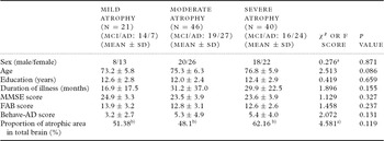
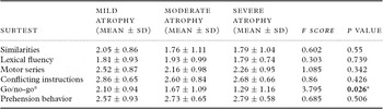
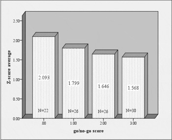
One-hundred-and-seven subjects were divided into the following three groups according to their Z-scores: (1) mild atrophy group (n = 21; MCI/AD: 14/7), (2) moderate atrophy group (n = 46; MCI/ AD: 19/27), and (3) severe atrophy group (n = 40; MCI/AD: 16/24). Table 1 shows the demographics of these groups. When the demographics of the three atrophy groups were compared, no significant differences in the age (F = 2.513; df = 2; P = 0.086), sex ratios (χ2 = 0.276; df = 2; P = 0.871), duration of illness (months) (F = 1.896; df = 2; P = 0.155), education (years) (F = 0.419; df = 2; P = 0.659), Behave-AD scores (F = 2.072; df = 2; P = 0.131), MMSE total scores (F = 1.129; df = 2; P = 0.327), FAB total scores (F = 1.458; df = 2; P = 0.237), or proportion of atrophy area in total brain (χ2 = 4.581; df = 2; P = 0.101) were observed among the three groups. Among the six subtest scores of the FAB, only the go/no-go score differed significantly among the three groups (F = 3.795; df = 2; P = 0.026), and a Tukey post-hoc test showed a significant difference between the mild and severe atrophy groups (P = 0.021) (Table 2). To confirm the almost negative correlation between the Z-score and the go/no-go score (r = −0.259, p = 0.008) using Spearman's correlation coefficient, we determined the average Z-score for each individual go/no-go score in the chart (Figure 1). On the other hand, memory registration (F = 0.785; df = 2; P = 0.459), memory delayed recall (F = 1.385; df = 2; P = 0.255), attention and calculation (F = 0.11; df = 2; P = 0.989), and 3-stage commands (F = 0.249; df = 2; P = 0.780) did not differ significantly among the three groups. Moreover, we investigated the association between the go/no-go score and such MMSE subtests using Spearman's correlation coefficient but did not find any significant correlations (memory registration: r = −;0.011, p = 0.909; memory delayed recall: r = 0.143, p = 0.148; attention and calculation: r = 0.127, p = 0.198; and 3-stage commands: r = 0.019, p = 0.846).
Subject characteristics (Mean ± SD)

FAB = Frontal Assessment Battery; MMSE = Mini-mental State Examination.
The sex ratio was analyzed using a χ2 test. The proportion of the atrophic area in the total brain (%) was analyzed using the Kruskall-Wallis test.
aχ2 score. bAverage rank score.
FAB subtest scores (mean ± SD)

aTukey post-hoc test: mild atrophy vs. severe atrophy: P < 0.05 (P = 0.021).
*P < 0.05.
Bar graph showing the average Z-score for each go/no-go score. The go/no-go score and the Z-score were almost significantly correlated (r = −0.259, P = 0.008). The statistical analysis was performed using the Spearman correlation coefficient.

Association of go/no-go score with hippocampal atrophy and diagnosis (AD or A-MCI)
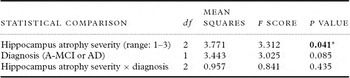
A generalized linear model examined (one variable) whether the severity of hippocampal atrophy (mild, moderate or severe) and diagnosis (AD or A-MCI) were associated with the go/no-go scores. As a result, we found that the go/no-go scores were significantly associated with the severity of hippocampal atrophy (F = 3.312; df = 2; P = 0.041) but not with the diagnosis (F = 3.025; df = 1; P = 0.085) (Table 3). A significant interaction between the severity of hippocampal atrophy and diagnosis (F = 0.841; df = 2; P = 0.435) was not detected.
Association between go/no-go, the severity of hippocampus atrophy and diagnosis

Generalized linear models were used to investigate whether the severity of hippocampus atrophy and the diagnosis (A-MCI or AD) were associated with the go/no-go scores.
*P < 0.05; R2 = 0.133.
Discussion
The present results suggested that atrophy of the entorhinal cortex and parahippocampal gyrus in patients with early AD or A-MCI might influence one of the executive functions – the go/no-go task of the FAB – independently of the diagnosis (A-MCI or early AD). In addition to memory cognitive impairments, the subjects with A-MCI also exhibited executive dysfunction, similar to previously reported patients with AD (Baudic et al., Reference Baudic, Barba, Thibaudet, Smagghe, Remy and Traykov2006; Traykov et al., Reference Traykov2007; Hanyu et al., Reference Hanyu, Sato, Takasaki, Akai and Iwamoto2009). To eliminate the influence of atrophy or disruption in other cerebral areas and cognitive impairments (attention deficits and memory disorders) other than executive dysfunction during the course of the disease, this study enrolled AD patients with a comparatively early stage of dementia (CDR of 0.5 or 1.0 and MMSE score of 15 or more points) and mild atrophy in the total brain. As a result, we were able to compare subjects with a homogeneous condition, other than executive function, in each of the hippocampal atrophy groups. Moreover, a significant influential interaction between the severity of hippocampal atrophy and the diagnosis (A-MCI or AD) could not be shown.
The FAB consists of six main cognitive components that are reportedly associated with different anatomical frontal regions (Dubois et al., Reference Dubois, Slachevsky, Litvan and Pillon2000). Of the six components, the go/no-go is known as “inhibition control” – i.e. conduct which inhibits inappropriate responses and regulates impulsions (Drewe et al., Reference Drewe1975; Dubois et al., Reference Dubois, Slachevsky, Litvan and Pillon2000). Some previous studies have reported that the go/no-go task was impaired in patients with an early stage of AD or A-MCI (Amieva et al., Reference Amieva, Phillips, Della-Sala and Henry2004; Hanyu et al, Reference Hanyu, Sato, Takasaki, Akai and Iwamoto2009). Some authors have suggested that this phenomenon is associated with selective attention deficit and psychomotor speed decrement, rather than errors in inhibition control (Langley et al., Reference Langley, Overmier, Knopman and Prod'Homme1998; Amieva et al., Reference Amieva, Phillips, Della-Sala and Henry2004). Collette et al. (Reference Collette, Schmidt, Scherrer, Adam and Salmon2009) implied that impaired performance on suppression tasks in patients with AD might represent a directed forgetting among working memory processes, rather than a specific alteration of some inhibitory resolving system. In the present study, however, the go/no-go score was not correlated with the MMSE subtest score reflecting attention deficits and memory impairment. If another neuropsychological test reflecting attention deficits or memory impairment had been used in the present study, a significant association might have been observed, possibly supporting the findings and hypotheses of these previous reports.
In neuroimaging studies using functional MRI (fMRI) and enrolling healthy controls, the inhibition control task was associated with the activation of the right orbitofrontal cortex and the inferior frontal cortex (Chikazoe et al., Reference Chikazoe2009). While some studies reported that patients with AD and MCI had impaired executive function since an early stage (Baudic et al., Reference Baudic, Barba, Thibaudet, Smagghe, Remy and Traykov2006; Traykov et al., Reference Traykov2007), several PET studies have shown that executive function is associated not only with the frontal lobe, but also with the tempo-parietal lobe or cingulate gyrus (Kessler et al., Reference Kessler, Mielke, Grond, Herholz and Heiss2000; Collette et al., Reference Collette, van der Linden, Delrue and Salmon2002). Moreover, in the neuroimaging studies researching inhibition tasks in patients with AD, some reports have presented a negative viewpoint in which the dysfunction is localized in the frontal lobe (Kessler et al., Reference Kessler, Mielke, Grond, Herholz and Heiss2000; Collette et al., Reference Collette, van der Linden, Delrue and Salmon2002). A previous neurophysiological study and a review also stated that the disruption of neural networks between the anterior and posterior cerebral areas, known as disconnection syndrome, during the initial stage of AD causes executive dysfunction, including inhibition control (Leuchter et al., Reference Leuchter, Newton, Cook, Walter, Rosenberg-Thompson and Lachenbruch1992; Delbeuck et al., Reference Delbeuck, Van Der Linden and Collette2003).
From a neuropathological view, the entorhinal cortex includes both afferent and efferent linkages between the hippocampus and the neocortical areas, respectively, and plays conjunctive roles (Leuchter et al., Reference Leuchter, Newton, Cook, Walter, Rosenberg-Thompson and Lachenbruch1992; Gómez-Isla et al., Reference Gómez-Isla, Price, McKeel, Morris, Growdon and Hyman1996; Mega et al., Reference Mega1997). Delbeuck et al. (Reference Delbeuck, Van Der Linden and Collette2003) stated that the deposition of neurofibrillary tangles (NFTs) in the entorhinal cortex may disrupt neural networks and cause disconnection syndrome in patients with early-stage AD. Thus, whether such neuropathological backgrounds indirectly or directly influence executive dysfunctions, including the go/no-go task, should be investigated in the future.
The present study had some limitations. First, we used only the FAB to confirm the patients' executive dysfunction in this study. Many other neuropsychological test batteries for evaluating executive functions exist, and these test batteries might have provided useful information. However, the FAB is one of the easiest tests to administer and can be completed at bedside without requiring any tools or instruments; we believe that the simplicity of this test makes it a valuable tool. Although some previous studies have reported that hippocampal atrophy was associated with memory disorders, the present study did not observe such an association between hippocampal atrophy and the MMSE subtest scores. Therefore, the Wechsler Memory Scale or the Rivermead Behavioral Memory Test might have been more useful, especially for the evaluation of memory disorder, rather than only using the MMSE subtests in the present study. Secondly, no associations between the other five FAB subscores and the severity of hippocampal atrophy was observed, although this result might represent a type II error because of the relatively small sample size in this study. Thirdly, the VSRAD system was developed to measure the total atrophy in the bilateral parahippocampal gyrus and entorhinal cortex. Ferreira et al. (Reference Ferreira, Diniz, Forlenza, Busatto and Zanetti2009) reported that in a VBM study enrolling subjects with A-MCI, the reduction in the gray matter volume of the left parahippocampal gyrus was the most consistent neuroanatomical biomarker for predicting the conversion from A-MCI to AD. Schroeter et al. (Reference Schroeter, Stein, Maslowski and Neumann2009) indicated that the bilateral hippocampus volume was significantly reduced in patients with AD, compared with normal controls, in a quantitative meta-analysis study. Thus, the association between executive function and both or each hippocampal volume reduction should be investigated in the future. Finally, while memory disorder and executive dysfunction are frequently observed in patients with early AD, apraxia symptoms as focal signs should be quantitatively assessed, as subjects with severe ideational apraxia cannot carry out sequences of action to achieve an intended purpose in the correct order.
In conclusion, to investigate the cognitive significance of hippocampal atrophy (including the entorhinal cortex and parahippocampal gyrus) in patients with early AD, we focused on the impairment of executive functions as a non-memory function, which is a core symptom of early AD. Accordingly, we found a significant association between go/no-go (inhibition control) and hippocampal atrophy in the present study. An association between hippocampal volume and executive function has not been previously reported, and our results may contribute to a better understanding of the actual pathogenesis of this disease. Furthermore, elucidating the neural network connecting the posterior and anterior areas in patients with AD will be an important task in the future.
Conflict of interest
None.
Description of authors' roles
Tomoyuki Nagata designed this study, examined the subjects, and wrote the paper. Shunichiro Shinagawa gave advice, including the analysis method, and reviewed the paper. Yusuke Ochiai, Ryo Aoki and Hiroo Kasahara examined the patients with AD at the Jikei University School of Medicine, Kashiwa Hospital. Kazutaka Nukariya, and Kazuhiko Nakayama reviewed and commented on the final paper.