Congenital primary pulmonary lymphangiectasia is a rare disorder consisting of greatly dilated lymphatic ducts throughout the lung. Reference Kliegman, Stanton, St Geme and Schor1 In 1970, Jacqueline Noonan described the three main pathological types: 1) as part of a generalised lymphangiectasia; 2) secondary to pulmonary venous or lymphatic obstruction; and 3) isolated. Reference Noonan, Walters and Reeves2
Apart from Noonan’s classification, pulmonary lymphangiectasia can also be divided into primary and secondary forms. Cardiovascular and lymphatic obstructive causes constitute the secondary group. Entities such as hypoplastic left heart syndrome, pulmonary veins atresia, congenital mitral stenosis, cor triatriatum, obstructive total anomalous pulmonary venous connection, and thoracic duct agenesis are known causes of secondary pulmonary lymphangiectasia. Reference Bellini, Boccardo, Campisi and Bonioli3 Pulmonary hypertension is a common finding, especially in the secondary forms of the disease. Reference Noonan, Walters and Reeves2,Reference Bellini, Boccardo, Campisi and Bonioli3
Diagnosis is usually suggested by thoracic CT or cardiac catheterisation with angiography, but the precise diagnosis is only possible via lung biopsy. Reference Kliegman, Stanton, St Geme and Schor1 However, fetal lung MRI has successfully shown the association between the “nutmeg” pattern and pulmonary lymphangiectasia. Reference Herrmann, Irons and Mascio4 In the secondary forms, treatment is aimed at the underlying condition or by resecting the affected part of the lung. Reference Kliegman, Stanton, St Geme and Schor1,Reference Nogarol5
Prognosis may be favourable if the condition is diagnosed beyond infancy. Reference Barker, Esther, Fordham, Maygarden and Funkhouser6
Case report
A non-syndromic baby girl with antenatal diagnosis of complete atrioventricular septal defect had successful complete surgical correction at the age of 8 months. The pregnancy was otherwise uneventful. Serial fetal echo scans did not identify any significant lung parenchymal abnormality hence no additional exam was considered.
She started diuretics shortly after birth. Aside from poor weight gain, she did not have severe heart failure symptoms nor cyanosis with oxygen saturations sitting on 98%. Medical management was optimised; however, due to this weight gain impairment she underwent corrective surgery only at the age of 8 months with a weight centile <3 (4450 g), according to WHO growth charts. Reference Onis, Onyango, Borghi, Siyam and Pinol7
The procedure and early post-operative period where uncomplicated with proper intracardiac pressures after surgical correction (35 mmHg right ventricle versus 80 mmHg left ventricle). On discharge she had mild pulmonary arterial hypertension based on mild right atrioventricular valve regurgitation Doppler analysis (right ventricular systolic pressure estimated in 45 mmHg), albeit less than the pre-operative estimated pulmonary arterial pressure. Nevertheless, during follow-up, it became evident that the pulmonary arterial pressure continued to rise and she was admitted at 11 months of age with persistent respiratory distress and failure to thrive, despite adequate diuretic and nutritional support.
Positive findings were mild anaemia, eosinophilia, mild respiratory acidosis (despite routine use of furosemide), and an Escherichia coli urinary tract infection. The transthoracic echocardiogram revealed right ventricular enlargement with mild left and right atrioventricular valve regurgitation. An estimated pulmonary systolic pressure of 60 mmHg and a new turbulent flow on colour Doppler in the right upper pulmonary vein with peak systolic velocity of 2.0 m/s were present.
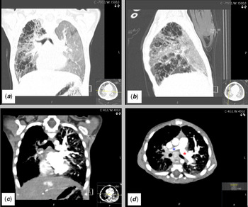
A chest CT angiogram was carried out that showed asymmetry of the pulmonary artery branches (right 5.2 mm versus left 9.4 mm) and reduced vascular markings in the right lung. The venous drainage of the right lung was not evident. Apart from the vascular findings, an area of increased mediastinal fatty tissue heterogeneity and density in the main lymphatic ganglion regions were described, as well as multiple “cystic-like” lesions affecting the right lung (Fig 1).
( a + b ) Chest CT angiogram: coronal ( a ) and sagittal ( b ) views showing cystic-like lesions in the right lung; ( c ) coronal view showing reduced vascular markings in the right lung; ( d ) axial view showing asymmetrical branches of the pulmonary artery. “Blue asterisk” – right pulmonary artery; “Red asterisk” – left pulmonary artery.

Further investigation excluded some possible related causes such as tuberculosis, Loeffler syndrome, Swyer-James syndrome, cystic fibrosis, emphysema associated with alpha-1-antitrypsin deficiency, sarcoidosis, primary and acquired immunodeficiency, metabolic conditions, or malignant diseases.
Despite nutritional and adequate diuretic management, the patient’s condition failed to improve and the echo-Doppler estimated pulmonary systolic pressure (110 mmHg) continued to rise to supra-systemic levels leading to RV dysfunction. Concomitantly, the measured flow jet in the right upper pulmonary vein increased to 3 m/s. Some weeks later, a new chest CT angiogram revealed an increase in size and number of “cystic-like” lesions in the right lung.
Owing to the severe pulmonary hypertension and cardiac deterioration, sildenafil was added to the therapeutic regimen without any success. A lung biopsy and eventual total right lung pneumectomy were considered; however, the patient´s clinical condition rapidly worsened leading to multi-organ failure, from which she did not recover.
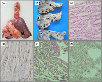
The post-mortem pathological examination revealed a post-operative cardiac status without sequelae. Tissue samples were formalin-fixed in a 10% formalin solution and paraffin embedded. Concerning the respiratory system, it was possible to differentiate a right superior, middle, and a portion of the inferior lobes affected by lymphangiectasic lesions, confirmed histologically.
The histologic examination showed the presence of spindle cells in the arteries and veins, findings suggestive of pulmonary arterial hypertension, and interlobular venules with a significant reduction of their size (Fig 2). No localised stenotic lesions were found in the pulmonary veins. There were no other congenital malformations.
( a ) Thoracic block including the right lung showing cystic-like lesions in its superior and middle lobes; it can also appreciate a normal inferior lobe and left lung. ( b ) Cut section of right lung after fixation shows the existence of multiple cavities in the lung parenchyma. ( c + d ) On histology, the cavities correspond to a single layer of bland lymphatic endothelium, confirmed with podoplanin immunostaining; ( c ) HE, 200×; ( d ) podoplanin, 200×). ( e + f ) Veins from right lung and left lung, respectively, showing significant reduction of diameter on the right lung (elastin, 200×).

Discussion
When the follow-up of surgically corrected congenital heart conditions, after exclusion of possible related sequelae, does not follow the expected outcome, other rarer, possibly linked entities should be sought.
Our patient had a good surgical outcome without early post-operative complications. Pulmonary artery and right ventricular systolic pressures were normal immediately after the procedure and rose steadily the following three months with progressive clinical deterioration.
The challenge regarding this case was whether the vascular pulmonary lesions were a primary event, or secondary to the right lower pulmonary vein stenosis or other causes, namely infectious, metabolic, autoimmune, or malignant diseases.
Primary pulmonary lymphangiectasia is an extremely rare condition, usually presenting at birth. Our patient had a later onset with a localised form of the disease (right lung) and presented with respiratory distress and failure to thrive. However, cough and chylothorax are usually described as well, but were absent. Reference Nogarol5,Reference Sant’Anna, Nogarol, Fátima, Leal, Madi and Alves8
Secondary forms of pulmonary lymphangiectasia are associated with CHD, particularly in left-sided obstructive lesions, but have also been linked to other forms, as reported by Noonan et al, Reference Noonan, Walters and Reeves2 where a membranous ventricular septal defect was described with this entity. When suspected antenataly, the “nutmeg” pattern seen on lung MRI can be diagnostic. Reference Herrmann, Irons and Mascio4
The association of shunt lesions and pulmonary vein stenosis was hypothesised by Drossner et al, Reference Drossner, Kim, Maher and Mahle9 which related the shear stress due to increased pulmonary vein blood flow to the synthesis of vasoactive substances, such as vascular endothelial growth factor, ultimately leading to intimal vascular lesions and secondary lymphagiectasia. Although theoretically sound, it does not explain the progressive nature of the condition in our patient, particularly, as the defect had been successfully corrected. Conversely, progressive primary lymphangiectasic lesions could compress pulmonary veins along their route with progressive flow acceleration.
Regardless of its rarity or clinical presentation, if a thoracic CT angiogram shows the evidence of diffuse or localised cystic-like lesions and/or interstitial infiltrates, in the context of CHD, pulmonary lymphangiectasia should be considered, and all efforts should be done to promptly diagnose this entity.
Acknowledgements
None.
Financial support
This report received no specific grant from any funding agency, commercial, or not-for-profit sectors.
Conflicts of interest
None.