Introduction
According to the World Health Organisation (WHO), cardiovascular disease (CVD) is a major cause of human death globally. According to the American Heart Association (AHA) 2019 report, in 2016, ~17.6 million deaths were caused by CVD globally (Ref. Reference Benjamin1). The foremost modifiable risk factor for CVD morbidity and mortality is a hypertension (HTN) (Refs Reference Rapsomaniki2, 3). The AHA 2017 guidelines define HTN as a systolic blood pressure (BP) of >130 mmHg or a diastolic BP of >80 mmHg, which raises the incidence of HTN globally (Ref. Reference Whelton4). High BP causing HTN affects the function of certain organs and leads to the term target organ damage (TOD). TOD comprises cardiac hypertrophy (CH), retinopathy, renal failure and vascular and haemorrhagic stroke (Ref. Reference Nadar5).
Briefly, CH is an increase in the mass of the heart. Development of the postnatal myocardium is very coordinated with its workload. Contrast to normal conditions, the myocardium works harder because of the elevation in workload such as HTN, causing a pressure overload. To compensate for the prolonged upsurge in the pressure of cells, the myocytes in the myocardium swell, leading to a gain of mass and size. The rise in myocardium mass is generally because of a rise of the weight of the ventricles (Ref. Reference McMullen6).
The myocardium comprises cardiac myocytes (muscle cells), non-myocytes (endothelial cells, fibroblasts, vascular smooth muscle cells and mast cells) and the adjacent extracellular matrix. Although, the ventricular myocytes contributes merely one-third of the total count of the myocardium's mass; it accounts for 70–80% of the mass of the heart (Ref. Reference Popescu7). The molecular mechanisms that initiate CH have been of widespread concern because CH and heart failure (HF) are closely associated. Presently, a cure for HF is lacking, and the rate of survival following HF remains poor (Ref. Reference McMurray8). Hence, various studies have been conducted with an aim to ascertain the molecular mechanisms that are linked with CH and the switch that activates HF in order to reveal novel therapeutic sites to either avert or reduce CH and HF.
Different types of CH
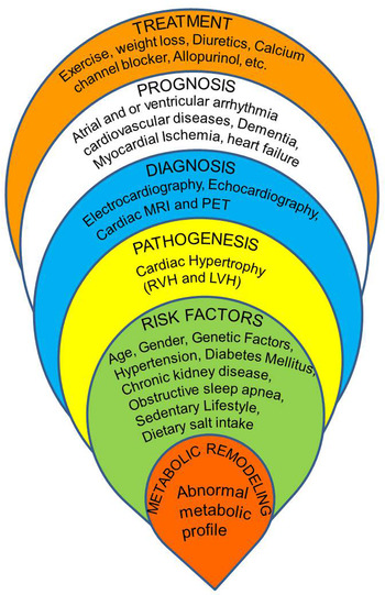
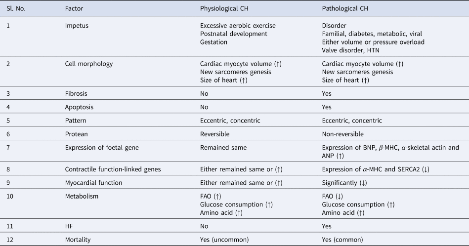
Briefly, CH can be classified into two forms: (i) physiological (normal) and (ii) pathological (detrimental). Physiological CH arises causing normal postnatal growth following prolonged physical exercise and during pregnancy. In contrast, pathological CH arises after a period of persistent, prolonged pressure or volume overload with an abnormal condition such as HTN, valvular cardiac disease, coronary artery disease (myocardial infarction or ischaemia), diabetes or congenital gene mutations. Both types of CH are linked with an augmentation in the size of the heart. Physiological CH is manifested with normal cardiac structure, reversible and normal or enhanced cardiac function; however, pathological CH is manifested with irreversible, myocytic necrosis or apoptosis, fibrosis, abnormal cardiac function and a high chance of HF and abrupt end of life (Table 1) (Ref. Reference McMullen6). A typical illustration of overview of CH is explained in Figure 1.
Typical illustration of the overview of CH.

Factors and their effects of physiological and pathological CH

↑; increase, ↓, decrease.
Both hypertrophies are subdivided into concentric and eccentric. These divisions are on the basis of different forms of remodelling, which are based on the instigating stimulus (Ref. Reference Pluim9). Concentric hypertrophy indicates a rise in comparative wall thickness and the mass of the heart, along with either slight lessening or unaltered volume of the chamber. It comprises a parallel arrangement of sarcomere addition, causing an augmentation in the width of cardio-myocytes compared with the normal condition (Fig. 2). Eccentric hypertrophy indicates a rise in cardiac mass along with augmented volume of the chamber, that is, dilated chamber. It contains relatively normal, reduced or higher wall thickness and comprises the addition of sarcomeres in a series that causes an augmentation in the length of the cardio-myocytes (Fig. 2).
Typical presentation of effect of changing cardiac phenotype. Left ventricle of (a) healthy heart, (b) LV concentric hypertrophy heart and (c) LV eccentric hypertrophy heart with their respective wall thickness in short-axis as well as long-axis and changes in sarcomere.

A pathological fillip such as HTN and aortic stenosis leads to pressure overload and creates an augmentation in systolic wall stress that incurs concentric hypertrophy (Ref. Reference Grossman10). On the contrary, a fillip such as aortic regurgitation, arterio-venous fistulas leads to volume overload and generates an augmentation of diastolic wall stress that incurs eccentric hypertrophy (Refs Reference Pluim9, Reference Grossman10) (Fig. 2). Scientific observations have suggested that eccentric CH triggered by a pathological fillip poses a higher risk compared with concentric CH (Ref. Reference Berenji11).
Concentric and eccentric hypertrophy can also be generated by a physiological fillip. Aerobic exercise and pregnancy can lead to volume overload and eccentric hypertrophy (Refs Reference Pluim9, Reference Eghbali12). Eccentric hypertrophy is typically indicated by an enlargement of the chambers and a relative adjustment of wall thickness. Strength exercises such as weight lifting, wrestling and heavy objects throw wavers into the cardiac pressure load and generate concentric hypertrophy (Ref. Reference Pluim9) (Table 1).
In spite of a relative enlargement of cardiac size, CH is linked with distinctive anatomical, functional, molecular and metabolic aspects.
Cardiac anatomical and functional aspects
In brief, pathological CH is linked through structural modification of constituents of the ventricular walls to entertain proliferations in the size of the myocytes, comprising alterations in the angiogenesis and fibrillar collagen system. Pathological hypertrophy occurs with cell death (necrosis and apoptosis) and cardiac loss of myocytes is switched with enormous type 1 fibrillar collagen (aka cardiac fibrosis). Enormous accretion of collagen hardens the ventricles, which ruins relaxation and contraction, ruins the electrical connections of cardiac myocytes with proteins of extracellular matrices and lessens the density of the capillaries. Cardiac myocyte fibrosis and diminished density of capillaries upsurge the oxygen diffusion spaces, lead towards myocardial ischaemia and probably contributes to the shift from CH to HF (Ref. Reference Gunasinghe and Mann13).
Cardiac molecular and biochemical aspects
In brief, pathological CH is linked with unique changes in contractile proteins of cardio-myocytes (α- and β-myosin heavy chain [MHC]), over-expression of foetal genes (α-skeletal actin, B-type natriuretic peptides [BNP]) and atrial natriuretic peptides [ANP]) as well as reduced expression of calcium-handling protein sarcoplasmic reticulum Ca2+-ATPase 2a (SERCA2a) (Ref. Reference Iemitsu14). Several thoroughly detailed readouts are available describing about the various signalling proteins that play specific roles in regulating CH. The current review will address, in brief, the role of the key nutrient, mTORC1, and the adenosine monophosphate-triggered protein kinase (AMPK) signalling pathways of CH.
The development of cardiomyocytes is displayed by raised protein fabrication or diminished deprivation of proteins. The mTOR comprises two different serine/threonine kinase complexes, mTORC1 and mTORC2, that are required to harmonise amino acid as nutrient and growth factor signals' accessibility with cell metabolism and development through regulating protein fabrication and deprivation (Ref. Reference Saxton15). In the response of signals of metabolic and mechanical factors, the activity of mTORC1 that is augmented in the evolution of both cardiac hypertrophies.
The mTORC1 encourages ribosomal protein synthesis by strict control of S6Ks and by impeding eIF4E-binding protein 1 (4EBP1), and permits uncontrolled cap-dependent translation by eIF4E (Ref. Reference Sciarretta16). Although enhancing the protein production and the quality control of mitochondria by mTOR are crucial, robust mechanisms for the myocardium in acute pressure overload (Ref. Reference Zhang17), continuous stimulation of mTOR is inimical, partly as a consequence of the abolition of autophagy and the subsequent degradation of the original trait of protein regulation mechanisms.
AMPK impedes mTORC1, and therapeutic abolition of mTORC1 suppresses angiotensin II persuaded and pressure-overload-stimulated pathological hypertrophy and HF (Refs Reference Sadoshima18, Reference Shioi19). Taking together the findings of various studies (Refs Reference Shioi19, Reference McMullen20) suggests that S6Ks are not crucial for the stimulation of either physiological or pathological hypertrophy. Possibly, 4EBPs play a major role in facilitating mTORC1-activated CH (Ref. Reference Zhang17). Noteworthily, mTORC2 is too triggered in reply to pressure overload, with impeding cell death by attenuation of the pro-apoptotic mammalian STE20-like protein kinase 1 (MST1; aka STK4) (Ref. Reference Sciarretta21). Hence, mTORC1 and mTORC2 behaviours in pathological hypertrophy are opposite to each other.
The harmonisation of heart development with cellular metabolism is crucial for a supple retaliation. AMPK is delineated as the focal point power sensor in all cells that harmonise upsurges in metabolic yield with the consumption of nutrients. AMPK is a complex protein that comprises three subunits: (i) the catalytic α subunit, (ii) the β subunit that connects the α and γ subunits and ties glycogen and (iii) the γ subunit that ties AMP, adenosine diphosphate or adenosine triphosphate (ATP) in a jointly unique approach (Ref. Reference Horman22).
The AMP connects with AMPK guides its instigation over geometrical alterations that permit phosphorylation of the α subunit through the upstream kinases liver kinase B1 (LKB1; aka STK11), calcium/calmodulin-dependent protein kinase kinase β (CaMKK β; also famous as CaMKK2) and transforming growth factor-β activated kinase 1 (TAK1; aka MAP3K7). Reduced energy contents are directed to AMPK induction by AMP that supports ATP synthesis through activating glycolysis and β-oxidation, along with lessening of ATP-controlled procedures as transcription and translation (Ref. Reference Horman22). Hence, AMPK is essential for stability and equilibrium of cardiac performance.
Various readouts indicate that chronic impediment of AMPK intensifies pathological hypertrophy, directing to HF, whereas sporadic AMPK induction possibly acts as a cardioprotective. For instance, in brief, AMPK activity reduced in mice heart null for Adipoq (an adipose tissue-secreted hormone) was stimulated to pressure overload persuaded hypertrophy (Ref. Reference Shibata23). Mouse heart null for Prkaa2, accounted for encrypts AMPKα2, and presented an intensified pathological retort to the hypertrophic impetus (Refs Reference Zarrinpashneh24, Reference Zhang25). Excitingly, knock-out of Stk11 accounted for the encryptions of LKB1 in the young animal's heart and headed to a shortened size of ventricular cardiomyocyte, and advising a positive character of AMPK in allowing accurate postpartum growth of heart (Refs Reference Sakamoto26, Reference Ikeda27). Nonetheless, reduced AMPK activity in older animals influenced hypertrophy, which was neutralised with rapamycin or with the help of constructive, robust AMPK mutant treatment (Ref. Reference Ikeda27).
To induce AMPK, treatment with the AMPK inducer, metformin, enhanced the cardiac performance after ischaemia–reperfusion lesion in the mouse by a mechanism comprising stimulation of endothelial nitric oxide synthase and the peroxisome proliferator-initiated receptor-γ co-activator 1α (PGC1α) (Ref. Reference Gundewar28).
Taking together, all gleaned readouts highlight the significance of the metabolic reformation in the hypertrophic reply to ensure appropriate alteration in the growth of physiological heart. Hence, considering pathological cardiac milieus, augmenting AMPK activity using particular biochemicals might be an appealing curative route to probe.
Cardiac metabolic aspects
In brief, in the healthy heart, 60–70% of ATP production is maintained by fatty acid oxidation (FAO) and the remaining ≃30% of ATP synthesis is controlled by glucose and amino acid metabolism (Refs Reference Akki29, Reference Gupta30). The heart is proficient in changing energy substrate constituents, subjecting them to workload, which reflects an adaptive approach that permits the heart to generate an uninterrupted resource of ATP in different milieus such as exercise, resting and fasting. Pathological CH is allied with drops in FAO and escalations in glucose metabolism (Refs Reference Gupta30, Reference Gupta31). This change in energy substrates consumption may be a defensive approach, letting the heart generate extra ATP per molecule of oxygen utilised (Ref. Reference vanBilsen32).
Appraisal of CH
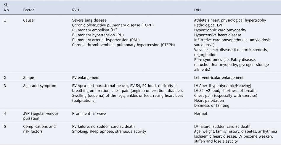
CH has two siblings: right ventricular hypertrophy (RVH) and left ventricular hypertrophy (LVH) (Table 2). RVH is a relatively rare subtype of hypertrophy that primarily affects the right ventricle. Abnormal or pathological increases in the muscles of the right ventricle occur in response to pressure overload. The size and function of the right ventricle are adversely affected by pulmonary HTN with or without left ventricular dysfunction, and by tricuspid regurgitation. The most common cause of RVH is severe lung disorders that induce pulmonary HTN and secondary RVH that includes pulmonary arterial hypertension (PAH) and chronic thromboembolic pulmonary hypertension (Ref. Reference William33). LVH is the most common complication of high BP and is associated with an increased risk of cardiovascular complications. LVH is an inadequate response to chronic pressure overload. Chronic pressure overload is believed to activate left ventricular myocardial growth and neurohormonal stimuli, which then trigger and sustain a hypertrophic response (Ref. Reference William33). LVH is a crucial target for HTN-based TOD.
Comparisons of RVH and LVH

The key diagnostic modalities for the appraisal of CH anatomical and functional changes are echocardiography, electrocardiography, positron emission tomography, and cardiac magnetic resonance imaging (Ref. Reference Greenland34). All these techniques, however, have several inherent limitations. Noteworthily, these modalities can detect only geometrical and functional changes of LVH and RVH. A study suggested that molecular alterations comes early and induce anatomical changes of the heart (Ref. Reference Maslov35), which lead to HTN and subsequent manifestations of anatomical changes of the heart.
Metabolic reprogramming and CH: opportunities of metabolomics
The intricacies of the molecular mechanisms of LV remodelling are still under debate. Moreover, early stages of LV remodelling are commonly asymptomatic, particularly in adult ages. System biology applies comprehensive tactics that aim at guiding and elucidating the molecular intricacies of diseases at early stages. This discipline comprises so-called omics such as genomics, transcriptomics, proteomics and metabolomics. System biology offers profound information about molecular alterations even in a single cell. Metabolomics is the science of omics where indexes of low-molecular-weight (molecular mass <1500 Da) molecules reflect perturbations in the genome, transcriptome and proteome of a single cell, tissue and an organism as well. Several worthwhile studies have been conducted using different biological matrices. The specific pattern of metabolites in biological specimens is able to decode the type of micro-organisms (Ref. Reference Gupta36) and specific diseases (Refs Reference Kumar37, Reference Gupta38) using metabolomics. Owing to its great potential for discovering biomarkers in various cardiac diseases (Refs Reference Griffin39, Reference McGarrah40), metabolomics is one of the most frequently applied approaches to understand the intricate molecular mechanisms of LVH. Non-targeted and targeted tactics of molecular analysis of metabolites seem to be an important approach to understand the molecular mechanisms of LVH. Tissue (cellular or local) metabolomics seems to be important, as the local cells are the first place where alterations of metabolites' level, triggered by the milieu of ailments, are expressed. Simultaneously systemic (blood or global) metabolomics can offer a broader spectrum of information regarding particular molecules for specific disease conditions for clinical usefulness.
Potential of metabolomics of CH
As mentioned above, the alteration in cardiac metabolism leads to cardiac remodelling and causes CH (Ref. Reference Maslov35). In this context, the metabolomic changes appear before the anatomical changes; that is why the role of metabolomics is so important. In this panorama, the current review will encompass local metabolomics (cardiac tissue-based observations) and global metabolomics (serum/plasma-based observations) with special emphasis on using nuclear magnetic resonance (NMR) spectroscopy and mass spectrometry (MS) comprising liquid chromatography (LC-MS) and gas chromatography (GC-MS) as an analytical technique. There are many detailed, in-depth reviews comprising the comprehensive methodologies of these analytical platforms and study design of different biofluids with coverage of the metabolome, cost, throughput, advantages, disadvantages and application of various statistical approaches to generate precise outcomes in metabolomics (Refs Reference McGarrah40–Reference Dunn42); hence, to reduce the reiteration, the current review will not address this part in detail.
In brief, by virtue of some specific atomic nuclei – namely 1H, 13C, 15N and 31P – have intrinsic spin on their own axis that generates a tiny magnetic field called the magnetic moment. An NMR spectrometer properly connects to a superconducting magnet, a magnetic field-gradient system, a committed computer and a radiofrequency (RF) coil. The concept of NMR is to apply RF pulse to stimulate the magnetic moment of spins of a specific nucleus in the biological samples in the presence of an external magnetic field and then record the RF response released as a consequence of nuclei relaxation in the form of free induction decay, which converts into interpretable NMR spectra using ‘Fourier transformation’ calculation. The specific frequency of resonance called chemical shift depends on the chemical properties of the nucleus and the strength of the magnetic field. An NMR spectrum comprises signal splitting patterns as a consequence of coupling of adjacent nuclear spin in the microenvironment of the compound (Refs Reference Gupta36, Reference McGarrah40).
Briefly, the MS depends on the ionisation of compounds and then on gauging of the amounts of the ions generated, which is determined according to mass-to-charge ratios (m/z). The metabolites can be identified using m/z ratio spectra and data fragmentation information. MS comprises three key components: (i) the ion source; (ii) the mass analyser and (iii) the detector. MS classically leads by front-end segregation, generally LC or GC. Chromatographic exclusion augments the precision and reduces the vital ion pre-saturation influences in the mass spectrometer through lessening the numeral of co-eluting the metabolic contents that enter the mass spectrometer at a particular time for further analysis (Refs Reference McGarrah40, Reference Dunn42).
The term metabolomics was coined in the late 1990s to describe the development of approaches to measure all metabolites present in a cell, tissue or organism during genetic modification or physiological stimulus (Refs Reference Oliver43, Reference Nicholson44). The first cardiac metabolomic study was reported in 1976 using 31P NMR in cardiac tissue (Ref. Reference Gadian45). In another study, 1H-NMR was also used to identify metabolomic biomarkers for LVH secondary to arterial HTN using blood samples (Ref. Reference Evaristi46). A recent animal study has carried out metabolomic analysis of PAH-induced rats' serum by 1H-NMR spectroscopy; the results showed metabolic shifts during the progression of vascular remodelling in RVH (Ref. Reference Lin47). As an integral part of the circulatory system, blood is closely connected to particular organ tissues; hence, their metabolic changes are reflected in the systemic circulation, which can be analysed using the metabolomics approach (Ref. Reference Holmes48). Moreover, measuring the entire serum/plasma metabolome rather than a single metabolite in a single assay makes the biomarker search process more convenient and the results more robust because metabolomics can simultaneously reveal direct and indirect biomarkers of underlying diseases (Ref. Reference Spratlin49).
Cellular metabolomics
Tissue metabolomics of RVH using NMR spectroscopy
Izquierdo et al. performed a metabolomics study on a PAH mouse model generated by hypoxia (HPX) exposure combined with Semaxanib (SU5416) administration (HPX + SU model). The results were compared with healthy, normoxic mice (NMX group) and healthy HPX mice. Intact heart tissues were analysed using high-resolution magic angle spinning (HR-MAS) NMR spectroscopy at 500 MHz (Ref. Reference Izquierdo-Garcia50). Metabolomics of RV tissues revealed augmented levels of glutamine, creatine phosphate, lactate, taurine and glycine; concomitantly, it revealed a reduced level of glucose in HPX + SU and HPX groups compared with NMX. Moreover, reduced levels of taurine, glycine and glutamate, and concomitantly augmented levels of free fatty acids (FAs) and alanine were observed in the HPX group compared with the HPX + SU cohort (Ref. Reference Izquierdo-Garcia50). Among vital metabolites, only glutamine and taurine exhibited significant Pearson's R-squared correlation with wall thickness of RV hypertrophy. Compared with the RV tissue sample, metabolomics of LV tissue samples exhibited considerably fewer acute metabolic influences. Only lactate metabolite was considerably augmented in LV tissue samples of HPX and HPX + SU groups compared with NMX. All other metabolites remained comparable in all three cohorts. In summary, authors concluded that glutamine and taurine can be used as a metabolic biomarker of human PAH and that it may be a valuable tool to test new targeting treatment strategies of underlying diseases (Ref. Reference Izquierdo-Garcia50).
Tissue metabolomics of RVH using LC-MS
Brittain et al.'s study revealed the systemic and cardiac FA metabolism in PAH patients using LC-MS (Ref. Reference Brittain51). In contrast to controls, PAH patients exhibited augmented levels of circulating free FAs and long-chain acylcarnitines. Cardiac RV exhibited enhanced long-chain FAs and concomitantly lessened long-chain acylcarnitines in PAH compared with controls. Further evidence, gleaned using in vivo 1H-MRS, revealed augmented levels of myocardial triglyceride in PAH patients compared with controls. Compared with controls, PAH RVs revealed higher levels of ceramide, a mediator of lipotoxicity. A mouse model of PAH revealed lessened FA oxidation via failure of palmitoylcarnitine to induce oxygen utilisation in the PAH RV. The authors summarised that impairments in FA metabolism can be identified using blood samples as well as cardiac PAH RVs and that the impairments are linked with in vivo cardiac steatosis and lipotoxicity. Moreover, an animal model revealed that lessening of FA oxidation may cause lipotoxicity (Ref. Reference Brittain51).
Tissue metabolomics of LVH using NMR spectroscopy
Jones et al. executed a solution state (tissue extract) as well as intact tissue metabolic profiling study to appraise the effects of a muscle LIM protein knock-out mouse model of LVH (MLPKO) with their control strains using NMR spectroscopy (Ref. Reference Jones52). Metabolomics-derived observations of tissue extract exhibited that citrate, phosphocholine, lysine, alanine, creatine, taurine and glutamate were the most perturbed metabolites in the MLPKO compared with control strain mice. Intact tissue metabolic profiling revealed augmented levels of taurine, creatine, lactate and lipids and reduced levels of phosphocholine/phosphatidylcholine and glutamate in the MLPKO compared with control strain mice. The authors expressed the study that despite the differences at the gene level, metabolomics can differentiate distinct metabolic profiles of each mouse model from its control strain. Hence, this approach can be a rapid technique of phenotyping mouse models of disease (Ref. Reference Jones52).
The creatine transporter over-expressing mice exhibited augmented levels of myocardial creatine [Cr] and preserved the heart in a milieu of ischaemia/reperfusion injury. In contrast, CH and dysfunction develops because of very high levels of [Cr]. Zervou et al. appraised these contrasting effects through the correlations of [Cr] level and metabolomics of LV tissue-derived metabolites (Ref. Reference Zervou53). This study revealed various strong correlations between myocardial [Cr] and cardiac metabolites such as positive correlations with α-glucose, acetyl-carnitine, glutamine and negative correlations with taurine, fumarate, aspartate, alanine and phosphocholine. This study further reveals the alterations in substrate utilisation and energy metabolic pattern of impaired energy generating pathways with high levels of [Cr] (Ref. Reference Zervou53).
Uddine et al.'s study determined that cardiac branched chain amino acid (BCAA) accumulation in dilated cardiomyopathy (DCM) patients was because of an impaired catabolism of BCAAs, and augmentation of cardiac function in failing heart mice through stimulation of cardiac BCAA oxidation (Ref. Reference Uddin54). NMR spectra of tissue extract exhibited that cardiac BCAA levels were considerably augmented in left ventricular biopsy of DCM patients compared with control biopsy samples. Isolated working mice hearts demonstrated significantly augmented cardiac BCAA oxidation in BT2-administered mice (3,6-dichloro-1-benzothiophene-2-carboxylic acid) compared with vehicles. BT2 administration also enhanced cardiac function in both sham and left ventricle constriction-transverse aortic constriction (TAC) failing mouse hearts. The authors summarised that impaired cardiac BCAA catabolism occurs in human HF, whereas stimulating BCAA oxidation can augment cardiac function in TAC mouse hearts (Ref. Reference Uddin54, Fig. 5).
Tissue metabolomics of LVH using LC-MS
Kato et al. appraised metabolic remodelling in compensated LVH and congestive heart failure (CHF) in male Dahl salt-sensitive (DS) rats (Ref. Reference Kato55). The glucose uptake was enhanced in the LVH stage, and further glucose uptake augmented and FA uptake lessened at the CHF stage. Glycolysis, FAO and mitochondrial function-related gene expression was well-maintained at the LVH stage but reduced at the CHF stage. The metabolomic analysis indicated activation of the pentose phosphate pathway (PPP) in CHF. Compared with the LVH stage, augmented pyruvate, lessened alanine and succinate were observed in the CHF stage. Administration of dichloroacetate (DCA) to CHF rat hearts exhibited improved cardiac function and the survival of animals via augmentation of glucose uptake and cardiac energy stores through the activation of PPP. DCA protected cardiomyocytes from oxidative stress-induced cell death, suggesting that PPP activation through increased glucose metabolism is a protective mechanism against CHF. The authors suggested that LVH or CHF is linked with discrete alterations in the metabolic profile of the heart. DCA can be used to attenuate the transition from LVH to CHF (Ref. Reference Kato55).
Kolwicz et al. performed an LC-MS/MS-based study focussed on pressure-overload an LVH-induced acetyl CoA carboxylase-2 (ACC2H−/−) mice model and addressed several key cardiac metabolic problems (Ref. Reference Kolwicz56). This study showed that cardiac-specific suppression of ACC2 decreased malonyl-CoA levels and increased FAO. In addition, ACC2 suppression maintained cardiac FAO and prevented increased glycolysis and anaplerosis (i.e. prevention of metabolic remodelling) during the development of LVH. Moreover, this metabolic animal model also exhibited preserved cardiac function and energetics with attenuated hypertrophy. These results suggest that modulation of mitochondrial substrate input and mitochondrial oxidative capacity during the development of heart disease is a viable approach for therapeutic interventions (Ref. Reference Kolwicz56).
Tissue metabolomics of LVH using GC-MS
Augmented expression of glucose transporter 1 (GLUT1) and glucose consumption is believed to be cardioprotective in pressure overload-induced hypertrophy (POH). In addition, lifelong overexpression of GLUT1 gene inhibits cardiac dysfunction following aortic constriction. Because of lifelong augmented glucose metabolism is a critical issue; hence, the importance of this genetic manipulation in clinical practice is uncertain. Pereira et al. examined whether a short-term escalation in GLUT1-mediated cardiac glucose uptake would still be considered cardio protective if overexpression has arisen at the beginning of POH (Ref. Reference Pereira57). Transgenic mice with cardiomyocyte-specific overexpression of a haemagglutinin (HA)-tagged GLUT1 (G1HA) and their controls were treated to TAC 2 days after transgene administration with doxycycline. Four weeks following TAC, GC-MS-based cardiac tissue extract exhibited augmented levels of glucose, glucose-6-phosphate, glucose-1-phosphate and lactic acid in G1HA compared with control mice. Moreover, control mice demonstrated reduction in mitochondrial function, ATP synthesis and mRNA expression of oxidative phosphorylation (OXPHOS) genes, and all these indexes were maintained in concert with increased glucose utilisation in G1HA mice. The authors summarised that short-term cardiac-specific escalation of GLUT1 at the beginning of POH reserves mitochondrial function and reduces pathological remodelling, and simultaneously provokes the hypertrophic phenotype, and is inadequate to avoid POH-induced cardiac impairment, probably because of a defect in calcium cycling (Ref. Reference Pereira57).
Systemic metabolomics
Serum/plasma metabolomics of RVH using NMR spectroscopy
Lin et al. conducted a serum-based metabolic profiling study of monocrotaline (MCT)-induced PAH in a Sprague–Dawley rat model at 600 MHz NMR spectroscopy (Ref. Reference Lin47). Their results indicated enhanced levels of betaine, glycerol, glucose, glycine and acetoacetate levels and reduced levels of creatine, NAC, acetate, choline and methionine in the first week (W1) of the study compared with controls. Moreover, glycerol, glucose, betaine, low-density lipoprotein/very-low-density lipoprotein (LDL/VLDL), glycine, choline and acetone levels increased; lactate, taurine, pyruvate, valine and acetoacetate levels decreased in the early stage of PAH (W2–3). Subsequently, there were increased serum levels of lactate, creatine, pyruvate, betaine, glycine, choline, glycerol, isoleucine, leucine and carnitine, and, decreased levels of LDL/VLDL, glucose and taurine in the persistent PAH stage (W4). Among all perturbed metabolites, betaine, methionine and glycine were demonstrated as vital pathways through pathway enrichment analysis and suggested that an impairment of methionine metabolic pathway possibly plays an important role in the onset of PAH. Furthermore, augmentation of the choline, betaine and methionine pathways and energy metabolism might be significant pathological mechanisms in the development and progression of PAH (Ref. Reference Lin47).
Serum/plasma metabolomics of RVH using LC-MS
Lewis et al. used LC-MS-based human plasma metabolic profiling to determine the signature metabolites of pulmonary HTN and associated right ventricular pulmonary vascular (RV-PV) dysfunction (Ref. Reference Lewis58). Their results demonstrated very good associations of RV-PV dysfunction with circulating indoleamine 2,3-dioxygenase-dependent tryptophan metabolites, TCA cycle intermediates and purine metabolites, and advocated earlier defined associations with arginine–nitric oxide metabolic pathways. The authors summarised that metabolic profiling can be used to characterise RV-PV dysfunction through both new biomarkers and pathways (Ref. Reference Lewis58).
Rhodes et al. conducted a study of plasma metabolic profiling using LC-MS to differentiate PAH patients at high risk of early death and patients who responded well to treatment compared with controls (Ref. Reference Rhodes59). Metabolomics reveals that a group of 20 metabolites were able to discriminate PAH patients from controls and that a group of 36 metabolites can be used as prognostic biomarkers of PAH. Compared with controls, the patient group at high risk exhibited augmented levels of tRNA-specific modified nucleosides, malate, fumarate, glutamate, FA acylcarnitines, tryptophan and polyamine metabolites and reduced levels of steroids, sphingomyelins and phosphatidylcholines. Comparable metabolic profiling was observed in patients who responded to calcium channel blocker therapy and controls. The authors concluded that metabolic profiling can be used not only to correlate survival of PAH patients and characterisation of underlying ailments, but also to monitor the patients' response to therapy (Ref. Reference Rhodes59).
Zheng et al. performed a metabolomic study using LC-MS on plasma samples of MCT-induced rat model of PAH to reveal profound metabolic alterations (Ref. Reference Zheng60). Among various altered metabolites, only seven metabolites – adenosine monophosphate, urea, 4-hydroxyproline, ornithine, N-acetylornithine, O-acetylcarnitine and betaine – were significantly correlated with PAH and able to discriminate from controls. The authors summarised that metabolomics can identify profound changes of PAH pathogenesis and can be used for clinical utility (Ref. Reference Zheng60).
Attard et al. used high-throughput LC-MS-based serum metabolomics to identify PAH patients' metabolic pathways associated with RV adaptation to PH (Ref. Reference Attard61). Their results exhibited that six metabolites (augmented levels of tRNA-specific modified nucleosides and FA acylcarnitines, and decreased levels of sulphated androgen) were significantly associated with high wall tension. The authors suggested that wall stress affects PH outcomes and is linked to metabolic pathways that take into account energy metabolism and the response to cellular stress (Ref. Reference Attard61).
Similarly, Chen et al. examined plasma metabolic profiles to differentiate PAH from controls using LC-MS (Ref. Reference Chen62). Their results demonstrated that, among the various metabolic pathways alterations, a panel of metabolites from lipid metabolism and FAO (lysophosphatidylcholine, phosphatidylcholine, perillic acid, palmitoleic acid, N-acetylcholine-d-sphingomyelin, oleic acid, palmitic acid and 2-octenoylcarnitine metabolites) were closely associated with PAH. The authors showed the importance of those metabolites for the discovery of biomarkers and for the development of therapeutic strategies (Ref. Reference Chen62).
Serum/plasma metabolomics of RVH using combined GC-MS and LC-MS
Pulmonary embolism (PE) is a common CVD, and leads to PH and RVH. Diagnosis of PE is often not established until autopsy. Bujak et al.'s study revealed profound information about PE and PH by inducing acute PE in large pigs (Ref. Reference Bujak63). LC-MS and GC-MS-derived metabolomics of plasma samples before and after PE induction demonstrated augmented levels of citrate, malate, fumarate, α-ketoglutarate, tryptophan, phenylalanine, acetoacetate, α-hydroxybutyrate, β-hydroxybutyrate, β-alanine, lactate, pyruvate, 2-keto-isocaproic acid, galacturonate, hypoxanthine, pyroglutamate and leukotriene C4 after PE induction. Arginine, creatine, sphingomyelin, ceramide-1-phosphate, phospholipids, lysophospholipids, dodecadienoic acid, docosapentaenoic acid and docosatetraenoic acid were reduced in PE. The authors summarised that metabolomics might be useful for understanding and explaining the pathogenesis of PE that leads to PH and RVH (Ref. Reference Bujak63).
Serum/plasma metabolomics of LVH using NMR spectroscopy
Evaristi et al. identified plasma-based metabolomic biomarkers of LVH using 600 MHz 1H-NMR to provide new diagnostic tools for the rapid detection of LVH in hypertensive populations (Ref. Reference Evaristi46). The signals of the methylene (–CH2–) and methyl (–CH3) fragments of the aliphatic chains of plasma lipids were identified as discriminating variables on the basis of the 1H-NMR-based metabolomic profiling. The –CH2–/–CH3 ratio, which indicates the mean length of the aliphatic lipid chains, was significantly higher in the LVH group compared with that in the hypertensive group without LVH and in controls. The authors summarised that this study revealed the methylene/methyl ratio in plasma as a metabolic biomarker for the detection of left ventricular remodelling in hypertensive patients (Ref. Reference Evaristi46).
Serum/plasma metabolomics of LVH using LC-MS
Li et al. conducted an LC-MS-based study to examine metabolic alterations in plasma samples from spontaneously hypertensive rats (SHR) during the early development of HTN and compared them with control Wistar–Kyoto rats (Ref. Reference Li64). Their results revealed an increase in pyruvate, lactate, acyl-carnitines and BCAAs (leucine, isoleucine and valine) at the onset of HTN development (1 and 2 months old) before the impairment of cardiac function and LVH. The authors established that metabolic abnormalities precede and coincide with impaired cardiac function and the development of LVH (Ref. Reference Li64). In the same line, Bravo et al. investigated the metabolomic disorders associated with left ventricular diastolic dysfunction (LVDD) associated with HIV infection (Ref. Reference Bravo65). LC-MS-based metabolomic profiling was performed on plasma samples from HIV-infected women (43 with LVDD and 82 without LVDD) and HIV-uninfected women (9 with LVDD and 26 without LVDD). Their results indicated that LVDD was significantly associated with higher concentrations of diacylglycerol, triacylglycerols and acylcarnitine, and lower concentrations of phosphocholine. There was no evidence of alteration of these relationships by HIV status. The authors concluded that women with or at risk of HIV with LVDD presented altered metabolic profiles and their potential alteration could offer new tactics to avert impairment of cardiac function (Ref. Reference Bravo65).
Serum/plasma metabolomics of LVH using combined GC-MS and LC-MS
The cardiac myosin-binding protein C gene (MYBPC3) mutations are the extremely common genetic cause of CH. Jørgenrud et al. explored the metabolic profiles of mutation carriers with CH-causing MYBPC3-Q1061X mutation with and without LVH and controls (Ref. Reference Jørgenrud66). LC-MS- and GC-MS-derived plasma metabolomics revealed that levels of BCAAs, triglycerides and ether phospholipids were augmented in mutation carriers with CH-causing MYBPC3 mutations with LVH compared with controls and without LVH. The authors summarised that BCAAs, triglycerides and ether phospholipids play a major role in remodelling and dysfunction of LV (Ref. Reference Jørgenrud66).
Summary of metabolomics of RVH and LVH
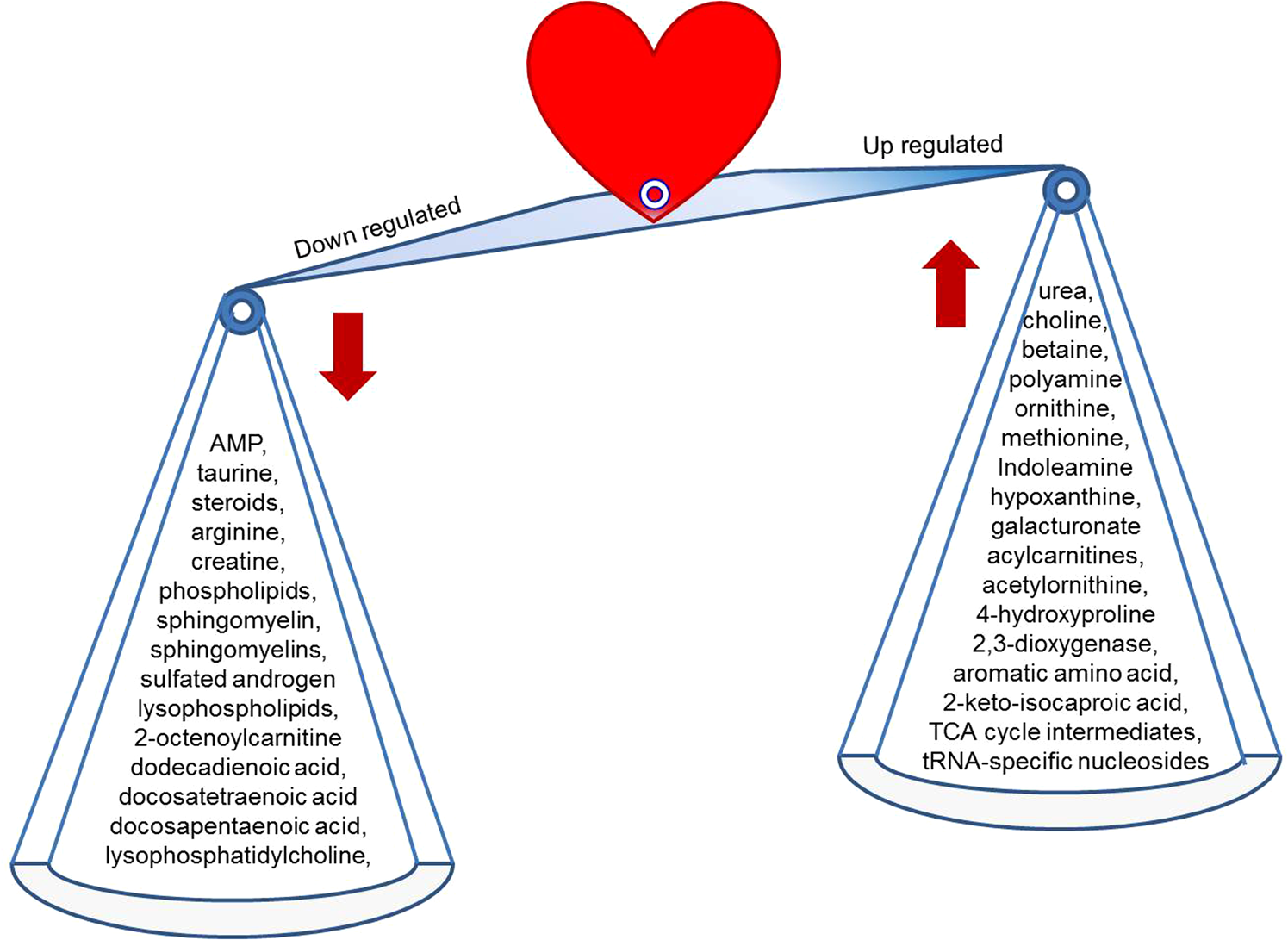
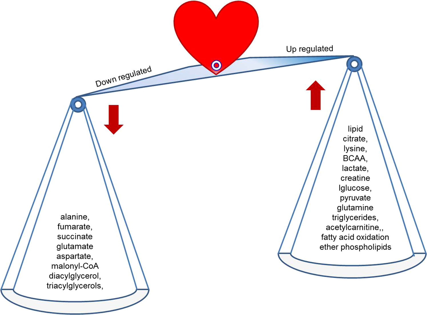
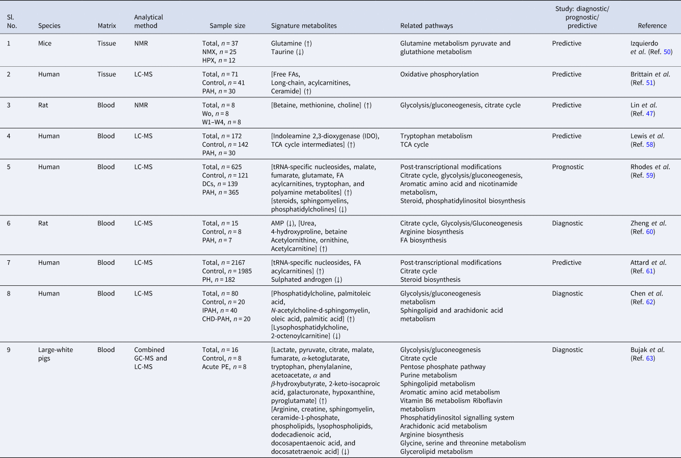
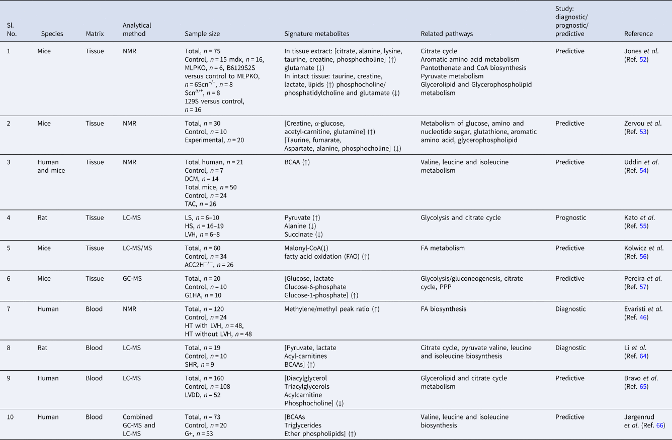
On the basis of the aforementioned readouts, it is apparent that CH is a metabolic disease and strictly associated with the harmonic imbalance of glycolysis, citric acid cycle, amino acids and lipid metabolism. To date, 19 readouts comprising nine studies with RVH (two cellular and seven systemic metabolomics) (Table 3, Fig. 3) and 10 studies with LVH (six cellular and four systemic metabolomics) are available (Table 4, Reference WheltonFig. 4), which are summarised as follows:
Summary of down-regulated and up-regulated selected metabolites in RVH observed in the gleaned literature.

Summary of down-regulated and up-regulated metabolites in LVH observed in the gleaned literature.

Cellular and systemic metabolomics-derived signature metabolites of right ventricle hypertrophy and related pathways

(↑), increase; (↓), decrease; NMX, normoxic mice; HPX, hypoxic mice; PAH, pulmonary arterial hypertension; Wo, control; W, week; DCs, disease control; PH, pulmonary hypertension; IPAH, idiopathic PAH; CHD-PAH, congenital heart disease-associated PAH; PE, pulmonary embolism; AMP, adenosine monophosphate.
Cellular and systemic metabolomics-derived signature metabolites of left ventricle hypertrophy and related pathways

(↑), increase; (↓), decrease; mdx, Duchenne muscular dystrophy; MLPKO, muscle LIM protein knock-out; Scn–/+, heterozygous gene deletion of the cardiac sodium channel; ScnΔ/+, heterozygous gain in function for the previous gene deletion; [Cr], creatine; WT, wild type; NFC, non-failing control; DCM, dilated cardiomyopathy; TAC, transverse aortic constriction; LS, low salt; HS, high salt; PPP, pentose phosphate pathways; BCAA, branched chain amino acids; ACC2H−/−, cardiac-specific deletion of acetyl CoA carboxylase-2; G1HA, haemagglutinin (HA)-tagged GLUT1 transgene; HT, hypertensive; LVH, left ventricle hypertrophy; WKY, Wistar–Kyoto rats; SHR, spontaneously hypertensive rats; LVDD, left ventricular diastolic dysfunction; G+, mutation carrier group.
The available one readout of NMR-based cellular metabolomics of RVH demonstrated that augmented levels of glutamine and taurine could be used as signature metabolites to detect human PAH (Ref. Reference Izquierdo-Garcia50). In the same line, the readout of LC-MS-based cellular metabolomics of RVH claimed that enhanced long-chain FAs and ceramide simultaneously reduced long-chain acylcarnitines can be used to identify PAH patients (Ref. Reference Brittain51).
In contrast to cellular metabolomics, NMR-based systemic metabolomics of RVH revealed that choline, betaine and methionine metabolic pathways play not only as an imperative role in the onset of PAH, but also in the development and progression of PAH (Ref. Reference Lin47). The LC-MS-based systemic metabolomics of the RVH study presented enhancement of circulating free FAs and long-chain acylcarnitines in PAH compared with controls (Ref. Reference Brittain51). In the same path, another study advocated that tryptophan derivatives and the TCA cycle, as well as purine pathways intermediate are able to identify RV-PV dysfunction (Ref. Reference Lewis58). At the next level, the LC-MS-based plasma metabolomics study differentiated PAH patients who were at high risk of early death and patients who responded well to treatment. This study advised that a group of 36 metabolites can be used as prognostic biomarkers of PAH and that metabolic profiling can be used to monitor the response to therapy as well (Ref. Reference Rhodes59). Similarly, an experimental study claimed that a group of seven metabolites can discriminate PAH from control rats using LC-MS-based plasma metabolomics (Ref. Reference Zheng60). Another LC-MS-based human serum metabolomics study established that six metabolic pathways are associated with RV adaptation to PH and respond to cellular stress (Ref. Reference Attard61). Moreover, another similar study identified PAH at an early stage and advised that a panel of lipid metabolism and FAO metabolites were closely linked with PAH (Ref. Reference Chen62). A combined approach of LC-MS- and GC-MS-based plasma metabolomics revealed that this tactic can explain the pathogenesis of PE that leads to PH and RVH (Table 3, Figs 3 and 5) (Ref. Reference Bujak63).
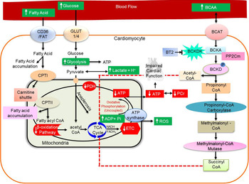
Overview of cardiac metabolism in the hypertrophic heart. Background with green colour represents augmented levels and background with red colour represents decreased levels of cascade components.

NMR-based cellular metabolomics of an MLPKO animal model of LVH exhibited that this is a rapid tactic for phenotyping of mouse models of underlying disease (Ref. Reference Jones52). Subsequently, correlations of cardiac [Cr] levels and metabolomics of LV tissue metabolites revealed that changes in substrate consumption and metabolic patterns of perturbed energy engendering pathways are highly significant at augmented levels of [Cr] (Ref. Reference Zervou53). Another NMR-based LV tissue metabolomics study exhibited the role of BCAA catabolism in human HF and indicated that induced BCAA oxidation was helpful in augmenting LV cardiac function in the TAC mouse heart (Ref. Reference Uddin54).
LC-MS-based tissue metabolomics studies have discriminated LVH and CHF and have established that both episodes are associated with distinct changes of metabolism of the heart. Moreover, DCA can be used as a therapeutic agent to attenuate the transition of LVH to CHF (Ref. Reference Kato55). On the same path, another LC-MS-based tissue metabolomics study was able to prove that ACC2 can be used to reduce LVH with the help of modulation of mitochondrial substrate input and mitochondrial oxidative capacity (Ref. Reference Kolwicz56). On the contrary, GC-MS-based tissue metabolomics has suggested that short-term cardiac-specific augmentation of GLUT1 at the onset of POH reserves mitochondrial function and diminishes pathological remodelling, concurrently aggravates the hypertrophic phenotype, and is insufficient to circumvent POH-induced cardiac impairment (Ref. Reference Pereira57).
In contrast to cellular metabolomics, NMR-based systemic metabolomics has identified the methylene/methyl ratio in plasma as a signature metabolite for rapid identification of LVH in hypertensive populations (Ref. Reference Evaristi46). The LC-MS-based systemic metabolic profiling study exhibited that an imbalance in harmonics of glycolysis, carnitine and BCAA metabolism precede and coincide with impaired cardiac function and with the development of LVH from HTN (Ref. Reference Li64). In the same line, another LC-MS-based plasma metabolomics study demonstrated that women with or at risk of HIV with LVDD showed perturbed metabolic profiles and suggested that these vital alterations may be used to avert metabolic impairment of LVH (Ref. Reference Bravo65). At the next level, a combined GC-MS- and LC-MS-derived systemic metabolomics of LVH revealed and suggested that BCAAs, triglycerides and ether phospholipids play an important role as central metabolic regulatory mechanisms in LVH (Table 4, Figs 4 and 5) (Ref. Reference Jørgenrud66).
Clinical relevance
Briefly, although some studies (Refs Reference Kato55, Reference Kolwicz56, Reference Chen62) have claimed that metabolomics-derived outputs can be used as therapeutic interventions of CH, such as DCA may be considered as a therapeutic agent to suppress the translation of LVH to CHF (Ref. Reference Kato55), the application of ACC2 suppression may be a robust tactic for therapeutic interventions of LVH (Ref. Reference Kolwicz56), and lipid metabolism intermediates can be good target agents for therapeutic applications of PAH (Ref. Reference Chen62). However, even after extensive efforts, clinically accepted unequivocal metabolic biomarkers for the management of CH are still lacking. Also lacking are clinical research studies with common metabolic biomarkers that disclosed from different platforms (NMR and MS) and different biological samples, such as tissue and blood. This raises the question of the significance of on-going progress and the auxiliary modification of metabolomics-derived findings. Although highly sensitive MS and highly specific NMR techniques have been applied to engender metabolic causes that lead to CH and subsequently to HF, it seems that the above-mentioned studies are just initial steps. These studies are not enough to conclude thorough biochemical aspects that may lead to CH. We need further extensive and well-defined studies to develop the actual molecular causes of CH as well as target agents for molecular therapeutics of CH.
Limitations of metabolomics for clinical application and overcoming barriers
Effective translation from-bench-to-bedside involves extensive appreciation of the underlying science, reception of ideal working measures, schooling in the understanding of outcomes and the availability of trustworthy metabolomics data archives. For instance, the transformation from a research technique to a feasible clinical approach involves auxiliary logistics through an array of developments at an indigenous level. Concerns are presently associated with the design and size of apparatus, exploratory justification, uniformity of approaches and explainable in a sincere and reproduce manner.
Translation pivots on numerous barriers. The first barrier is the uniformity of exploratory processes and apparatuses, principally over the acceptance of approved operational processes (Ref. Reference Emwas67). The robustness of the data needs normalisation. Considering this scenario, the NIHR-MRC National Phenome centre has computed the normalisation of specimen collection and examination (Ref. 68). Targeted metabolomics is based on analysis and gauging of a panel of a few, well-identified metabolites. However, the indemnity of the metabolic deployment is restricted; the lower charge means that huge groups may be identified. This may guide us to important insightful information regarding the aetiology of the underlying disease. Additionally, because of its economical price and its uniform procedure, these kit-derived tactics have earned recognition.
Two service providers, Biocrates and Metabolon, deal rapid quantification of a plethora of metabolites using a semi-quantitative approach. Siskos et al. have revealed 7.6% of inter-laboratory coefficient of variation by the application of different apparatuses with the same procedure (Ref. Reference Siskos69). The study also revealed profound information of the error model of instrument. This information is valuable mainly for data scientists involved in meta-analysis and in executing the amalgamation of datasets. Various consortia have chosen to use metabolomics approach for phenotyping with several participants. For instance, Jourdan et al. conducted a study with KORA S4 (aged 54–75 years, n = 1614 participants) and KORA F4 (aged 31–82 years, n = 3061 participants) to compare their serum metabolomics and to establish the association between the body fat-free mass index and branched-chain amino acids (Ref. Reference Jourdan70). Similarly, Lotta et al. led a metabolomics study with approximately 10 000 patients to examine the association between the impaired metabolism of the branched-chain amino acids and the risk of type-2 diabetes (Ref. Reference Lotta71). Draisma et al. conducted a study with 7500 patients using Biocrates profiling to determine the genetic variants contributing to serum metabolite variations (Ref. Reference Draisma72). These readouts demonstrate how metabolomic studies may be applied proficiently to assist the interpretation of underlying disease as well as profound mechanisms of the disease and how this novel approach can advise clinical recommendations in regular practice.
The second crucial obstacle is in the precise acquisition and analysis of data. The above-mentioned NMR and MS are two of the most important analytical techniques; both comprise considerably distinct prerequisites. Originally, in order to conduct precise collection of data, there are complaints with sample size and time. The processing of the acquired data is tricky and comprises fixing of experimental artefacts, apparatus variations, fixing of batch and normalisation. This begins with huge variations in the standardisation of data analysis. Compared with MS, NMR is more inherently highly reproducible with standard milieus, in part because the detector does not come into physical contact with the biological specimens. MS, in spite of its utmost sensitivity, endures with imperfect reproducibility because of different machine responses over time and different operators or laboratory management (Refs Reference Katajamaa73, Reference Wu74). Substantial hard work is obligatory in MS to acquire the robust data required for transformation to clinical practice.
The third barrier is the understanding of data, which entails cutting-edge chemometric statistical tactics such as machine learning. An important stumbling hindrance is the high level of competency needed to fit and decode these replicas vigorously at an indigenous level for this expertise to progress into regular clinical practice. In recent years, huge advances have taken place in both considering the necessity for elucidation of data in this field (e.g. noise removal, normalisation and indefinite tactics to data analysis) and in the inception of novel algorithms for detection and prediction of metabolites, mainly from machine-learning approaches (Refs Reference Takis75–Reference Alakwaa77). In certain circumstances, such as the iKnife, a technique applied to a binary verdict: tumour or not tumour. In other presentation (e.g. urine specimens) will undoubtedly entail uniform submission of computational channels that ensue in robust and understandable outputs. This is one of the key objectives of the EU-2020 agenda of PhenoMeNal – a European e-infrastructure that delivers compatible tools and procedures for contemporary and comprehensive clinical data of metabolomics (Ref. 78). Besides the integration of machine learning, attaining a biochemically persistent note in untargeted metabolic profiling remains a main issue in metabolomics. The present benchmark for precise characterisation of a metabolite pivots around analogous distinguished features to a commercially available, certified compound, which has been achieved following comparable approaches and similar apparatuses. This stiffness rationally leads to various pragmatic issues during attempts to use this approach on colossal datasets. A broadly applied method to address this issue is to apply spectral archives, which are usually partial, or substitute computational approaches, which may have several analogous characteristics with solitary features (Ref. Reference Alden79). Apparently, this is an additional critical difficulty in the broad circulation of the technology in our current epoch.
The fourth barrier concerns the cybernetic and e-infrastructural assistance that is highly necessary. Numerous data of human metabolomics are, at present, imperfect and are stored amidst shattered arrays of data banks. The project per se, PhenoMeNal intention to offer biomedical investigators through proven and regenerable, operational, routine tests of clinical specimens. This comprises the algorithm-based high-tech aggregation of metabolomics data for the underlying disease foliation and procedures for biological pathway enhancements. Moreover, PhenoMeNal also offers readily reachable cyber infrastructures that may support broad investigation through adequate mathematics and reservoir reserves.
The transformation of novel metabolomics information into routine clinical habits will entail the improvement of an ethical context that confirms obedience and implementation of the rights of participating subjects. Efforts regarding PhenoMeNal (http://phenomenal-h2020.eu/home/), Elixir (https://www.elixireurope.org/) and Tryggve (https://neic.no/tryggve) are paving the way for leading-edge, extensible, trans-border e-infrastructures for effective, secure and an e-amenable reservoir of delicate, individual and metabolomics-derived phenotyping information. These efforts are also heavily contributing to demonstrate informative power to ensure Findable, Accessible, Interoperable, Re-usable (FAIR) information ethics execution in the evaluation, exchange and reworking of delicate individual information. As long as it has been duly regulated, the eventual path of these contexts will permit next-level large data evaluation and artificial intelligence competencies to merge and harmonise the advanced evaluation of these metabolic information datasets.
Conclusions
Metabolomics is a hypothesis generating branch of biomedical research. It is able to demonstrate with evidence that biochemical changes generate and manifest earlier then histological cell abnormalities that lead to anatomical changes of the heart. Studies that provide proof-of-evidence-based meta-analysis comprising highly specific, sensitive, reproducible analytical platforms and cellular as well as systemic biological matrices with the same qualitative and quantitative measures are lacking. Metabolomics facilitates investigators' use of various analytical platforms and biological matrices to delineate real-time molecular mechanisms that are accountable for mediating different forms of pathologies of cardiovascular system. It can reveal the biochemical differences between pathological and physiological CH that can be used to open new possibilities for novel drug development. Metabolomics-derived detection of inimitable signature metabolites can be used as regulators of CH and towards the development of novel pharmacotherapies in the clinical management of HF.
Metabolomics is a highly spirited and futuristic division of clinical research that exhibits the utmost power to ascertain the signature metabolic pathways at an initial stage before the onset of CH. All the above-mentioned studies are based on a single healthcare centre with limited sample size. In order to move on to the next level of this potential technology, multicentre and multi-country studies with personalised and appraisal of global cohorts are now essential. The investigations that amalgamate multi-omics may contribute to verifying the clinical utility of metabolomics results and to possibly illuminate the signature metabolic biomarker step along the path for clinical applications. The promising aftermaths of the above-mentioned gleaned readouts exhaustively recommend that metabolomics is an extremely powerful technology for not only CH screening but also for monitoring of the transition from HTN to CH, which can be a highly beneficial aspect for clinical management of CH and reduce the risk of HF.
Future opinions
The decades after the genomic epoch have furnished the opportunity to reveal profound information of pathogenesis of CVD. Since most cardiac diseases share impairments in cardiac metabolism, metabolomics specifically has innovative hypothesis-generating potential to understand the molecular underpinnings of underlying diseases. Contrast to other omics approaches, metabolomics offers functional profound comprehensive information regarding upstream genetic, transcriptomic and proteomic differences, along with environmental stimulus; hence, metabolomics reflects particular metabolic pathways more closely to pathology. Metabolomics data should not be evaluated individually because metabolic networks do not present individually in the biological system. All omics have extremely intricate and vigorous connectivity. The integration of metabolomics information along with other omics epitomises a powerful and emerging tactic to reveal a highly comprehensive aspect of the intricate connectivity of CH. Innovative tactics are escalating scientific facility to define the metabolome and to measure flux via particular metabolic trails to trace the providences of specific substrates. On the contrary, progress in the advancement of computational tactics should also remain to permit the incorporation of omics datasets in order to conclude a general opinion of the molecular underpinnings of cardiac ailments. As the actual mechanisms of the metabolic causes of cardiac diseases remain to be revealed, metabolomics will lead to the scientific innovation in this field. Metabolomics can be used to extract key molecular mechanisms that cause the initiation and progression of pathological concentric and eccentric CH. Moreover, combined cellular and systemic studies can demonstrate the profound understanding of the mechanisms that cause the transition from HT to CH and eventually to HF. It would be extremely imperative for the development of novel and next level therapeutics of CH with high capacity to improve cardiac function of the failing heart in contrast to delaying the advanced stages of underlying disease.
Author contributions
S.P.S. and A.G. searched data for this review article. A.G. and S.P.S. wrote the manuscript and generated figures and tables. R.S., S.P.S. and S.K.S. edited the manuscript. R.S. suggested and wrote the clinical part of the manuscript. All authors made substantial contributions to discussions of content and reviewed and edited the manuscript before submission.
Financial support
The authors thank Indian Council of Medical Research, New Delhi, India for providing financial supports of student fellowship (5/3/8/3/ITR-F/2020) and Ad hoc-grant (5/3/8/39/2020-ITR).
Conflict of interest
The authors have no relevant affiliations or financial involvement with any organisation or entity with a financial interest in or financial conflict with the subject matter or materials discussed in the manuscript. This includes employment, consultancies, honoraria, stock ownership or options, expert testimony, grants or patents received or pending or royalties.