Chagas disease is an infection caused by Trypanosoma cruzi. It is transmitted to humans by blood-feeding hemipterans of the Triatominae subfamily and less frequently by transfusion or oral transmission. Transplacental congenital transmission is its main cause in the paediatric population. It affects six to seven million people in Latin America,1 but migration has made it a concern in many countries.2,Reference Moscatelli, García Bournissen and Freilij3 Historically considered a regional disease, Chagas disease has now become a global disease, reaching regions such as Europe, USA, Australia, and Japan.Reference Moscatelli, García Bournissen and Freilij3–Reference Montgomery, Starr and Cantey7
The acute stage of Chagas disease is usually followed by a chronic oligosymptomatic stage. After many years, cardiac manifestations may appear in approximately 30% of patients.Reference Prata8 Multiple mechanisms may be responsible for the development of cardiac lesions in Trypanosoma cruzi infection. The presence and persistence of the parasite is a critical factor, which triggers a specific immune response, inducing vascular endothelial cell damage. This may play an important role in the pathogenesis of Chagas disease cardiac involvement.Reference Dávila, Rossell and de Bellabarba9 At tissue level, a diffuse cellular infiltrate with microcirculation alterations affects the parasympathetic innervations, producing a relative increase of sympathetic activity and further cardiac damage and arrhythmia.Reference Bonney and Engman10 Electrocardiogram alterations are early signs of cardiac involvement. Some electrocardiogram signs of Chagas disease are complete right bundle branch block, QRS complex widening or fragmentation, and sinus bradycardia. In more advanced stages, damage of the cardiac conduction system leads to second and third-degree atrioventricular block, atrial arrhythmias, ventricular premature beats, or ventricular tachycardia.Reference Prata8,Reference Rojas, Glisic and Pletsch-Borba11 In later stages, cardiac alterations in adults may include segmental dysfunctions in the wall and ventricular aneurysms or global alterations, such as dilated cardiomyopathy associated with valve insufficiency.Reference Marin-Neto, Cunha-Neto and Maciel12 It is very rare to find echocardiographic alterations in paediatric Chagas disease patients, although cases have been described.Reference Salazar-Schettino, Cabrera-Bravo and Vazquez-Antona13
Two-dimensional speckle-tracking echocardiography is a relatively new technique that allows detailed analysis of left ventricle movement and deformation. Regional strain measures the percentage of deformation between two ventricle sites during contraction, and strain rate is the speed at which this deformation occurs.Reference Geyer, Caracciolo and Abe14 These methods allow early detection of segmental alterations.Reference Acquatella, Asch and Barbosa15
During the acute phase of the disease, parasites may be found in blood and infect cardiomyocytes, but are not found in blood thereafter. Persistence of the parasite in cardiomyocytes appears to be fundamental in the development of cardiomyopathy,Reference Zhang and Tarleton16 so effective treatment during the acute phase is important, however it is not yet clear whether treatment prevents development of cardiologic complications.
The aim of this study was to use new echocardiographic techniques to determine whether paediatric patients treated for Chagas disease show delayed alterations of cardiac function.
Material and method
Doppler and speckle tracking echocardiograms were obtained from 84 patients and 27 controls between August 2017 and July 2019. All patients were treated for Chagas disease with benznidazole (6.5 mg/kg/day) or nifurtimox (10 mg/kg/day), and followed up in the Parasitology and Chagas unit at the Ricardo Gutierrez Children’s Hospital. The study included patients at least 6 years after treatment completion. The control subjects were relatives of the infected persons with negative serologies.
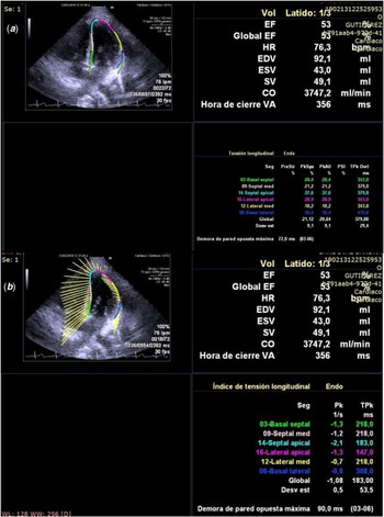
Cardiac function was evaluated with a Siemens® Acuson SC 2000 echograph and a 2–5 MHz cardiac and three-dimensional matrix transducer. An electrocardiographic record was also obtained in all patients. All images and clips were stored on the hard disc for later reproduction and analysis. A single operator collected the measurements. Patient’s values were plotted in z-score charts for age and compared to values obtained from control subjects. Left ventricular diameters and fractional shortening were assessed from a ventricular short axis view using two-dimensional M-mode method. Mitral and tricuspid annular plane systolic excursion (apical four-chamber view), were obtained using two-dimensional M-mode at speed of 100 mseconds. Doppler evaluation of mitral valve E wave (rapid filling) and A wave (presystole) in metres per second, and tissue Doppler, e’, a’, and s’ waves in centimetres per second, were also measured in apical four-chamber views and at a speed of 100 mseconds. The E/e’ ratio was calculated for all patients. Estimated right ventricle systolic pressure was calculated by assessing tricuspid regurgitation in four- chamber views. Tei index was measured in five-chamber views. Three-dimensional left ventricle ejection fraction and volume were automatically calculated using Siemens® software, in four-chamber views. Two-dimensional apical four-chamber view was used to measure global left ventricle peak systolic longitudinal strain and longitudinal systolic strain rate. At least four beats were recorded for later measurements. Encirclement of the ventricle was manually done at endocardiac level (Fig 1).
Example of measurement of longitudinal peak systolic strain (a) and longitudinal systolic strain rate (b) from apical 4-chamber view.

Standard apical four-chamber views were acquired with a frame rate of 30 to 60 f/seconds. For motion-mode images of the left ventricle and pulsed wave Doppler images of the left ventricle, frame rate was above 150 f/seconds.
Reproducibility
Inter- and intra-observer variabilities of myocardial deformation parameters were assessed in 30 randomly selected studies following the Bland and Altman method.Reference Bland and Altman17 To assess intra-observer variability, the same observer who performed all the measurements (A.G.), re-measured the same segments at least 1 month later. To assess inter-observer variability, deformation parameters were measured by one independent and blinded observer (C.M.). Reproducibility was expressed as bias (average difference between the paired samples) and its limits of agreement, calculated as +1.96 and −1.96 times the standard deviation of the difference of paired samples. Moreover, the coefficient of variation was calculated as the standard deviation of the difference of paired samples (absolute values) divided by the average of the paired samples and multiplied by 100.
Inclusion criteria: Chagas disease patients that have completed treatment with benznidazole or nifurtimox at least 6 years before the current evaluation.
Exclusion criteria: Patients with congenital heart disease. Patients with chronic kidney, liver and neurological diseases.
Ethical consideration: The protocol was registered at clinical trials.gov # NCT04090489.
Statistical analysis: Data in all tables are mean ± standard deviation unless stated otherwise in the legend. All comparisons between two groups were made with Student’s t-test. p < 0.05 was considered statistically significant. IBM SPSS® 20 statistical software for Windows was used.
Results
Patients
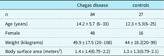
The mean age of Chagas disease patients (n = 84) and healthy controls (n = 27) was 14.2 years (6–33) and 12.3 years (6–25) respectively. The number of female subjects was 48 in the Chagas disease group and 16 in the healthy group. All subjects were born in the Argentine Republic and lived in the Buenos Aires Metropolitan Area during the whole follow up period (since treatment for Chagas disease finished after diagnosis). The route of infection was mainly congenital. Mean follow-up after treatment was 10 years, which explains the presence of adult patients in the protocol. Average weight at the time of the studies was 49.9 kg (20–108) in Chagas disease patients and 44 kg (20–99) in control subjects (Table 1). All subjects were asymptomatic at the time of the studies.
Patient demographics

Mean values ± Standard deviation (range).
Analysis of left and right ventricular systolic function by two-dimensional and M-mode echocardiography
Mean diastolic diameter was 42.1 ± 5.1 mm (32–54) in Chagas disease patients and 40.2 ± 6.1 mm (27–54) in controls (p = 0.11). Shortening fraction, which was assessed in the short ventricular axis using M-mode, was 41.5 ± 5.3% (31–53) and 42.7 ± 4.4% (32–56) in patients and healthy subjects, respectively (p = 0.3). Mitral annular plane systolic excursion was on average 16 ± 2.1 mm (12–22) in Chagas disease patients and 16 ± 2.1 mm (11–20) in control subjects (p = 0.97), and tricuspid annular plane systolic excursion was 20 ± 3.4 mm (11–29) and 21 ± 3.3 mm (15–27) respectively (p = 0.34). No significant differences were found between Chagas disease patients and controls. All measurements were correlated with percentiles for age and were normal.
Analysis of left ventricular systolic and diastolic function by Doppler echocardiography
Mean E wave was 0.85 m/second in Chagas disease (range, 0.6 to 1.1 m/second) and 0.92 m/second in controls (range, 0.7–1.2 m/second) (p = 0.01), while A wave was 0.5 m/second in both groups (range, 0.3 to 0.7 m/second for Chagas disease patients and 0.3 to 0.6 m/second for controls) (p = 0.06). The tissue Doppler evaluation provided an average of 16.2 cm/second (10 to 23) and 17.2 cm/second (13 to 23) for e’ wave (p = 0.10), 7 cm/second (4 to 10) and 6.9 cm/second (5 to 10) for a’ wave (p = 0.72), and 10.5 cm/second (6 to 16) and 10 cm/second (6 to 15) for s’ wave (p = 0.26), for patients and control subjects respectively. The average Tei index was 0.3 in both groups (range 0.15 to 0.51 and 0.18 to 0.46 for controls) (p = 0.1), but in one patient with Chagas disease a value higher than 0.5 was obtained. The E/e’ ratio returned a mean of 5.4 (3 to 10) and 5.5 (3.9 to 7.7), respectively (p = 0.92), but three patients presented values above eight. The values were normal and no differences were found between Chagas disease patients and control subjects. Estimated right ventricle systolic pressure measured by tricuspid regurgitation was normal in all cases.
Left ventricle ejection fraction analysis by three-dimensional echocardiography
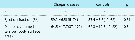
Ejection fraction was measured by three-dimensional echocardiography in 56 patients with Chagas disease and in 17 controls. Mean ejection fraction was 59.2% (45 to 74) and 57.4% (49 to 68) respectively. Average left ventricle diastolic volume was 64.5 ml/body surface area (37 to 122) and 62.3 ml/body surface area (40 to 82). No significant differences were found between both groups (Table 2). An ejection fraction lower than 50% was found in four patients with Chagas disease and one control subject, while left ventricle volume in six Chagas disease patients presented values equal or greater than 90 ml/body surface area.
Three-dimensional echocardiogram of left ventricle
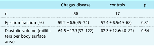
Mean values ± Standard deviation (range).
Strain and strain rate analysis by two-dimensional speckle tracking echocardiogram
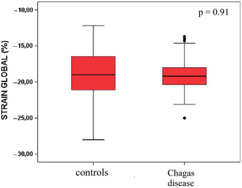
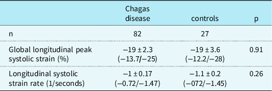
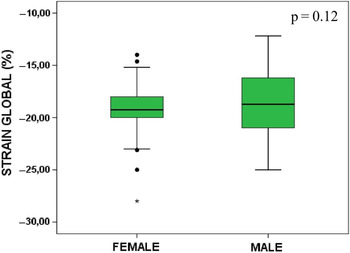
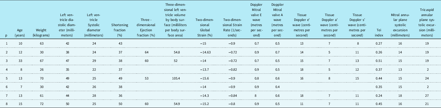
This evaluation could be carried out in 82 patients and 27 controls. Mean four-chamber global longitudinal peak systolic strain was −19% (range, −13.7 to −25 and −12.2 to −28 for controls) in both groups while two-dimensional longitudinal systolic strain rate was −1/seconds (−0.72 to −1.47) and −1.1/seconds (−0.72 to −1.45) for Chagas disease patients and control subjects respectively (Fig 2 and Table 3). The median for longitudinal strain were −19.1 and −19% for patients and controls respectively, while for strain rate it was −1/seconds in both groups. There were no statistically significant differences between groups. No gender differences were found in global longitudinal strain in Chagas disease patients (p = 0.12) (Fig 3). Values were above −16% (i.e., less negative) in eight patients with Chagas disease (Table 4) and three controls. Regarding strain rate, values above −0.8/seconds were observed in three Chagas disease patients and two control group subjects.
Left ventricle global peak systolic longitudinal strain. Box and whisker plots for controls (n = 27) and Chagas disease (n = 82).

Two-dimensional strain measurements of the left ventricle

Mean values ± Standard deviation (range).
Left ventricle global peak systolic longitudinal strain and gender. Box and whisker plots for female (n = 48) and male (n = 36).

Profile of patients with lower limit two-dimensional global peak systolic longitudinal strain of the left ventricle

Reproducibility of speckle tracking echocardiography was assessed in 30 randomly selected subjects (Table 5). For global longitudinal peak systolic strain, the results are in good agreement with those reported by others,Reference Koopman, Rebel and Gnanam18 with a coefficient of variation of less than 10% both for intra- and inter-observer measures. Of note, for two-dimensional longitudinal systolic strain rate, the coefficient of variation was high probably as a consequence of the low frame rates that could be used in our clinical setting study.
Intra- and inter-observer variability of left ventricle two-dimensional strain measurements

Discussion
Physiopathology of chronic myocardial damage in Chagas disease is complex and multifactorial.Reference Tarleton and Zhang19 Experimental studies conclude that in early stages of myocardial dysfunction the combination of inflammation, necrosis, and fibrosis in focal areas produces alterations in segmental mobility of the left ventricle wall that could lead in later stages to global ventricular dysfunction and cardiomyopathy.Reference De Oliveira, Romano and De Carvalho20 Bearing this in mind, we speculated that strain and strain rate measurements with speckle tracking echocardiography could reveal early myocardial dysfunction in the patients analysed in the present study. The main finding of the present study is that global longitudinal strain is a reproducible measure useful for the follow up of paediatric Chagas disease patients, which show no evidence of early myocardial dysfunction when assessed several years after completing treatment.
Although there is currently consensus on using global longitudinal strain and strain rate measurements to assess early myocardial dysfunction in paediatric patients, such as cardiotoxicity in paediatric patients who underwent chemotherapy,Reference Plana, Galderisi and Barac21,Reference Thavendiranathan, Poulin and Lim22 to our knowledge, there were not any previous reports of these measurements in paediatric patients treated for Chagas disease. Normal global longitudinal strain values in children vary subtly according to the echocardiographic methodology used and depending on whether just one view (four chambers view) or an average of several views are taken into consideration.Reference Levy, Machefsky and Sanchez23 According to Jashari et al, who made a meta-analysis, mean value is −20.5% with a range of −12.9 to −26.5%.Reference Jashari, Rydberg and Ibrahimi24 In another meta-analysis Levy et al concluded that mean two-dimensional global longitudinal strain value measured in four chambers view was −20.4% with a range of −15.1 to −24.8%. The value of the review by Levy et al is that it highlights the differences introduced by the different vendor platforms and echocardiographic views.Reference Levy, Machefsky and Sanchez23 Using the same Philips platform used in the present study, in 103 healthy paediatric patients, Koopman et al obtained mean values in four-chamber views of −20.6% with percentile 5 of −25.2% and percentile 95 of −16.8%.Reference Koopman, Rebel and Gnanam18 Finally, in 238 patients Kuebler et al found a mean of −21.6% (−12.9%/−29.6%) with differences for age, where older patients had less negative values.Reference Kuebler, Ghelani and Williams25 In our study of 82 young patients, the average global longitudinal strain of −19% is well within the range considered normal in recent literature,Reference Koopman, Rebel and Gnanam18,Reference Levy, Machefsky and Sanchez23–Reference Kuebler, Ghelani and Williams25 and even the highest values observed fell within the normal range. We looked in more detail the data of eight patients with global longitudinal strain values above −16% and found that their other studied variables were within the normal range, except for one patient who presented a Tei index above 0.5 (0.51) and another patient with left ventricle volume slightly above Z+2 (105.4 ml/body surface area). Regarding the Tei index, in an investigation of 289 healthy children, Cui et al concluded that its normal value is <0.5, with a mean of 0.36 ± 0.07.Reference Cui and Roberson26 In our study, the patient with a Tei of 0.51 had a global strain of −14% (Table 4). Three control subjects also showed global longitudinal strain values above −16%, but none presented other alterations. With no statistically significant between-group differences and all patient values falling within the normal range reported by previous studies, we considered global longitudinal strain to be normal in the Chagas disease patients.
Ferferieva et al concluded that strain rate depends less on cardiac load conditions than global longitudinal strain.Reference Ferferieva, Van den Bergh and Claus27 This feature of strain rate may allow a better evaluation of segmental contractility. Normal two-dimensional strain rate values are very heterogeneous and in the meta-analysis published by Levy et al of 889 paediatric patients, involving 19 studies using four-chamber view, an average of −1.2/seconds with a range of −0.41 to −2.59/seconds was found. This dispersion of values did not depend on age, gender, heart rate and blood pressure.Reference Levy, Machefsky and Sanchez23 Using the Philips® platform, Koopman et al obtained a mean of −1.4 ± 0.30/seconds in 103 patients.Reference Koopman, Rebel and Gnanam18 Finally, in another meta-analysis by Jashari et al the average strain rate was −1.3/seconds (−0.9 to −2.7) and changed with age.Reference Jashari, Rydberg and Ibrahimi24 In our sample, the mean was −1/seconds, coinciding with the average values in other publications. We analysed in more detail three patients who presented values above −0.8/seconds and two with a global longitudinal strain above −16%, including the one having a Tei index of 0.51, however, they were asymptomatic and the other measurements were normal. Since our strain rate data showed a high intra- and inter-observer variability, further studies with a higher frame rate should be necessary to assess its utility in the follow-up of Chagas disease patients.
In paediatric patients with Chagas disease, clinical and echocardiographic alterations are infrequent, even in untreated patients.Reference Da Cunha, Daud and Rimmel28 A large retrospective study performed in Brazil that included all medical records obtained at a University Hospital between 1965 and 1984 and more than 18000 autopsies, revealed 19 Chagas disease patients under 18 years old with positive serology and findings compatible with chronic Chagasic heart disease.Reference de Araujo, Bessetti, Godoy and Oliveira29 In a seroepidemiological study performed in México, where more than 3000 untreated individuals younger than 18 years old living in a risk area were tested, Salazar-Schettino et al found 14 paediatric patients with septal and/or wall hypertrophy, ejection fraction below 57% and increased systolic pulmonary artery pressure.Reference Salazar-Schettino, Cabrera-Bravo and Vazquez-Antona13 Although average ejection fraction in our study was 59.2% and there were no differences with the control group, four patients with Chagas disease and one control subject presented values below 50%, without any other pathological finding. These patients are being followed-up to determine whether these changes predict the emergence of cardiomyopathy. There is a debate concerning which is the lowest ejection fraction that can be considered normal in children assessed with three-dimensional echocardiography. Krell et al reported mean and standard deviation ejection fraction values of 61.5 ± 5% and 62.7 ± 6%, for 370 healthy paediatric subjects, depending on the software used for analysis.Reference Krell, Laser and Dalla-Pozza30 Kuebler et al suggested 51% as a limit and this value correlates better with calculations based on magnetic resonance imaging.Reference Kuebler, Ghelani and Williams25 Lastly, a meta-analysis published by Bucchieri et al reports 61% as a minimum.Reference Buccheri, Costanzo and Tamburino31 The values below 50% observed in some of our patients are below the normal limits suggested by all these studies.
Concerning our three-dimensional echocardiographic measures of left ventricle diastolic volume, six patients with Chagas disease showed values above normal limits (volume equal or greater than 90 ml/body surface area), if we refer to Krell et al age- and gender-adjusted body surface area-indexed percentiles.Reference Krell, Laser and Dalla-Pozza30 However, if we take the work of Kuebler et al, who calculated z-scores based on body surface area and ventricle volume, regardless of age and gender, only two patients had volumes above Z+2 (105 and 123 ml/body surface area respectively).Reference Kuebler, Ghelani and Williams25 These patients presented no other pathological echocardiographic findings. Strikingly, the only parameter studied that showed a statistically significant between groups difference was the mitral valve E wave, but the change was not accompanied by any other indication of diastolic dysfunction and was not considered of clinical relevance in the present study.
A limitation of the present study is the limited number of controls. Additionally, frame rate should be increased in future studies to improve strain rate measurement reproducibility. Moreover, subsequent studies could include segmental strain analysis, not just global left ventricle analysis, to increase the chances of detection of early wall-motion alterations.
In summary, in our case control study of echocardiographic alterations in Chagas disease paediatric patients assessed on average 10 years post-treatment, no statistically significant clinically relevant between group differences were found. Follow up studies of the few patients showing values outside the normal range will indicate whether strain analysis is useful in predicting the emergence of delayed cardiac alterations. Taking into account the long latency between infection and emergence of cardiac alterations in Chagas disease, the main clinical implication of our study is that the assessment of left ventricle strain, ejection fraction and diastolic volume should be included in the long-term follow-up of paediatric Chagas disease patients, in an aim to detect early myocardial dysfunction and thus be able to start treatment.
Conclusions
Paediatric patients treated for Chagas disease do not present significant echocardiographic alterations in a follow-up study performed several years after completion of treatment with both conventional techniques and new tools to evaluate ventricular function.
Acknowledgements
None
Financial support
This research received no specific grant from any funding agency, commercial or not-for-profit sectors.
Conflicts of interest
None to report
Ethical standards
The study is in accordance with the 1964 Declaration of Helsinki and was approved by the ethics committee of the Ricardo Gutierrez Children’s Hospital. Written informed consent by the patient and healthy controls and/or parents was obtained for all patients according to age and local regulations. The protocol was registered at clinical trials.gov # NCT04090489