Salmonella is a genus of Gram-negative bacteria in the family Enterobacteriaceae. Salmonella includes two species: Salmonella enterica and Salmonella bongori. Salmonella enterica (S. enterica) is divided into six subspecies and 2500 serotypes. Salmonella species are intracellular pathogens; some serotypes cause disease (Grimont, Reference Grimont2008) and S. enterica has a worldwide impact on public health as it is one of the most important aetiological factors causing gastroenteritis (serovar Typhimurium or Enteritidis) and typhoid fever (serovar Typhi: Dougan and Baker, Reference Dougan and Baker2014; Garai et al., Reference Garai, Gnanadhas and Chakravortty2012). S. enterica is the serotype most commonly isolated from humans and is the second most common etiological agent of human gastrointestinal infections after Campylobacter spp (Agbaje et al., Reference Agbaje, Begum, Oyekunle, Ojo and Adenubi2011). Regarding S. enterica transmission to humans, the primary vector for transmission of this bacterium to cause disease in humans is food, a significant health problem in developed and underdeveloped countries (Lamas et al., Reference Lamas, Miranda, Regal, Vazquez, Franco and Cepeda2018). S. enterica can be found in animals consumed by humans, such as poultry, sheep, cattle and pigs. Farm animals are the most commonly infected, at rates of between 17.6 and 28%, due to mishandling (Evangelopoulou et al., Reference Evangelopoulou, Kritas, Govaris and Burriel2014; Rodriguez et al., Reference Rodriguez, Pangloli, Richards, Mount and Draughon2006).
Thediarrhea cases due to Salmonella (serotype enterica the most prevalent) are estimated to exceed 150 million, with 55,000 deaths annually (Alba et al., Reference Alba, Leekitcharoenphon, Carfora, Amoruso, Cordaro, Di Matteo, Ianzano, Iurescia, Diaconu, Study Group, Pedersen, Guerra, Hendriksen, Franco and Battisti2020; Besser, Reference Besser2018; Kwambana-Adams et al., Reference Kwambana-Adams, Darboe, Nabwera, Foster-Nyarko, Ikumapayi, Secka, Betts, Bradbury, Wegmüller, Lawal, Saha, Hossain, Prentice, Kampmann, Anderson, Dalessandro and Antonio2015). This high prevalence is due to virulence factors (for example, toxins, flagellae, capsules, adhesion proteins, among others), species variability (high numbers of serotypes), the capacity to contaminate food and antibiotic resistance, this being one of the most important (Huang et al., Reference Huang, Chen, Wong, Chiu and Chu2019; Jajere, Reference Jajere2019). Several studies have isolated S. enterica from different sources, such as livestock, raw meat and seafood, and have shown that it has high antibiotic resistance (Gharieb et al., Reference Gharieb, Tartor and Khedr2015; Vinueza-Burgos et al., Reference Vinueza-Burgos, Cevallos, Ron-Garrido, Bertrand and De Zutter2016). This represents a significant problem for public health, because foodborne infections are caused by multidrug-resistant (MDR) S. enterica strains. Infectious diseases caused by MDR strains increase the probability of treatment failure, complicating the patient or even causing death (Mills and Marchaim, Reference Mills and Marchaim2021). S. enterica uses several mechanisms to increase antibiotic resistance, however, biofilm formation is one of the most important. Biofilms are structured populations of microorganisms attached to a surface and embedded in an extracellular matrix consisting mainly of exopolysaccharides, proteins and extracellular DNA. They act as barriers to avoid the effects of antibiotics and immune system cells (Abraham WR, Reference Abraham2006; Panlilio and Rice, Reference Panlilio and Rice2021). Bacteria within biofilms could be 1000 times more resistant than other bacteria because they survive in conditions of low nutrients, different atmospheres, heat, acid pH and low temperatures (Lamas et al., Reference Lamas, Miranda, Vazquez, Cepeda and Franco2016).
Biofilms, coupled with the antibiotic resistance that bacteria have developed, are closely related to the indiscriminate use of antibiotics not only in clinical practice but also in agriculture, aquaculture and veterinary practice (Aslam et al., Reference Aslam, Khurshid, Arshad, Muzammil, Rasool, Yasmeen, Shah, Chaudhry, Rasool, Shahid, Xueshan and Baloch2021; García-Vello et al., Reference García-Vello, González-Zorn and Setsoafia Saba2020; Manyi-Loh et al., Reference Manyi-Loh, Mamphweli, Meyer and Okoh2018). Therefore, there is a need to develop new strategies to combat infectious diseases. Innate immune proteins and peptides with antimicrobial activity, such as lactoferrin, are under investigation. Lactoferrin (LF) is a cationic monomeric glycoprotein that has demonstrated antimicrobial activity against Gram-positive and Gram-negative bacteria by direct (bactericidal/cell membrane interaction) or indirect (bacteriostatic/iron sequestering) mechanisms (Acosta-Smith et al., Reference Acosta-Smith, Viveros-Jiménez, Canizalez-Román, Reyes-Lopez, Bolscher, Nazmi, Flores-Villaseñor, Alapizco-Castro, de la Garza, Martínez-Garcia, Velazquez-Roman and Leon-Sicairos2018; Bai et al., Reference Bai, Teng, Tian, Zhu, Yang and Wang2010; Baker and Baker, Reference Baker and Baker2009). Like other proteins, bLF is hydrolyzed by gastrointestinal enzymes, resulting in the release of bLF-derived peptides, which also have antibacterial activity (Nibbering et al., Reference Nibbering, Ravensbergen, Welling, van Berkel, van Berkel, Pauwels and Nuijens2001; Xu et al., Reference Xu, Xiong, Hu, Zuo, Shao, Lan, Lu, Xu and Xiong2010; Zarzosa-Moreno et al., Reference Zarzosa-Moreno, Avalos-Gómez, Ramírez-Texcalco, Torres-López, Ramírez-Mondragón, Hernández-Ramírez, Serrano-Luna and de la Garza2020). Because of the ability of these peptides to combat infectious diseases, bD-LFcin17-30, bD-LFampin265-284, bLFcin17-30, bLFampin265-284 and bLF-chimera have been synthesized in the laboratory (Bolscher et al., Reference Bolscher, Nazmi, van Marle, van ‘t Hof and Veerman2012). The bLF-peptides have shown antibacterial activity against Gram-positive and Gram-negative bacteria (Flores-Villaseñor et al., Reference Flores-Villaseñor, Canizalez-Román, Reyes-Lopez, Nazmi, de la Garza, Zazueta-Beltrán, León-Sicairos and Bolscher2010; Leon-Sicairos et al., Reference Leon-Sicairos, Angulo-Zamudio, Vidal, Lopez-Torres, Bolscher, Nazmi, Reyes-Cortes, Reyes-Lopez, de la Garza and Canizalez-Roman2014), however, there are no data on the antibacterial activity of the bD-LFcin17-30 and bD-LFampin265-284 peptides. The importance of bovine lactoferrin (bLF) and its derived peptides lies in their mechanism of action to lyse bacteria and inhibit biofilm formation or disaggregate it, such as Streptococcus pneumoniae (Angulo-Zamudio et al., Reference Angulo-Zamudio, Vidal, Nazmi, Bolscher, Leon-Sicairos, Antezana, Canizalez-Roman and León-Sicairos2019; Leon-Sicairos et al., Reference Leon-Sicairos, Angulo-Zamudio, Vidal, Lopez-Torres, Bolscher, Nazmi, Reyes-Cortes, Reyes-Lopez, de la Garza and Canizalez-Roman2014).
In summary, S. enterica strains resist antibiotics and can form biofilms, a significant public health problem. Therefore, it is necessary to look for alternatives to treat infections caused by S. enterica. This work evaluated the bactericidal activity of bLF and bLF peptides against planktonic S. enterica to determine whether bLF and bLF peptides have anti-biofilm activity against preformed biofilms of S. enterica on abiotic surfaces.
Material and methods
Lactoferrin and peptides
Bovine lactoferrin (bLF), 20% iron saturated, was purchased from NutriScience (USA). The synthetic bovine lactoferrin peptides bLactoferricin17-30, bD-Lactoferricin17-30, bLactoferrampin265-284, bD-Lactoferrampin265-284 and bLF-chimera were obtained by solid-phase peptide synthesis using Fmoc-protected amino acids chemistry with a MilliGen 9050 synthesizer, and purified with RP-HPLC to purity at 95%, while the ion trap mass spectrometry with a LCQ Deca XP was used to confirm the authenticity of the peptides (Bolscher et al., Reference Bolscher, Nazmi, van Marle, van ‘t Hof and Veerman2012).
Bacteria and culture media
This study used S. enterica ATCC 14028, incubated in brain and heart infusion (BHI) agar medium (Becton Dickinson, USA) at 37°C overnight for all assays. Bacteria were then inoculated into BHI broth, incubated at 37°C, and grown to approximately 4 × 10⁴ CFU/ml.
Growth inhibition of planktonic cultures in the presence of bLF and bLF-peptides
To assess whether bLF and bLF-peptides have a bactericidal effect on Salmonella, we used the methodology of Leon-Sicairos et al. (Reference Leon-Sicairos, Angulo-Zamudio, Vidal, Lopez-Torres, Bolscher, Nazmi, Reyes-Cortes, Reyes-Lopez, de la Garza and Canizalez-Roman2014) with some modifications. Approximately 4 × 10⁴ CFU/ml of S. enterica ATCC 14028 were seeded in 24-well plates (Corning, USA) containing BHI broth medium. Cultures were treated with 40 or 20 µM of bLF or with 10 and 5 µM of bLFcin17-30, bD-LFcin17-30, bLFampin265-284, bD-LFampin265-284 or bLF-chimera. Untreated S. enterica was used as a positive growth control, and 20 µM gentamicin was used as an inhibition control. Plates containing cultures and treatments were incubated for 2 hours at 37°C with constant shaking. To determine the viability of the cultures at 1 and 2 h after treatment, samples were taken to measure cell viability by counting CFU/ml on BHI agar medium using 10-fold serial dilutions prepared in 1X PBS. Colonies were counted using an electronic counter (CountTM, Heathrow Scientific, USA).
Establishment of abiotic surface preformed biofilms of salmonella enterica
To standardize the formation of biofilms of S. enterica ATCC 14028 on the abiotic surface, the number of bacteria within the preformed biofilms of S. enterica were determined per CFU/ml as described below, and then approximately 4 × 10⁴ CFU/ml bacteria were inoculated into 24-well plates (Corning, USA) containing BHI broth medium. The bacteria were incubated at 37°C for 2, 4, 8 and 12 hours until biofilm formation. After time incubation, planktonic cells were removed, and 1X PBS was added to sonicate for 15 s in a Bransonic ultrasonic water bath (Branson, Danbury, CT, USA). The biofilms were washed 20 times with PBS. Finally, viable cells were obtained as CFU/ml from 10-fold serial dilutions prepared in diluted PBS and seeded on BHI agar plates.
Determination of the antibiofilm activity of bLF and bLF peptides against abiotic surface preformed biofilms of S. enterica
To evaluate the effect of bLF and its bLF-peptides on pre-formed biofilms, we followed the methodology of Angulo-Zamudio et al. (Reference Angulo-Zamudio, Vidal, Nazmi, Bolscher, Leon-Sicairos, Antezana, Canizalez-Roman and León-Sicairos2019) with some modifications. We first pre-formed biofilms on the abiotic surface as described above. They were incubated for 8 or 12 hours to form the biofilms. Once the biofilms were formed, they were washed once with 1X PBS and then fresh BHI broth medium was added along with the treatments: 40 µM of bLF, 10 µM of bLFcin17-30, D-bLFcin17-30, bLFampin265-284, D-bLFampin265-284 or bLF-chimera and 10 µM of gentamicin for 4 or 6 hours at 37°C. To determine the antibiofilm activity of treatments, viable bacteria were counted in biofilms as described below: planktonic cells were removed from biofilms, then they were sonicated for 15 s in a Bransonic ultrasonic water bath (Branson, Danbury, CT, USA), the biofilms were washed 20 times with PBS and finally, viable cells were obtained as CFU/ml from 10-fold serial dilutions prepared in diluted PBS and seeded on BHI agar plates. Colonies were counted using an electronic counter (CountTM, Heathrow Scientific, USA).
Determination and visualization of biofilm viability
A biofilm-specific viability kit was used to confirm the effect of bLF and LF peptides. Approximately 4 × 10⁴ CFU/ml bacteria were inoculated into 24-well plates (Corning, USA) containing BHI broth medium to form biofilms; bacteria were incubated at 37ºC for 8 hours until biofilm formation. Planktonic cells were then removed, biofilms were washed once with 1X PBS, and new BHI broth was added together with the treatments: 40 µM of bLF, or 10 µM of bLF-chimera, or 10 µM of gentamicin for four h at 37ºC. Biofilms were washed once, and the live/dead biofilm viability kit (ThermoFisher Scientific, USA) was added for 30 min at 37°C according to the manufacturer's instructions. Biofilms were washed once with PBS and visualized by fluorescence microscopy and fluorescence quantification using a Fluroskan Ascent fluorescence plate reader (Thermo Scientific, USA).
Statistical analysis
Experiments were performed in triplicate, and means as well as standard deviations were calculated. Statistical analysis was performed with the ANOVA test using Sigma plot software (version 12.0; Systat Software Inc., USA). A P-value < 0.05 was considered to indicate a significant difference.
Results
Bovine lactoferrin and its peptides inhibit planktonic cultures of salmonella enterica
To evaluate whether bLF, bLFcin17-30, bD-LFcin17-30, bLFampin265-284, bD-LFampin265-284 and bLF-chimera inhibit the growth of planktonic S. enterica ATCC 14028, we performed growth kinetics with different concentrations of bLF and bLF-peptides for 1 or 2 h (Figures 1 and 2). At lower concentrations (20 and 5 µM), we observed that only bLF, bD-LFcin17-30 and bLF-chimera showed inhibitory activity at 1 h (Figure 1A), when approximately 60% of S. enterica growth was inhibited (P < 0.05). At 2 h (Figure 1B), we found no growth inhibitory effect on S. enterica cultures. At higher concentrations (40 and 10 µM), bLF and bLF-peptides inhibited up to 70-80% of microbial viability (P < 0.05; Figure 2A). However, after 2 h of treatment only bLF, bLFcin17-30 and bLF-chimera maintained the growth inhibition effect of around 80% in S. enterica cultures (P < 0.05). The best effect after 2 h was presented by bLF-chimera (Figure 2B); this peptide inhibited more than 85% of bacterial growth compared to untreated bacteria (P < 0.05). Notably, gentamicin inhibited more than 95% of S. enterica cultures in all assays (P < 0.05).
Growth inhibition of S. enterica ATCC 14028 planktonic cultures. S. enterica cultures (4 X10⁴ cfu/ml) were incubated in a 24-well plate containing BHI broth and treated with 20 µm blf or five µm blfcin17-30, bd-lfcin17-30, blfampin265-284, bd-lfampin265-284 or blf-chimera (panels A and B). bacteria grown in BHI broth were used as positive growth controls. Gentamicin 5 µm was used as a control for growth inhibition. Cultures were incubated for 1 and 2 h at 37 °C with constant shaking and monitored by counting viable cells in cfu/ml obtained from 10-fold serial dilutions prepared in BHI. ‘untreated bacterial counts were set to 100% viability and used to calculate the viability of treated planktonic bacteria. Experiments were performed in triplicate, and the mean and standard deviation were given. Statistical significance (*) was determined by the ANOVA test for p-values less than 0.05.

Growth inhibition of planktonic S. enterica ATCC 14028. S. enterica cultures (4 X10⁴ cfu/ml) were incubated in a 24-well plate containing BHI broth and treated with 40 µm blf or ten µm blfcin17-30, bd-lfcin17-30, blfampin265-284, bd-lfampin265-284 or blf-chimera (panels A and B). untreated bacteria grown in BHI broth were used as positive growth controlS. Gentamicin 10 µm was used as a control for growth inhibition. Cultures were incubated for 1 and 2 h at 37 °C with constant shaking and monitored by counting viable cells in cfu/ml obtained from 10-fold serial dilutions prepared in BHI. Untreated bacterial counts were set to 100% viability and used to calculate the viability of treated planktonic bacteria. Experiments were performed in triplicate, and the mean and standard deviation were given. Statistical significance (*) was determined by the ANOVA test for p-values less than 0.05.

Standardization of salmonella enterica biofilms formation on abiotic surface
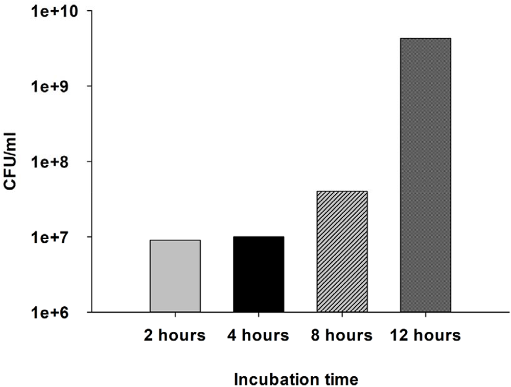
The first step in evaluating the effect of bLF and LF peptides on S. enterica biofilms was to standardize the formation of S. enterica biofilms. Biofilms are characterized by incubation time and bacterial density within the biofilms. We tested four incubation times (Figure 3). After 8 hours of incubation, the S. enterica biofilms recruited about 1 × 10⁸ CFU/ml, whereas, after 12 hours of incubation, the planktonic S. enterica increased to more than 5 × 10⁹ CFU/ml (Figure 3). We used these biofilm incubation times in the subsequent experiments.
Biofilm formation by S. enterica. Cultures of S. Enterica (4 x10⁴ cfu/ml) were inoculated into a 24-well plate containing BHI and incubated at 37ºC for 2, 4, 8, and 12 hours. The planktonic cells were removed, and the biofilms were washed once to enumerate their contained bacteria. Biofilms were washed 20 times with PBS, and viable cells in cfu/ml were obtained from 10-fold serial dilutions prepared in diluted PBS and seeded on BHI agar plates. Experiments were performed in triplicate, and the mean was given.

Bovine lactoferrin and bLF-chimera disaggregated pre-formed Salmonella enterica ATCC 14028 biofilms on abiotic surface
Biofilms are complex structures consisting mainly of a matrix of exopolysaccharides that confer high protection to the bacteria. To evaluate whether bLF, bLFcin17-30, bD-LFcin17-30, bLFampin265-284, bD-LFampin265-284, and bLF-chimera affect biofilms of S. enterica ATCC 14028, we treated pre-formed biofilms formed on abiotic surface of S. enterica for 8 and 12 h with bLF and bLF-peptides. In biofilms formed for 8 h and treated for 4 h (Figure 4A), bLF disrupted more than 60% of S. enterica biofilms compared to untreated biofilms (P < 0.05). Still, more importantly, bLF-chimera reduced about 80% of S. enterica biofilms, similar to gentamicin (P < 0.05: Figure 4A). In biofilms treated for 6 h, bLF and bLF-chimera maintained their effect, reducing more than 50% and 80% of biofilms, respectively (P < 0.05). However, the bLFampin265-286 peptide disrupted approximately 70% of biofilms formed during 8 h and treated for six h (P < 0.05: Figure 4B). Furthermore, in biofilms formed for 12 h and treated for 4 h, only bLF maintained its effect, as this peptide reduced 50% of the S. enterica biofilms (P < 0.05). No bLF-peptide showed a significant effect against these S. enterica biofilms (Figure 5A).
Bovine lactoferrin and peptides disrupted pre-formed biofilms of S. enterica for eight hours. Cultures of S. Enterica (4 x10⁴ cfu/ml) were inoculated into a 24-well plate containing BHI and incubated at 37ºC for eight hours (until biofilm formation). the planktonic cells were then removed, and the biofilms were washed once and treated for four hours (panel A) or six hours (panel B) with 40 µm blf or ten µm blfcin17-30, bd-lfcin17-30, blfampin265-284, bd-lfampin265-284 or blf-chimera and ten µm gentamicin. Treated biofilms were harvested, serially diluted 10-fold, and plated on BHI agar plates to obtain viable biofilm counts (cfu/ml). the untreated control biofilm counts were set as 100% viability and used to calculate the viability of the treated biofilms. The experiments were repeated at least three times. The standard deviation of the mean is shown. Asterisks indicate statistical significance calculated by ANOVA test (P < 0.05).

Bovine lactoferrin peptides disrupted pre-formed biofilms of S. enterica for 12 hours. S. enterica cultures (4 x10⁴ cfu/ml) were inoculated into a 24-well plate containing BHI and incubated at 37ºC for 12 hours (until biofilm formation). the planktonic cells were then removed, and the biofilms were washed once and treated for four hours (panel A) or six hours (panel B) with 40 µm blf or ten µm blfcin17-30, bd-lfcin17-30, blfampin265-284, bd-lfampin265-284 or blf-chimera or ten µm gentamicin. Treated biofilms were harvested, serially diluted 10-fold, and plated on BHI agar plates to obtain viable biofilm counts (cfu/ml). the untreated control biofilm counts were set as 100% viability and used to calculate the viability of the treated biofilms. The experiments were repeated at least three times. The standard deviation of the mean is shown. Asterisks indicate statistical significance calculated by ANOVA test (P < 0.05).

In biofilms pre-formed for 12 h and treated for 6 h, neither bLF nor bLF-peptides had an antibiofilm effect, although gentamicin did (Figure 5B). To confirm the effects of bLF and bLF-chimera on S. enterica biofilms, biofilms pre-formed for 8 hours and treated for 4 hours were stained with the live/dead biofilm viability kit. This kit stains live cells green, and the fluorescence displayed can be visualized and measured. After treatments, we found that bLF and bLF-chimera reduced the mass of biofilms by approximately 50% (P < 0.05: Figure 6B) compared to untreated biofilms (Figure 6A). Our results showed that bLF and bLF-peptides can inhibit the growth of planktonic S. enterica at different concentrations and times. On the other hand, bLF and bLF-chimera can also affect S. enterica biofilms.
Bovine lactoferrin and blf-chimera disrupted pre-formed biofilms of S. enterica for eight hours. Cultures of S. Enterica (4 x10⁴ cfu/ml) were inoculated into a 24-well plate containing BHI and incubated at 37°C for eight hours (until biofilm formation). the planktonic cells were removed and washed once. The planktonic cells were removed; the biofilms were washed once and treated for four hours with 40 µm blf or 10 µm blf-chimera or gentamicin. The treated biofilms were stained with the live/dead biofilm viability kit. The biofilms were then visualized by fluorescence microscopy (leica) (panel B) and analyzed using a fluorescence plate reader (panel A). the experiments were repeated at least three times. The standard deviation of the mean is shown. Asterisks indicate statistical significance calculated by ANOVA test (p < 0.05) – bar 20 nm.

Discussion
We have shown that bLF and its bLF-peptides reduced the growth of S. enterica cultures at different concentrations and that bLF and bLF-chimera could disrupt pre-formed biofilms. The ability of bLF to reduce S. enterica cultures has been observed in other work under different conditions and Salmonella species. Biernbaum et al. (Reference Biernbaum, Gnezda, Akbar, Franklin, Venturelli and McKillip2021) reduced the concentration of S. enterica and E. coli O157:H7 in raw milk, and the antibacterial activity of bLF was also observed in HEp-2 cells infected with S. enterica (Barreto Arce et al., Reference Barreto Arce, Contreras García, Durand Vara and Ochoa Woodell2016) as well as in different types of Salmonella, e.g., against planktonic S. Typhimurium and S. enteritidis (Del Olmo et al., Reference Del Olmo, Calzada and Nuñez2010; Quintero-Martínez et al., Reference Quintero-Martínez, Canizalez-Roman, Angulo-Zamudio, Flores-Villaseñor, Velazquez-Roman, Bolscher, Nazmi and Leon-Sicairos2024). This activity is not exclusive to bLF, as camel LF has also shown antimicrobial activity against S. enterica (Almehdar et al., Reference Almehdar, Abd El-Baky, Mattar, Albiheyri, Bamagoos, Aljaddawi, Uversky and Redwan2023, Reference Almehdar, El-Baky, Alhaider, Almuhaideb, Alhaider, Albiheyri, Uversky and Redwan2020). The ability of bLF to reduce S. enterica cultures may be due to the cationic charges of these molecules (Drago-Serrano et al., Reference Drago-Serrano, de la Garza-amaya, Luna and Campos-Rodríguez2012; Rosa et al., Reference Rosa, Cutone, Lepanto, Paesano and Valenti2017). This property confers the ability to interact with negative components of the cell membrane, such as the lipopolysaccharide of Gram-negative bacteria, e.g., S. enterica, or the membrane-localized teichoic acid of Gram-positive bacteria. This leads to the destabilization of the cell membrane, disruption of its selective permeability and, ultimately, bacterial lysis, which is the bactericidal effect (Flores-Villaseñor et al., Reference Flores-Villaseñor, Canizalez-Román, Reyes-Lopez, Nazmi, de la Garza, Zazueta-Beltrán, León-Sicairos and Bolscher2010). It has also been reported that LF indirectly impacts the pathogenic microorganism, as this protein can sequester iron metal, an element necessary for all cells to grow (Teraguchi et al., Reference Teraguchi, Shin, Ozawa, Nakamura, Fukuwatari, Tsuyuki, Namihira and Shimamura1995; Zarzosa-Moreno et al., Reference Zarzosa-Moreno, Avalos-Gómez, Ramírez-Texcalco, Torres-López, Ramírez-Mondragón, Hernández-Ramírez, Serrano-Luna and de la Garza2020). Some pathogens cannot obtain iron because it is sequestered by LF, so they die of nutritional deficiency (Leon-Sicairos et al., Reference Leon-Sicairos, Canizalez-Roman, de la Garza, Reyes-Lopez, Zazueta-Beltran, Nazmi, Gomez-Gil and Bolscher2009).
The direct effect of bLF and bLF-peptides has been demonstrated on various bacteria, such as Gram-positive bacteria, Streptococcus pneumoniae, Staphylococcus aureus (Flores-Villaseñor et al., Reference Flores-Villaseñor, Canizalez-Román, Reyes-Lopez, Nazmi, de la Garza, Zazueta-Beltrán, León-Sicairos and Bolscher2010; Leon-Sicairos et al., Reference Leon-Sicairos, Angulo-Zamudio, Vidal, Lopez-Torres, Bolscher, Nazmi, Reyes-Cortes, Reyes-Lopez, de la Garza and Canizalez-Roman2014), and other bacteria that use food as a vector, such as Escherichia coli, Vibrio cholerae and Vibrio parahaemolyticus (Leon-Sicairos et al., Reference Leon-Sicairos, Canizalez-Roman, de la Garza, Reyes-Lopez, Zazueta-Beltran, Nazmi, Gomez-Gil and Bolscher2009) as well as parasites such as Entamoeba histolytica, Giardia intestinalis and others (Aguilar-Diaz et al., Reference Aguilar-Diaz, Canizalez-Roman, Nepomuceno-Mejia, Gallardo-Vera, Hornelas-Orozco, Nazmi, Bolscher, Carrero, Leon-Sicairos and Leon-Sicairos2017; León-Sicairos et al., Reference León-Sicairos, Reyes-López, Ordaz-Pichardo and de la Garza2006). In addition, we have shown the effect of bLF to be concentration-dependent; the higher the concentration, the more effective the growth inhibition. The effect of bLF and bLF-peptides was also time-dependent, as the bactericidal effect decreased with increasing incubation time. Other work has also demonstrated the time and concentration-dependent effect of bLF and bLF-peptides (Barreto Arce et al., Reference Barreto Arce, Contreras García, Durand Vara and Ochoa Woodell2016).
As for bLF-peptides, there is little information on their antimicrobial activity against S. enterica. Huertas Mendez et al. (Reference Huertas Mendez, Vargas Casanova, Gomez Chimbi, Hernandez, Leal Castro, Melo Diaz, Rivera Monroy and Garcia Castaneda2017) have shown that different bLF-derived synthetic peptides (specifically bLFcinB) have different bactericidal activities against a wide range of bacteria, including S. enteritidis ATCC 13,076. In this study, the authors designed six different variants of bLFcinB peptides and found that the bLFcinB (20-15)2 peptide reduced the growth of S. enteritidis. It is particularly striking that the bLFcinB(20-15)2 variant was more effective than the native protein (Huertas Mendez et al., Reference Huertas Mendez, Vargas Casanova, Gomez Chimbi, Hernandez, Leal Castro, Melo Diaz, Rivera Monroy and Garcia Castaneda2017). Other studies demonstrated the antibacterial activity of LFcin17-30 against S. enterica MDR (Gourkhede et al., Reference Gourkhede, Bhoomika, Pathak, Yadav, Nishanth, Vergis, Malik, Barbuddhe and Rawool2020). In our work, we found that at higher concentrations in the first hour, all bLF-peptides showed antibacterial activity. This is the first work to report antibacterial activity of bD-LFcin17-30 and bD-LFampin265-284 against S. enterica or other bacteria. Regarding both types of LF-peptides (bLF-peptides and bDLF-peptides) used in this work, both are composed of the same amino acid sequence; however, bDLF-peptides (bD-LFcin17-30 and bD-LFampin265-284) have different spatial conformation that makes them more resistant to proteolysis.
We also observed that gentamicin could eliminate 100% of planktonic S. enterica. In comparison, when gentamicin faced pre-formed S. enterica 20% of bacteria survived against its effect. In this regard, the ability to form biofilms is one of the mechanisms by which bacteria develop antibiotic resistance (Koo et al., Reference Koo, Allan, Howlin, Stoodley and Hall-Stoodley2017). In this study, we evaluated the antibiofilm effect of bLF and bLF-peptides. The results showed that bLF, bLFcin17-30, and bLF-chimera could disrupt biofilms, but only bLF and bLF-chimera could reduce bacteria within S. enterica biofilms. Furthermore, in preliminary experiments, adding exogenous iron to S. enterica biofilms reduced the antibiofilm effects of bLF (data not shown). Recently, inhibition of antibiofilm formation by bLF and bLF-chimera and activity against pre-formed S. typhimurium biofilms formed on abiotic surfaces and HEp-2 cells was demonstrated, but neither bD-LFcin17-30 nor bD-LFampin2645-284 were evaluated (Barreto Arce et al., Reference Barreto Arce, Contreras García, Durand Vara and Ochoa Woodell2016; Quintero-Martínez et al., Reference Quintero-Martínez, Canizalez-Roman, Angulo-Zamudio, Flores-Villaseñor, Velazquez-Roman, Bolscher, Nazmi and Leon-Sicairos2024). In addition, several studies have assessed the antibiofilm effect of bLF, for example, in Escherichia coli, Klebsiella pneumoniae, Aggregatibacter actinomycetemcomitans and Streptococcus pneumoniae (Angulo-Zamudio et al., Reference Angulo-Zamudio, Vidal, Nazmi, Bolscher, Leon-Sicairos, Antezana, Canizalez-Roman and León-Sicairos2019; Leon-Sicairos et al., Reference Leon-Sicairos, Angulo-Zamudio, Vidal, Lopez-Torres, Bolscher, Nazmi, Reyes-Cortes, Reyes-Lopez, de la Garza and Canizalez-Roman2014; Ruangcharoen et al., Reference Ruangcharoen, Suwannarong, Lachica, Bolscher, Nazmi, Khunkitti and Taweechaisupapong2017; Sheffield et al., Reference Sheffield, Crippen, Poole and Beier2012).
The mechanism by which bLF and bLF-peptides prevent or disrupt biofilm formation is unclear. Still, we speculate that during the early steps of biofilm formation, the mechanism is likely to be consistent with the work reported by Singh (Reference Singh2004) in which bLF inhibited Pseudomonas aeruginosa contraction due to the iron chelating effects of bLF (Singh, Reference Singh2004). In S. enterica, we proposed that bLF and its peptides could affect the exopolysaccharide matrix, which is negatively charged, and then the bLF and LF peptides create pores in the biofilms. These pores could then be the tools that allow antibiotics to enter the biofilms and attack the bacteria (Ammons and Copié, Reference Ammons and Copié2013). Another possible mechanism by which bLF disrupts biofilms is through its DNase activity, as demonstrated by bLF against Streptococcus pneumoniae biofilms, where bLF degraded the extracellular DNA of pneumococcal biofilms, leading to destabilization of the biofilm structure to allow its disaggregation (Angulo-Zamudio et al., Reference Angulo-Zamudio, Vidal, Nazmi, Bolscher, Leon-Sicairos, Antezana, Canizalez-Roman and León-Sicairos2019). Further research is needed to clarify the potential mechanism of the antibiofilm action of bLF and bLF-peptides.
Only antibiotics are used to combat infectious diseases, regardless of whether infections are caused by biofilms and not by planktonic bacteria. This raises a problem. Antibiotics are designed to attack bacteria structures (internal or external) and not biofilm structures, which leads to a decrease in the effectiveness of antibiotics regardless of whether the bacteria in the planktonic state are resistant or not, as shown in this study (Rather et al., Reference Rather, Gupta and Mandal2021; Venkatesan et al., Reference Venkatesan, Perumal and Doble2015). Instead, proteins and peptides from the immune system, such as bLF and bLF-peptide, have multiple mechanisms of action against biofilms or planktonic bacteria. Nevertheless, biofilms not only develop resistance to antibiotics, they also can develop resistance to bLF and bLF-peptides. Some bacteria can increase the production of some cationic polysaccharides in their wall, as is the case of S. aureus, which increases the production of the intracellular polysaccharide responsible for adhesion, which increases the positive charge of the biofilm matrix and alters the process of interaction of biofilms with bLF and bLF-peptides by electrostatic attractions. This relative resistance of biofilms against cationic proteins and peptides could be solved by increasing the concentration of treatments or designing peptides with mechanisms that do not depend on electrical interactions (Band and Weiss, Reference Band and Weiss2015; Joo et al., Reference Joo, Fu and Otto2016; Moravej et al., Reference Moravej, Moravej, Yazdanparast, Heiat, Mirhosseini, Moosazadeh Moghaddam and Mirnejad2018).
We did not use crystal violet to quantify biofilms, but we quantified live bacteria in two different ways. The first was by disaggregating the biofilm mass and performing counts by serial dilutions. The second method we used was the live/dead viability kit. Both techniques allow the quantification of live cells, as opposed to crystal violet, where we do not know whether the bacteria within the biofilms are alive. The limitations of this work were that the biofilms were not tested with violet glass, the antibiofilm activity of bLF and bLF-peptide was not tested at longer S. enterica incubation times and the biofilms were not tested at longer S. enterica incubation times.
In conclusion, the results obtained in this work show that bLF and bLF-peptides have antibacterial and antibiofilm activity against S. enterica within a short incubation time. These results lay the foundation for future research in which bLF and bLF-peptides could be used as an alternative or complementary therapy to treat infections caused by S. enterica and other pathogenic bacteria.
Acknowledgements
We thank Loranda Calderon-Zamora, Tonatzin K. Domínguez-Ulloa, and Miriam K. Ortíz-Gonzalez for their efficient technical support. This research was supported by PROFAPI-UAS of the Universidad Autónoma de Sinaloa, grant number PROFAPI 2022 PRO-A3-037, and by CONAHCYT Mexico grant number CB-2014-236546, to Nidia Leon-Sicairos.