Introduction
The species Moenkhausia oligolepis, first described by Günther (1864), belongs to the genus Moenkhausia. Members of this genus present pre-maxillary teeth in two rows. The inner row consists of five multicuspid teeth, generally arranged in a lateral line with no sharp curvature (Marinho and Langeani, Reference Marinho and Langeani2016). The fire-eye tetra Moenkhausia oligolepis is popularly known as ‘piaba’ or ‘lambari’ (Reia, Reference Reia2018). This species is small sized, with a typical length of 10 cm for an adult male, but plays a large role in the aquarium industry; and therefore carries considerable commercial value (Froese and Pauly, Reference Froese and Pauly2018). Fire-eye tetra are widely distributed in the Amazon basin (Reia, Reference Reia2018), and have a reproductive period that typically occurs during the summer, between January and February (unpublished data). Only during this time can specimens be readily found in accessible streams. Even so, the limited knowledge of its reproductive biology hinders its reproduction capacity in captivity.
According to FAO (2018), captive fish production has increased considerably in recent decades (1980–2017). This is largely due to the frequent exploitation of natural stocks by predatory fishing practices. This drastically reduces the native population, and ultimately prevents the supply of natural-caught fish from meeting the demands of a growing, global population (Streit Jr. et al., Reference Streit, Moraes, Ribeiro, Cardozo and Moreira2002; Andrade and Yasui, Reference Andrade and Yasui2003; Miller and Spoolman, Reference Miller and Spoolman2015; Tavares-Dias and Mariano, Reference Tavares-Dias and Mariano2015).
While there are a number of different approaches to increase captive fish populations, all are based on artificial reproduction. This requires an understanding of both male and female gametes to succeed. Although there has been a growing body of research on male gametes, as exemplified by Dreanno et al. (Reference Dreanno, Suquet, Desbruyères, Cosson, Dellioud and Billarda1998), Streit Jr et al. (Reference Streit, Moraes, Ribeiro, Cardozo and Moreira2002); Paulino et al. (Reference Paulino, Murgas, Felizardo and Freitas2012), Silva et al. (Reference Silva, Galvão, Teixeira and Farias2014), Lenz et al. (Reference Lenz, Victorio, Lima, Prado, Paula, Meirinhos and Arnhold2018), Caldas and Godoy (Reference Caldas and Godoy2019) and Kowalski and Cejko (Reference Kowalski and Cejko2019), very little information is known about the semen of many native species, including the fire-eye tetra, M. oligolepis.
Studies on semen quality and sperm count (Rurangwa et al., Reference Rurangwa, Kime, Ollevier and Nash2004) are vital for the success and efficacy of artificial fertilization, captive reproduction, and the generation of viable larvae and juveniles for aquaculture purposes (Rurangwa et al., Reference Rurangwa, Kime, Ollevier and Nash2004). The stress that fish are subjected to in captivity during semen collection procedures can cause morphological changes to their sperm (Murgas et al., Reference Murgas, Felizardo, Ferreira, Andrade and Veras2011). This may affect the artificial fertilization process, and therefore the production of healthy embryos, larvae and juveniles. Rurangwa et al. (Reference Rurangwa, Kime, Ollevier and Nash2004) reported that the analysis of sperm morphology is critical for evaluative studies seeking to determine semen quality. This knowledge can then be employed during the artificial fertilization process, as each oocyte is fertilized by only one spermatozoa.
As fish demonstrate a high degree of variability in seminal characteristics (Andrade et al., Reference Andrade, Carvalho, Ferreira, Paula, Rodrigues, Felizardo, Reis Neto and Murgas2014) and reproductive strategies (Taborsky, Reference Taborsky1998), semen analysis is of paramount importance for the performance of artificial fertilization (Murgas et al., Reference Murgas, Felizardo, Ferreira, Andrade and Veras2011). Apart from this, such studies also provide valuable insight into the reproductive biology of native species, and contribute to the establishment of standardized semen conservation protocols (Silva et al., Reference Silva, Galvão, Teixeira and Farias2014).
In addition to a morphological evaluation, parameters such as the rate and duration of total sperm motility, sperm vigour and sperm concentration are frequently measured during seminal characterization (Streit Jr et al., Reference Streit, Ribeiro, Moraes, Mendez, Gallo, Digmayer and Povh2006; Murgas et al.; Reference Murgas, Felizardo, Ferreira, Andrade and Veras2011). Total sperm motility and sperm vigour are determined by movement and movement speed, respectively (Rurangwa et al., Reference Rurangwa, Kime, Ollevier and Nash2004). Sperm concentration is characterized by the number of sperm per millilitre of semen. These parameters are of great importance in semen characterization, providing general (total motility, vigour and sperm concentration) and specific, qualitative and quantitative (morphology) information. As such, this study aims to provide the seminal characterization of the M. oligolepis species.
Materials and methods
Place of collection
Specimen collection was performed at the Zoobotanical Foundation of Marabá, PA, in accordance with ICMBio rules and with proper authorization (no. 62027-1). This site has an area of 15650 m2. During the collections process, the perimeter was demarcated by point 1 (05°23′24.5″S, 049°03′03.9″W), point 2 (05°23′27.9″S, 049°03′07.6″W), point 3 (05°23′31.6″S, 049°03′12.3″W), point 4 (05°23′32.2″S, 049°03′13.3″W), point 5 (05°23′51.6″S, 049°04′08.0″W), and point 6 (05°23′36.0″S, 049°04′02.7″W).
The collection area was characterized by the presence of intermittent streams with a water regime that varies throughout the year according to weather. Some streams completely dry out in the dry months from May to October. However, heavy rainfall from November to April may result in the streams overflowing their banks. Moreover, some streams are lentic in nature, while in areas with high water circulation they are lotic. Streams also vary in depth (c. 0.5–1.5 m), turbidity, solar penetration (1.25–1.45 m, Secch disc), the amount of available organic matter, and in many among other factors that may influence water quality as well as fish reproduction and survival.
Animal sampling
M. oligolepis specimens were collected in the summer period, in which the streams water temperature was in average 25.8 ± 0.87°C. Two fishing nets (4.75 × 1.10 m) with 1.0 × 1.0 mm mesh were used. The animals were placed in 30-litre plastic bags with portable aerators (VIGOAR) and taken to the laboratory for the acclimatization period, which lasted a minimum of 15 days.
Experimental design
Hormonal induction
Three sexually mature males, with a mean weight of 2.47 ± 0.94 g and a mean total length of 5.2 ± 0.36 cm, were selected and submitted to pre-induction anaesthesia in a 600 ml water container with 2.5 ml of eugenol solution (20 ml eugenol in 180 ml 100% alcohol, biodinâmica). Hormonal induction was performed using crude carp pituitary extract, EHC (Danúbio Aquacultura), diluted in 0.9% sodium chloride solution (Laboratório Farmacêutico Arboreto Ltda). Each male was submitted to hormone induction with a single dose of 0.001 mg/g, injected into the peritoneal cavity using a 1 ml insulin syringe (SR Productos Pará La Salud S.A.). After injection, the fish were kept with females in a 13 litre aquarium with constant aeration, and a water temperature of 26.4°C, for a duration of 6 h.
Gamete collection
At 6 h after induction, the animals were anesthetized and submitted to semen collection. This was performed by antero-posterior abdominal pressure, with the aid of a 1000 µl micropipette (AGIPETTE). Initially, the male ventral region was cleaned and dried with paper towels to avoid contamination and early activation of sperm. Using the micropipette, an average volume of 2 µl of semen was collected from the urogenital papilla of each animal and emptied into a 1.5 ml microtube containing 10 µl of dibasic sodium phosphate extender solution. The semen solution was then stored in a styrofoam box with ice at 6°C.
Seminal evaluation
Analysis of total motility
Here, 1 µl of diluted semen was added to a histological slide previously covered with bovine albumin solution (3.8 g/dl, Interteck Katal). Following this, 10 µl of distilled water was added for gamete activation. The slides were evaluated under a ×400 magnification optical microscope (Physis), with a tablet PC attached. Videos were recorded for subjective analysis of sperm motility and vigour. This was accomplished by using the regression rate percentage of movement, and a sperm velocity score from 0 to 5, respectively. The motility parameters were defined from the moment of water addition until the total arrest of cell movement.
Sperm concentration analysis
Here, 0.5 µl of fresh semen was collected, fixed and diluted in 500 µl glutaraldehyde (2.5%) (Êxodo Científica). Following this, 10 µl of the diluted semen was added to a Neubauer chamber (Grid Optik) and analyzed using an optical microscope, under ×400 magnification, with a coupled tablet PC. The formula for sperm cell count per ml of semen was defined as the following: Concentration (spz/ml) = the total number of cells × dilution factor × 1000.
Sperm morphology analysis
Next, 20 µl of diluted semen was added to a histological slide, followed by 10 µl of distilled water and 40 µl of nigrosine–eosin dye (Botuvital). Six semipermanent slides were produced, two for each animal. These slides were sealed with colourless enamel and analyzed using an optical microscope (Biofocus Equipamentos) under ×1000 magnification, with an attached camera (Even) for photodocumentation. Finally, 100 spermatozoon from each male were selected and measured (flagella length and head diameter) using ImageJ software (v.1.52a). These measurements were used to identify the presence of primary abnormalities (macro and microcephaly, long, strongly long, curled, strongly curled, degenerated flagella and biflagellar cells) and secondary abnormalities (loose or isolated head and broken or folded flagella).
Statistical analysis
The experiment had three replicates, with each animal representing one replicate. Data on sperm concentration and morphology were expressed as mean ± standard error (mean ± SE). Qualitative variables (motility and sperm vigour) were submitted to an r squared test (r 2), with a significance threshold of 0.05, using R software (v.3.5.0).
Results
Analysis of sperm motility
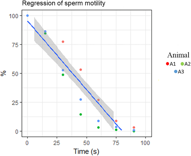
The progressive sperm motility in M. oligolepis is showed in Fig. 1. Regression rate began with 95.32% of total sperm motility, and decreased every 15 s to 85.3, 59.6, 31.7, 13.0, 4.6, and finally 1.2%. (Fig. 1).
Regression of sperm motility in M. oligolepis.

Analysis of sperm volume and concentration
The average sperm volume obtained was 2 µl, and the average sperm concentration for the fire-eye tetra was found to be 4.97 × 10⁹ ± 3.46 sperm/ml semen.
Analysis of sperm morphology
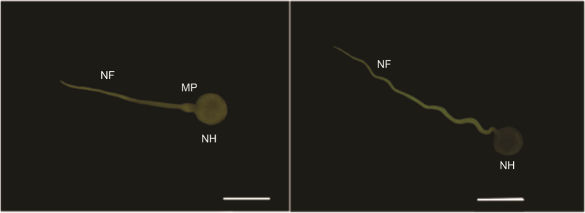
The fraction of sperm revealed to be dead with a ruptured plasma membrane by head staining was, on average, 20.33%, with the remaining 79.67% of sperm being alive and having an intact cell membrane (Fig. 2). Of the living sperm, 69% of the cells were normal, with an average total length of approximately 30 µm. This length is composed of the head diameter, having an average length of 4.488 ± 0.7 µm, and the flagellum plus mid-piece having an average length of 26.071 ± 12.4 µm (Fig. 2).
Normal sperm. MP, mid-piece; NH, normal head; NF, normal flagellum. Scale bars, 10 µm.

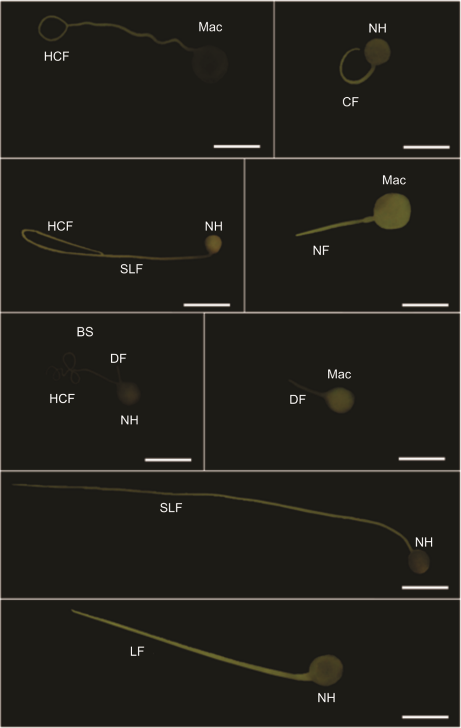
Within the remaining abnormal population (31%), primary and secondary abnormalities were detected. In cases with primary abnormalities, 100% presented macrocephaly. Regarding the flagella, 16% presented long flagella (between 39 µm and 50 µm); 8% presented strongly long flagella (> 50 µm); 38% presented rolled flagella; 6% presented strongly rolled flagella; and 18% presented degenerated flagella. A single biflagellate spermatozoon was found in the analyzed images (Fig. 3).
Sperm with primary abnormalities. BS, biflagellate sperm; CF, coiled flagellum; DF, degenerate flagellum; HCF, heavily coiled flagellum; LF, long flagellum; Mac, macrocephaly; NH, normal head; SLF, strongly long flagellum. Scale bars, 10 µm.

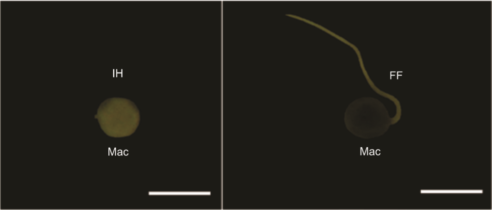
Secondary abnormalities were characterized by isolated heads and folded flagella. Of the abnormal sperm, 6% had a flagellum-free-head (isolated), and 14% had folded flagella (Fig. 4).
Sperm with secondary abnormalities. FF, folded flagellum; IH, isolated head; Mac, macrocephaly. Scale bars, 10 µm.

Discussion
Several factors are responsible for the success of fish reproduction. Among them are sperm quantity (sperm concentration) and seminal quality (motility, vigour and sperm morphology). These can determine reproductive outcomes of natural and artificial fertilization as well as spawning (Kowalski and Cejko, Reference Kowalski and Cejko2019). As reproduction is vital to growing captive populations, semen evaluation in aquaculture is necessary. The short-term preservation of semen to avoid repeated semen collection is an example of how seminal evaluation can lead to increased profits (Rurangwa et al., Reference Rurangwa, Kime, Ollevier and Nash2004). In addition to playing an important ecological role, these studies enable procedures such as cryopreservation and genetic banking to preserve endangered species, such as the European eel (Anguilla anguilla) and sturgeon (Acipenser sturio) (Kowalski and Cejko, Reference Kowalski and Cejko2019). Therefore, the seminal characterization of M. oligolepis presented here can contribute to further studies with this species both for commercial and conservation purposes.
Unlike mammals, most externally fertilized fish have immobile sperm suspended in seminal plasma. This immobility is due to the presence of potassium ions (K+), which inhibit the onset of motility (Alavi et al., Reference Alavi, Cosson, Bondarenko and Linhart2019). However, motility may be activated once the spermatozoa enter an aqueous medium (Rurangwa et al., Reference Rurangwa, Kime, Ollevier and Nash2004). This induction is brought about by the osmolarity difference between the seminal plasma and the external medium. In freshwater fish, seminal plasma osmolarity is greater than that of the external environment (Morisawa and Suzuki, Reference Morisawa and Suzuki1980). Therefore, the sperm of these fish initiate flagellar beating once introduced into a hypo-osmotic medium (Morisawa and Suzuki, Reference Morisawa and Suzuki1980; Alavi et al., Reference Alavi, Cosson, Bondarenko and Linhart2019). The typical duration of motility is short, ranging from one to 2 min (Dreanno et al., Reference Dreanno, Suquet, Desbruyères, Cosson, Dellioud and Billarda1998; Poleo et al., Reference Poleo, Denniston, Reggio, Godke and Tiersch2001). This short duration, also observed in M. oligolepis, is related to rapid energy expenditure after the onset of motility (Caldas and Godoy, Reference Caldas and Godoy2019). This also manifests in decreased sperm vigour with time (Murgas et al., Reference Murgas, Felizardo, Ferreira, Andrade and Veras2011). The sperm motility rate observed with the fire-eye tetra was close to the maximum rate of 100%. This is higher than values obtained with another species from the Moenkhausia genus. The sperm motility rate for M. sanctaefilomenae was just over 50% (Domínguez-Castanedo et al., Reference Domínguez-Castanedo, Toledano-Olivares and Ávalos-Rodríguez2015). The use of a hormonal inducer may have elevated the motility rate of M. oligolepis sperm, as observed by Ferreira et al. (Reference Ferreira, Nuñer, Luz, Tataje, Esquivel and Restrepo2001) with the jundiá, Rhamdia quelen.
In mammals, like cattle, horses and pigs, insemination doses are standardized with regards to semen volume and sperm concentration. However, in fish no ideal sperm concentration for the fertilization of a given oocyte is known (Bombardelli et al., Reference Bombardelli, Mörschbächer, Campagnolo, Sanches and Syperreck2006) for species such as dourado (Salminus brasiliensis) (Sanches et al., Reference Sanches, Bombardelli, Baggio and Souza2009), piracanjuba (Brycon orbignyanus) (Felizardo et al., Reference Felizardo, Murgas, Drumond and Silva2010) and tambaqui (Colossoma macropomum) (Leite et al., Reference Leite, Melo, Oliveira, Pinheiro, Campello, Nunes and Salmito-Vanderley2013). The failure to use a minimum inseminating dose for species reproduction results in decreased fertilization rates (Rurangwa et al., Reference Rurangwa, Kime, Ollevier and Nash2004). The sperm concentration found in the studied species is similar to other species from the same genus. M. sanctaefilomenae was reported to have an average sperm concentration of 6.8 × 10⁸ sperm per ml semen (Domínguez-Castanedo et al., Reference Domínguez-Castanedo, Toledano-Olivares and Ávalos-Rodríguez2015). However, variations in seminal characteristics are common in fish, both between different species and individuals of the same species (Silva et al., Reference Silva, Murgas, Felizardo, Pereira, Navarro and Mello2009; Murgas et al., Reference Murgas, Felizardo, Ferreira, Andrade and Veras2011). Significant variation has even been reported within the same individual (Billard, Reference Billard1983), either as a result of the time of year (Borges et al., Reference Borges, Siqueira, Jurinitz, Zanini, Amaral, Grillo, Oberst and Wassermann2005), successive seminal collections (Kavamoto et al., Reference Kavamoto, Mainardes-Pinto, Andrade-Talmelli and Campos1997) or diet. With regards to seminal characteristics and diet, Domínguez-Castanedo et al. (Reference Domínguez-Castanedo, Toledano-Olivares and Ávalos-Rodríguez2015) studied the species M. sanctaefilomenae, and found significantly positive changes in sperm concentration and spermatozoa size as a result of an astaxanthin carotenoid diet.
Studies related to sperm morphology have become an important tool in seminal evaluation. This is especially true with regards to cryopreservation, as some morphological abnormalities can be caused by cryoprotectant, the storage temperature and thawing time (Baulny et al., Reference Baulny, Le Vern, Kerboueuf and Maisse1997; Paulino et al., Reference Paulino, Murgas, Felizardo and Freitas2012). Plasma membrane damage, like that observed with the sperm cells of this species, results in mitochondrial injuries that affect ATP production and, consequently, sperm motility (Baulny et al., Reference Baulny, Le Vern, Kerboueuf and Maisse1997). Although, there is no established mortality rate for fish sperm, the cell mortality rate obtained for the fire-eye tetra was similar to that observed with semen of the freshwater rainbow trout Oncorhynchus mykiss (Baulny et al. Reference Baulny, Le Vern, Kerboueuf and Maisse1997). The same is true for the pathology-free and total living cell rate. When compared with species of the same family, Brycon opalinus (85% live sperm) (Narahara et al., Reference Narahara, Andrade-Talmelli, Kavamoto and Godinho2002) and P. mesopotamicus (40.21% normal) (Streit Jr et al., Reference Streit, Oliveira, Ribeiro, Sirol, Moraes, Galo and Digmayer2009), the rate of total and free live sperm in M. oligolepis was similar. However, due to large variations in the seminal characteristics of fish species, this result does not safely express the oocyte fertilization capacity. Conservatively speaking, such abnormalities may be indicators of poor fertilization outcomes. However, there is no standard level of abnormality that indicates this in fish. As a result, the rate established for mammals is normally used as a benchmark. According to Paulino et al. (Reference Paulino, Murgas, Felizardo and Freitas2012), the Brazilian College of Animal Reproduction does not recommend the use of semen with abnormal sperm counts higher than 30% for cattle and horses, or 20% for pigs. Given this information, it can be inferred that the amount of abnormal sperm found in M. oligolepis would not result in unsuccessful fertilization.
However, the specific assessment of primary and secondary abnormalities provides more accurate and reliable information on the semen’s fertilization capacity. Primary abnormalities are associated with spermatogenesis, while secondary abnormalities are caused by environmental factors (Herman et al., Reference Herman, Mitchell and Doak1994). In this sense, primary abnormalities are considered more severe in both natural and captive environments. For example, degenerate flagellum can cause an oscillating motion, therefore making oocyte fertilization difficult (Kavamoto et al., Reference Kavamoto, Barnabe, Campos and Andrade-Talmelli1999). Secondary abnormalities may be considered minor as they are often caused by handling of samples (Paulino et al., Reference Paulino, Murgas, Felizardo and Freitas2012) and can be avoided or minimized. That said, folded flagellum sperm are less successful in fertilizing due to their oscillation (Kavamoto et al., Reference Kavamoto, Barnabe, Campos and Andrade-Talmelli1999) and non-progressive circular motion.
According to Taborsky (Reference Taborsky1998), the variation in sperm cell size and structure is due to competition between co-specific individuals with external fertilization. Taborsky (Reference Taborsky1998) also stated that those variations are derived from adaptive evolutionary processes, aimed at improving the reproductive success of a species. Large sperm are related to non-territorial species, in which energy investment is mainly focused on gonadal and gamete development (Taborsky, Reference Taborsky1998). In contrast, territorial species have a greater energy investment for the development of secondary sexual characteristics and robustness. The fire-eye tetra’s behaviour in captivity is peaceful, which may explain the percentage of long sperm found in the species. Sperm length is a major determinant of sperm motility and vigour. However, the longevity of long sperm is reduced compared with shorter sperm (Stockley et al., Reference Stockley, Gage, Parker and Moller1997). This is probably due to the higher consumption of ATP required for movement. Although, Taborsky (Reference Taborsky1998) argued that spermatozoa presenting long flagellum are effective for fertilizing the oocyte over large distances in the aquatic environment.
In conclusion, the sperm concentration found in the studied species is higher than in the other studied species of the same genus and the rate of morphological abnormalities observed in the M. oligolepis spermatozoa is considered normal and does not impair the fertilization capacity of its semen, enabling its sperm to be used in artificial reproduction protocols and can also inform taxonomical studies, as the classification of this species is still uncertain, as Benine et al. (Reference Benine, Mariguela and Oliveira2009) consider M. oligolepis to be a multitude of coexisting species. Such confusion can result in an incorrect definition of its state of conservation.
Acknowledgements
The author express thanks to the Zoobotanical Foundation of Marabá in which the species were collected and to the Laboratory of Neuroscience and Behavior ‘Frederico Graef’ (LaNeC) for its support and structure.
Ethical Standards
Specimen collection was performed at the Zoobotanical Foundation of Marabá – PA, in accordance with ICMBio rules and with proper authorization (no. 62027-1).
Financial Support
This study was funded by ‘Conselho Nacional de Desenvolvimento Científico e Tecnológico’ CNPq, which awarded a PIBITI grant (EDPROPIT04).
Conflict of interest
The authors declare that they have no conflict of interest.