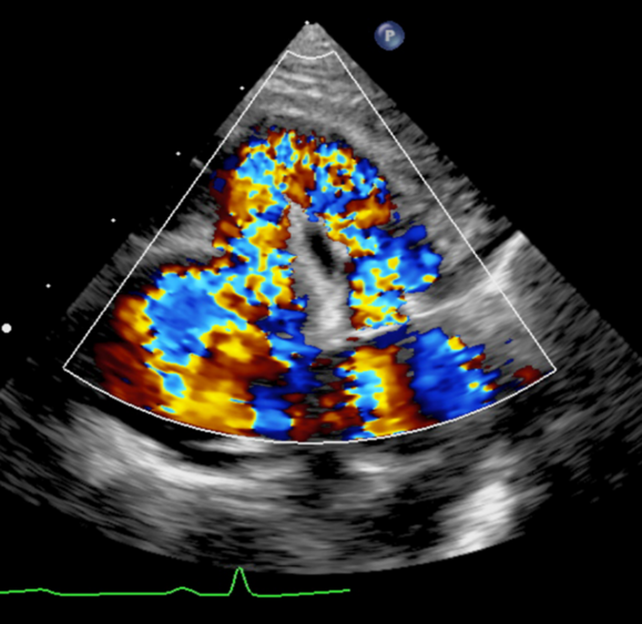
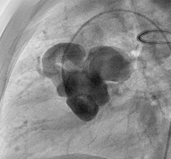
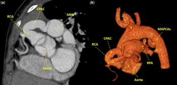
A 4-year-old girl from Mali was referred to our department as part of a humanitarian mission for surgical repair of her congenital heart disease. Her working diagnosis was severe Tetralogy of Fallot with pulmonary blood supply augmented either via a patent ductus arteriosus or a major aortopulmonary collateral artery, diagnosed at the age of 2 years during hospitalisation for pneumonia. Her oxygen saturation was 75% in room air. She was not dysmorphic, but 22q11 deletion syndrome was not ruled out. An electrocardiogram showed no myocardial ischemia. Echocardiography showed Fallot-type pulmonary atresia and ventricular septal defect, absent left pulmonary artery, right aortic arch, and a large collateral from the aortic root supplying the main pulmonary artery (Fig 1). Cardiac catheterisation confirmed the large fistulous connection between the aortic root and a single right pulmonary artery (Fig 2). A computed tomography scan demonstrated more clearly a right coronary artery to pulmonary artery collateral, an absent left pulmonary artery, and two major aortopulmonary collaterals to the right lung, one of which was severely stenotic. An atretic ampulla of a probable patent ductus arteriosus arising from the innominate artery that had possibly supplied an isolated distal left pulmonary artery was also shown (Fig 3 and Supplementary video S1).
Transthoracic echocardiography (parasternal short-axis view with colour Doppler) showing blood flow in a large collateral originating from the aortic root and supplying the main pulmonary artery.

Aortic root angiogram (straight lateral view) showing a dilated right coronary ostium with a large collateral from the right coronary artery to the main pulmonary artery.

Computed tomography angiogram ( a ) and 3D volume rendering ( b ) showing a right coronary artery to pulmonary artery collateral, an absent left pulmonary artery, two major aortopulmonary collateral arteries, and an atretic ampulla at the base of the innominate artery (asterisk) where a patent ductus arteriosus supplying an isolated distal left pulmonary artery possibly originated (CPAC: coronary artery to pulmonary artery collateral; MAPCAs: major aortopulmonary collateral arteries; MPA: main pulmonary artery RCA: right coronary artery; RPA: right pulmonary artery).

The patient underwent surgical repair with ventricular septal defect closure, placement of a valved right ventricle-to-right pulmonary artery conduit, coronary to pulmonary artery collateral artery ligation, and unifocalisation of the major aortopulmonary collaterals to the right lung. No significant source of pulmonary blood supply to the left lung, including a vestigial left pulmonary artery, was found. The pulmonary arterial pressure was acceptable at half-systemic post-operatively, so an intracardiac fenestration was not performed.
Coronary artery to pulmonary artery collaterals are present in about 10% of patients with pulmonary atresia and ventricular septal defect. Reference Amin, McElhinney and Reddy1 They should be systematically sought as a source of pulmonary blood supply.
Acknowledgements
None.
Financial support
This research received no specific grant from any funding agency, commercial or not-for-profit sectors.
Conflicts of interest
None.
Ethical standards
The authors assert that all procedures contributing to this work comply with the ethical standards of Swiss ethics and with the Helsinki Declaration of 1975, as revised in 2008, and has been approved by the institutional committee of Lausanne University Hospital.
Supplementary material
To view supplementary material for this article, please visit https://doi.org/10.1017/S1047951120003315