Introduction
The pathophysiological basis of schizophrenia is poorly understood. Neuroimaging studies have shown that the disorder is associated with relatively small reductions in gray matter volume (GMV) in many different regions of the brain. Thus, magnetic resonance imaging (MRI) studies of schizophrenia have reported findings in the frontal, cingulate, temporal and parietal cortices, the striatum and the thalamus (Ellison-Wright et al. Reference Ellison-Wright, Glahn, Laird, Thelen and Bullmore2008). Functional neuroimaging studies in schizophrenia have described differential activity and activation in a similarly wide set of regions (Koch et al. Reference Koch, Wagner, Nenadic, Schachtzabel, Schultz, Roebel, Reichenbach, Sauer and Schlosser2008), and diffusion tensor imaging (DTI) studies indicate that the integrity of the white matter (WM) in schizophrenia is altered in most of the major fasciculi that connect these cortical and subcortical areas (Ellison-Wright & Bullmore, Reference Ellison-Wright and Bullmore2009). Collectively, these data are consistent with the notion that the brain changes associated with schizophrenia are distributed rather than focal, involving interconnected networks of areas as opposed to single regions (Minzenberg et al. Reference Minzenberg, Laird, Thelen, Carter and Glahn2009; Ragland et al. Reference Ragland, Laird, Ranganath, Blumenfeld, Gonzales and Glahn2009).
Neuroimaging data also suggest that schizophrenia involves alterations in the structural and functional connectivity between different cortical and subcortical regions (Shenton et al. Reference Shenton, Dickey, Frumin and McCarley2001; Wright et al. Reference Wright, Rabe-Hesketh, Woodruff, David, Murray and Bullmore2000). Theoretical models of the disorder have independently linked dopamine dysfunction in schizophrenia to changes in the interaction between the prefrontal cortex and the subcortical dopamine system (Howes & Kapur, Reference Howes and Kapur2009), and glutamate dysfunction in schizophrenia to alterations in thalamocortical connections (Sharp et al. Reference Sharp, Tomitaka, Bernaudin and Tomitaka2001). These two neurochemical changes have been linked in a model that suggests that medial temporal glutamate dysfunction drives increased striatal activity in schizophrenia (Lodge & Grace, Reference Lodge and Grace2008). Many of the neuropsychological impairments associated with schizophrenia have been attributed to changes in frontostriatal interactions (Robbins, Reference Robbins1990; Pantelis et al. Reference Pantelis, Barnes, Nelson, Tanner, Weatherley, Owen and Robbins1997). A common feature of these models is the involvement of cortical and subcortical areas, particularly the prefrontal and medial temporal cortex, and the thalamus and striatum. Findings from animal models implicate cortico-subcortical dysfunction in the development of psychotic symptoms and cognitive disturbances (Andreasen et al. Reference Andreasen, Paradiso and O'Leary1998), and recent studies on cortical thickness and functional connectivity suggest cortical–subcortical abnormalities and dysfunction in schizophrenia patients (Rimol et al. Reference Rimol, Hartberg, Nesvag, Fennema-Notestine, Hagler, Pung, Jennings, Haukvik, Lange, Nakstad, Melle, Andreassen, Dale and Agartz2010; Zhang et al. Reference Zhang, Guo, Hu, Li, Zhao and Liu2012).
Thus, schizophrenia was considered as a dysconnection disorder (Stephan et al. Reference Stephan, Baldeweg and Friston2006, Reference Stephan, Friston and Frith2009; Pettersson-Yeo et al. Reference Pettersson-Yeo, Allen, Benetti, McGuire and Mechelli2011). In the past decade, connections in the brain have been studied at micro-, meso- and macroscopic resolutions (Sporns et al. Reference Sporns, Tononi and Kotter2005). Structural and functional brain connectivity has been examined by neuroimaging methods, mostly at macroscopic resolution. Region-of-interest (ROI) and voxel-wise mapping approaches have been used for characterizing pair-wise interactions between various brain regions. Dysfunction between regions, both intra- and inter-regionally, has been observed (Glantz & Lewis, Reference Glantz and Lewis2001; Harrison & Weinberger, Reference Harrison and Weinberger2005; Walterfang et al. Reference Walterfang, Wood, Velakoulis and Pantelis2006; Zalesky et al. Reference Zalesky, Fornito, Seal, Cocchi, Westin, Bullmore, Egan and Pantelis2011). The latest graph comparison techniques have been used to reveal alterations in prefrontal cortex connectivity with the cerebellum, and parietal and occipital cortices, and especially frontotemporal dysconnectivity (Liu et al. Reference Liu, Liang, Zhou, He, Hao, Song, Yu, Liu, Liu and Jiang2008; Zalesky et al. Reference Zalesky, Fornito and Bullmore2010; Fornito et al. Reference Fornito, Yoon, Zalesky, Bullmore and Carter2011).
Based on the neurodevelopmental paradigm, brain regions that form a specific network may present a covariation in volume or density (Bullmore et al. Reference Bullmore, Woodruff, Wright, Rabe-Hesketh, Howard, Shuriquie and Murray1998; Zhang & Sejnowski, Reference Zhang and Sejnowski2000; Mechelli et al. Reference Mechelli, Friston, Frackowiak and Price2005). This was examined by testing the covariation in volume of putative correlated regions in patients with schizophrenia (Bullmore et al. Reference Bullmore, Woodruff, Wright, Rabe-Hesketh, Howard, Shuriquie and Murray1998) and autism (McAlonan et al. Reference McAlonan, Cheung, Cheung, Suckling, Lam, Tai, Yip, Murphy and Chua2005), and in a healthy population (Mechelli et al. Reference Mechelli, Friston, Frackowiak and Price2005), with traditional manual tracing of the ROI. However, even within the same structure (e.g. the thalamus), where different nuclei are connected to different regions of the brain (Afif & Bergman, Reference Afif and Bergman2005), the density or volume of subregions may covary with the other proportions of the networks they belong to, because connected neurons mutually benefit from the trophic effects of glutamatergic synapsis or common experience-related plasticity (Draganski et al. Reference Draganski, Gaser, Busch, Schuierer, Bogdahn and May2004; Mechelli et al. Reference Mechelli, Crinion, Noppeney, O'Doherty, Ashburner, Frackowiak and Price2004).
The present study aimed to investigate the changes in regional GMV that are associated with schizophrenia both globally and inter-regionally. We compared a large sample of patients in the first episode of the disorder with a matching sample of healthy controls. The patients were antipsychotic naive, minimizing the risk that the findings were secondary to effects of previous treatment or chronic progression of the illness. Our first objective was to identify brain areas that showed robust alterations in GMV in patients relative to controls. We then sought to compare the covariation between regional GMVs in the patient and control groups. Our first prediction was that patients would show regional reductions in GMV in areas identified in meta-analyses of MRI studies in schizophrenia (Ellison-Wright et al. Reference Ellison-Wright, Glahn, Laird, Thelen and Bullmore2008), such as the prefrontal, insular and medial temporal cortex, the striatum and the thalamus. Our second hypothesis was that the pattern of GMV covariation within these regions would be significantly different in patients compared to controls, particularly in terms of the relationship between the volumes of cortical and subcortical regions.
Method
Subjects
In total, 172 right-handed subjects were studied, comprising 86 patients with first-episode schizophrenia (FES) and 86 age- and gender-matched healthy controls. In the recruited sample of 103 patients, nine patients did not give their consent to participate in the MRI procedures; five did not cooperate adequately with the MRI acquisition procedures; and three were lost to MRI data analysis procedures because of data format incompatibility.
The patients were recruited through the Mental Health Center of West China Hospital, Sichuan University. They were assessed by one of two psychiatrists (W.D. and M.L.) when they first presented to the mental health services, using the SCID-I/P (First et al. Reference First, Spitzer, Gibbon and Williams2002) and the Positive and Negative Syndrome Scale (PANSS; Kay et al. Reference Kay, Fiszbein and Opler1987). All met DSM-IV criteria for a first episode of schizophrenia. A volumetric MRI scan was acquired in all patients in 24 h after recruitment, before they were started on antipsychotic medication or any other form of treatment (e.g. electroconvulsive therapy, ECT).
Healthy volunteers were recruited from the same geographical community as the patients through poster advertisements. All volunteers were interviewed using the SCID-I/NP (non-patient version) to exclude subjects with any history of neuropsychiatric illness. Volunteers were also excluded if their first-degree relatives had suffered from any mental illness. All volunteers were naive to antipsychotic medication.
All participants were Han Chinese, and right-handed, as assessed using the Annett Handedness Scale (Annett, Reference Annett1970). For both groups, subjects with organic brain disorders, a history of alcohol or drug abuse, pregnancy, or severe physical illnesses were excluded. All participants provided written informed consent. The study was approved by the Institutional Review Broad (IRB) of West China Hospital, Sichuan University.
Acquisition of MRI data
Subjects were scanned using a 3-T MRI system (EXCITE, General Electric, USA), with a volumetric three-dimensional (3D) spoiled gradient recall (SPGR) sequence [repetition time (TR) = 8.5 ms, echo time (TE) = 3.4 ms, flip angle = 12°, slice thickness = 1 mm] with an eight-channel phase array head coil. Datasets were aligned to the anterior commissure–posterior commissure (AC–PC) line. A 24-cm2 field of view was used with an acquisition matrix comprising 256 readings of 128 phase-encoding steps that produced 156 contiguous coronal slices, with a slice thickness of 1.0 mm and in-plane resolution of 0.47 × 0.47 mm2.
Image processing and analysis
Image files in DICOM format were transformed to analyze files using MRIcro (Rorden & Brett, Reference Rorden and Brett2000). Images were manually aligned on the AC–PC line following a correction for intensity inhomogeneity with a non-parametric non-uniformity intensity normalization (N3), using the Medical Image NetCDF (MINC) software package (http://www.nitrc.org/projects/minc/). Optimized voxel-based morphometry (VBM; Good et al. Reference Good, Johnsrude, Ashburner, Henson, Friston and Frackowiak2001) was implemented in Matlab 7.0 (The MathWorks, USA) using Statistical Parametric Mapping (SPM2, Wellcome Department of Imaging Neuroscience, London, UK; www.fil.ion.ucl.ac.uk/spm). First, all 172 structural MRI scans (in native space) were segmented into GM, WM, cerebrospinal fluid (CSF), and other non-brain partitions. The GM images were affine normalized in the same stereotactic space (International Consortium for Brain Mapping, ICBM) based on Montreal Neurological Institute (MNI) GM templates (gray.mnc). The normalization parameters were then applied to the original structural images to maximize segmentation of the fully normalized images. Each structural MRI image (in stereotactic space) was segmented into GM, WM and CSF compartments; then smoothed [8-mm full-width at half-maximum (FWHM) isotropic Gaussian kernel] and averaged to create GM, WM and CSF templates in stereotactic space. Second, the images were normalized and segmented using an integrated generative model (unified segmentation) (Ashburner & Friston, Reference Ashburner and Friston2005). Unified segmentation involves alternating between segmentation and normalization to obtain local optimal solutions for each process. The customized templates were used to segment and normalize (affine and 16 iteration non-linear transformations) the images respectively. Then Jacobian determinants derived from the spatial normalization step were applied to the segmented images to correct voxel signal intensity for volume displacement during normalization and reflect the volume. Each optimally normalized, segmented, modulated image was smoothed with a 6-mm FWHM kernel. An estimate of volume of each tissue was collected from the smoothed images using custom Matlab (The MathWorks) code (www.cs.ucl.ac.uk/staff/G.Ridgway/vbm/get_totals.m).
Global GMV comparison
Between-group voxel-based comparisons of GMVs were performed. The preprocessed data were analyzed by fitting the general linear model (GLM) in SPM2. Age, sex and intracranial volume (Whitwell et al. Reference Whitwell, Crum, Watt and Fox2001) were used as covariates of no interest in an analysis of covariance (ANCOVA). Results at p < 0.05 at the voxel level, and p < 0.05 at the cluster level, corrected for multiple comparisons, using the family-wise error (FWE) rate, were considered significant. An additional extent threshold of 100 voxels was used to exclude small clusters.
Definition of ROIs
Sixteen spherical ROIs were defined to quantify GMV in specific areas (Bonilha et al. Reference Bonilha, Rorden, Castellano, Cendes and Li2005). We included 14 cortical, thalamic and striatal regions that showed significant GMV reductions in FES in a previous meta-analysis (Ellison-Wright et al. Reference Ellison-Wright, Glahn, Laird, Thelen and Bullmore2008), and two regions of GMV reduction detected in the current study, and we then located the former using the coordinates reported in the meta-analysis. To take the cluster size of regional GMV change into consideration (Ellison-Wright et al. Reference Ellison-Wright, Glahn, Laird, Thelen and Bullmore2008), we used a radius of 20 mm for the uncus/amygdala ROI, and 10 mm for the other ROIs. The stereotactic coordinates of the ROIs were originally reported in Talairach space (Ellison-Wright et al. Reference Ellison-Wright, Glahn, Laird, Thelen and Bullmore2008): for the present analysis we converted these to coordinates corresponding to MNI space, using MRIcro (http://cnl.web.arizona.edu/mricro.htm).
For the network analysis, two parallel means were taken. First, SPSS version 13.0 (SPSS Inc., USA) was used to detect the main effects of diagnosis in GMV in the ROIs using a multivariate ANCOVA, covarying for age, sex and intracranial volume, with a Bonferroni correction for multiple comparisons. Second, we defined associations in GMVs between 16 ROIs to construct an anatomical connection matrix, following the concept of morphometry-based connections (Worsley et al. Reference Worsley, Chen, Lerch and Evans2005; Lerch et al. Reference Lerch, Worsley, Shaw, Greenstein, Lenroot, Gledd and Evans2006), previously applied to measures of cortical thickness (He et al. Reference He, Chen and Evans2007; Chen et al. Reference Chen, He, Rosa, Germann and Evans2008). For each ROI, a linear regression was performed to regress out the effects of age, gender and intracranial volume, along with the effects of the other ROIs. The residuals of this regression were used to measure the statistical covariance in GMV between each pair of ROIs, the Pearson correlation coefficient for every pair of regions across subjects, creating a 16 × 16 group correlation matrix (n × n, where n is the number of ROIs). For the correlation analysis, performed for all 16 × 15/2 = 120 pairs of regions, it was necessary to conduct a multiple comparisons correction to test the significance of these correlations. We applied the Bonferroni correction procedure to correct the multiple comparisons at a corrected p value < 4.17 × 10⁻⁴ (0.05/120). To the correlation coefficient r with statistical significance, Fisher's r-to-z transformation was applied to obtain a comparable z value instead of the original r, and then a difference of any paired z value was calculated. |Δz| ⩾1.96 (p < 0.05, standard normal distribution, 95% confidence interval) was considered significant. In addition, anthropometric parameters were compared by t tests for analysis of variance. Pearson's correlations were used for assessing correlations between GMV (with residuals substituted for the raw GMV values) and disease duration and PANSS total score, and also associations with PANSS subscale scores and symptom dimension scores (thought disturbances, activation, depression, anergia, paranoid/belligerence and impulsive aggression/complementary) (Kay, Reference Kay1991; Huang et al. Reference Huang, Lui, Deng, Chan, Wu, Jiang, Zhang, Jia, Li, Li, Chen, Li and Gong2010). For correlations between GMV and PANSS subscale scores, the correlation coefficient with a Bonferroni-corrected p value < 0.0056 (0.05/9) was considered significant.
Results
Group differences in GMV
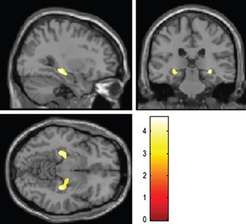
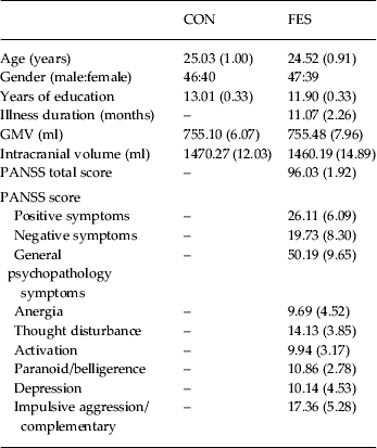
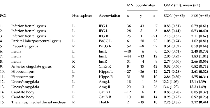
As shown in Table 1, there were no significant group differences in demographic variables, total GMV (p > 0.05) or intracranial volume (p > 0.05). The VBM analysis (Fig. 1) revealed that patients had significantly less GMV than controls in two clusters centered on the hippocampus bilaterally [cluster size: 1213 voxels on the right (MNI coordinates x = 31, y = −28, z = −10) and 879 voxels on the left hippocampus (MNI coordinates x = −27, y = −26, z = −12); p < 0.05, corrected for multiple comparisons]. The ROI analysis (Table 2) revealed that patients had smaller GMV than controls in the left inferior frontal gyrus (p = 0.012), bilateral hippocampus (p = 0.005 for the left, p = 0.002 for the right) and right thalamus (p = 0.015) (in a part corresponding to the medial dorsal nucleus) (p < 0.05, corrected).
Group differences in gray matter volume (GMV) revealed by voxel-based morphometry (VBM) analysis. Significant reduction in GMV in the hippocampus bilaterally in first-episode patients relative to controls, with 1213 voxels on the right (x = 31, y = −28, z = −10) and 879 voxels on the left hippocampus (x = −27, y = −26, z = −12) in cluster level, and threshold at p < 0.05 (corrected). Foci of reduced gray matter in patients were overlaid on brain slices spatially normalized into an approximation to the Talairach and Tournoux stereotactic atlas, displayed on the sagittal, coronal and transaxial planes.

Demographic and clinical data for all subjects

CON, Controls; FES, first-episode schizophrenia; GMV, gray matter volume; PANSS, Positive and Negative Syndrome Scale.
Values are given as number or mean (standard error).
No statistically significant differences between groups (p > 0.05).
Gray matter volume (GMV) in regions of interest (ROIs)

MNI, Montreal Neurological Institute; CON, controls; FES, first-episode schizophrenia; L, left; R, right; s.e., standard error.
Bold values indicate p < 0.05 (ANOVA, Bonferroni corrected for multiple comparisons).
Results of the covariant analysis
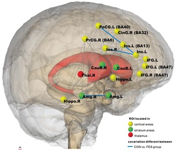
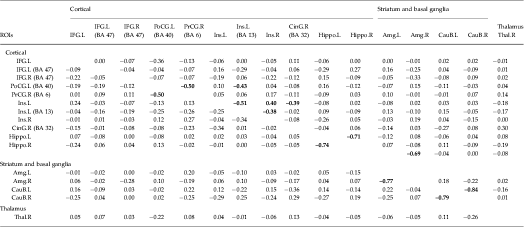
Following Fisher's r-to-z transformation, at a threshold of |Δz| ⩾1.96, the ROI pairs that were different between patients (FES) and controls (CON) predominantly involved the insula (Ins) and amygdala (Amg) bilaterally, in addition to the postcentral gyrus (PoCG), as follows: Ins.L [Brodmann area (BA) 13]–PoCG.L (BA 40) (r FES = −0.43, r CON = −0.25, Δz = –2.60), Ins.L (BA 13)–Ins.L (r FES = −0.51, r CON = −0.25, Δz = –3.97), Ins.R–Ins.L (r FES = −0.40, r CON = −0.04, Δz = 5.92) and Amg.L–Amg.R (r FES = −0.69, r CON = –0.77, Δz = −2.19) (Table 3, Fig. 2).
Covariation with a significant difference (|Δz| ⩾1.96, p < 0.05) was observed between four pairs (shown by blue lines) of regions of interest (ROIs) when the first-episode schizophrenia (FES) group was compared to the control (CON) group, including intra- and inter-insula, inter-amygdala and insula–parietal connections. The network is visualized with Pajek (http://vlado.fmf.uni-lj.si/pub/networks/pajek/). PANSS, Positive and Negative Syndrome Scale; BA, Brodmann area; IFG, inferior frontal gyrus; PoCG, postcentral gyrus; PrCG, precentral gyrus; Ins, insula; CinG, cingulate gyrus; Hippo, hippocampus; Amg, uncus/amygdala; CauB, caudate body; Thal, thalamus; L, left; R, right.

Correlation matrix for gray matter volume (GMV) in regions of interest (ROIs)

PANSS, Positive and Negative Syndrome Scale; BA, Brodmann area; IFG, inferior frontal gyrus; PoCG, parietal lobe/postcentral gyrus; PrCG, precentral gyrus; Ins, insula; CinG, anterior cingulate gyrus; Hippo, hippocampus; Amg, uncus/amygdala; CauB, caudate body; Thal, thalamus, medial dorsal nucleus; L, left; R, right.
Values given are Pearson correlation coefficients between ROIs for control (left of diagonal) and first-episode schizophrenia (right of diagonal) groups with residuals substituted for the raw GMV values after regressing out the effects of age, gender and intracranial volume.
Bold values indicate p < 0.05 (Bonferroni corrected for multiple comparisons).
Relationship between GMV and clinical variables
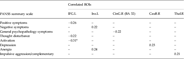
Within the FES group, the volume of the left inferior frontal ROI was negatively correlated with the scores for positive symptoms (r = −0.26, p = 0.02), thought disturbance (r = −0.22, p = 0.04) and activation factor of the PANSS (r = −0.31, p = 0.0038, corrected for multiple comparisons) (Table 4). The volume of the left insular ROI was positively correlated with the scores for negative symptoms (r = 0.22, p = 0.046) and anergia (r = 0.24, p = 0.03). The anterior cingulate ROI volume was negatively correlated with the general psychopathology score (r = −0.22, p = 0.04). The right caudate body ROI volume was positively correlated with the score for depression (r = 0.23, p = 0.04). The right thalamus GMV was correlated with the impulsive aggression/complementary score (r = 0.21, p = 0.049).
Association of PANSS subscale and gray matter volume (GMV) of ROIs in FES group

PANSS, Positive and Negative Syndrome Scale; ROI, region of interest; FES, first-episode schizophrenia; IFG, inferior frontal gyrus; Ins, insula; CinG, anterior cingulate gyrus; BA, Brodmann area; CauB, caudate body; Thal, thalamus, medial dorsal nucleus; L, left; R, right.
Values given are Pearson correlation coefficients with significance between ROIs (with residuals substituted for the raw GMV values after regressing out the effects of age, gender and intracranial volume) and PANSS subscale scores.
* p < 0.05 (Bonferroni corrected for multiple comparisons).
Discussion
In the present study, we examined a large sample of patients to identify neuroanatomical changes associated with schizophrenia. To exclude the potential effects of antipsychotic medication (Andreone et al. Reference Andreone, Tansella, Cerini, Rambaldelli, Versace, Marrella, Perlini, Dusi, Pelizza, Balestrieri, Barbui, Nose, Gasparini and Brambilla2007; Navari & Dazzan, Reference Navari and Dazzan2009; Ho et al. Reference Ho, Andreasen, Ziebell, Pierson and Magnotta2011) and chronicity of illness (Premkumar et al. Reference Premkumar, Fannon, Kuipers, Cooke, Simmons and Kumari2008) on GMV, all patients were in the first episode of psychosis and were naive to medication (or any other form of treatment). A VBM analysis revealed volume reductions in the hippocampus bilaterally, and an ROI analysis identified additional reductions in the left inferior frontal gyrus and right thalamus. A covariation analysis revealed an abnormal pattern of correlations between the GMV in several regions that normally participate in cortico-subcortical loops.
GMV loss in FES
Our finding of a reduction in hippocampal volume is consistent with data from previous MRI and post-mortem studies in schizophrenia (Nelson et al. Reference Nelson, Saykin, Flashman and Riordan1998; Wright et al. Reference Wright, Rabe-Hesketh, Woodruff, David, Murray and Bullmore2000), including studies in first-episode patients (Ellison-Wright et al. Reference Ellison-Wright, Glahn, Laird, Thelen and Bullmore2008). Reductions in hippocampal volume have also been reported in non-psychotic subjects at increased risk of the disorder, including the siblings of patients (Lawrie et al. Reference Lawrie, Whalley, Kestelman, Abukmeil, Byrne, Hodges, Rimmington, Best, Owens and Johnstone1999; van Erp et al. Reference van Erp, Saleh, Huttunen, Lonnqvist, Kaprio, Salonen, Valanne, Poutanen, Standertskjold-Nordenstam and Cannon2004) and people presenting with 'prodromal’ symptoms (Pantelis et al. Reference Pantelis, Velakoulis, McGorry, Wood, Suckling, Phillips, Yung, Bullmore, Brewer, Soulsby, Desmond and McGuire2003; Wood et al. Reference Wood, Yucel, Velakoulis, Phillips, Yung, Brewer, McGorry and Pantelis2005).
In the VBM analysis, the reduction in GMV was mainly in the middle part of the hippocampus on each side. In the ROI analysis, the region mainly included the anterior part of the hippocampus, close to the uncus, in addition to the body of the hippocampus. The absence of a significant reduction in the uncus suggests that the body of the hippocampus was particularly affected in this sample. Previous studies have localized volume reductions to the anterior part (Szeszko et al. Reference Szeszko, Goldberg, Gunduz-Bruce, Ashtari, Robinson, Malhotra, Lencz, Bates, Crandall, Kane and Bilder2003), but also to the mid- to posterior parts in schizophrenia (Narr et al. Reference Narr, Thompson, Sharma, Moussai, Zoumalan, Rayman and Toga2001, Reference Narr, van Erp, Cannon, Woods, Thompson, Jang, Blanton, Poutanen, Huttunen, Lonnqvist, Standerksjold-Nordenstam, Kaprio, Mazziotta and Toga2002). A shape analysis revealed mid- to anterior-lateral hippocampal volume changes in first-episode patients (Narr et al. Reference Narr, Thompson, Szeszko, Robinson, Jang, Woods, Kim, Hayashi, Asunction, Toga and Bilder2004). In follow-up studies, poor outcome in schizophrenia was linked to a smaller hippocampal tail volume (Bodnar et al. Reference Bodnar, Malla, Czechowska, Benoit, Fathalli, Joober, Pruessner, Pruessner and Lepage2010), whereas a reduction in body and tail volumes was associated with subsequent transition from the prodromal phase into psychosis (Witthaus et al. Reference Witthaus, Mendes, Brune, Ozgurdal, Bohner, Gudlowski, Kalus, Andreasen, Heinz, Klingebiel and Juckel2010). However, in a multicenter study on individuals at ultra-high risk, the parahippocampal cortex rather than the hippocampus was reported to be associated with the later onset of psychosis (Mechelli et al. Reference Mechelli, Riecher-Rossler, Meisenzahl, Tognin, Wood, Borgwardt, Koutsouleris, Yung, Stone, Phillips, McGorry, Valli, Velakoulis, Woolley, Pantelis and McGuire2011). Furthermore, by using VBM analysis, either the hippocampus or the parahippocampus have been reported with peak maxima of regional differences in FES patients (Schaufelberger et al. Reference Schaufelberger, Duran, Lappin, Scazufca, Amaro, Leite, De Castro, Murray, McGuire, Menezes and Busatto2007; Honea et al. Reference Honea, Meyer-Lindenberg, Hobbs, Pezawas, Mattay, Egan, Verchinski, Passingham, Weinberger and Callicott2008; Meisenzahl et al. Reference Meisenzahl, Koutsouleris, Bottlender, Scheucrecker, Jager, Teipel, Holzinger, Frodl, Preuss, Schmitt, Burgermeister, Reiser, Born and Moller2008; Leung et al. Reference Leung, Cheung, Yu, Yip, Sham, Li, Chua and McAlonan2011).
The ROI analysis revealed additional reductions in GMV in the left inferior frontal gyrus and right thalamus. Reduced inferior frontal volume is a robust finding in MRI studies of both chronic (Honea et al. Reference Honea, Crow, Passingham and Mackay2005) and first-episode schizophrenia (Ellison-Wright et al. Reference Ellison-Wright, Glahn, Laird, Thelen and Bullmore2008). Reductions in inferior frontal volume have also been described in people with prodromal symtoms of psychosis (Pantelis et al. Reference Pantelis, Velakoulis, McGorry, Wood, Suckling, Phillips, Yung, Bullmore, Brewer, Soulsby, Desmond and McGuire2003; Mechelli et al. Reference Mechelli, Riecher-Rossler, Meisenzahl, Tognin, Wood, Borgwardt, Koutsouleris, Yung, Stone, Phillips, McGorry, Valli, Velakoulis, Woolley, Pantelis and McGuire2011), and in the relatives of patients with schizophrenia (Job et al. Reference Job, Whalley, McIntosh, Owens, Johnstone and Lawrie2006). Altered inferior frontal activation has been reported in several functional neuroimaging studies in schizophrenia (Curtis et al. Reference Curtis, Bullmore, Brammer, Wright, Williams, Morris, Sharma, Murray and McGuire1998) and in prodromal subjects (Broome et al. Reference Broome, Matthiasson, Fusar-Poli, Woolley, Johns, Tabraham, Bramon, Valmaggia, Williams, Brammer, Chitnis and McGuire2009).
The reduction in thalamic GM in part corresponded to the medial dorsal nucleus. The thalamus has been described as a site of volume reduction in previous MRI studies of schizophrenia (Honea et al. Reference Honea, Crow, Passingham and Mackay2005), and the medial dorsal nucleus was specifically implicated in a meta-analysis of first-episode MRI data (Ellison-Wright et al. Reference Ellison-Wright, Glahn, Laird, Thelen and Bullmore2008) and in post-mortem studies of schizophrenia (Danos et al. Reference Danos, Baumann, Kramer, Bernstein, Stauch, Krell, Falkai and Bogerts2003; Danos et al. Reference Danos, Schmidt, Baumann, Bernstein, Northoff, Stauch, Krell and Bogerts2005). It has been hypothesized that the medial dorsal nucleus participates in filtering information from the prefrontal cortex to the striatum, which may contribute to cognitive disruption of schizophrenia (Vollenweider & Geyer, Reference Vollenweider and Geyer2001).
In the present study, the patients were in the first episode of the disorder and were treatment naive. The reductions in regional GMV that we detected are therefore unlikely to be secondary to effects of either illness progression or antipsychotic medication (Dazzan, Reference Dazzan2010; Ho et al. Reference Ho, Andreasen, Ziebell, Pierson and Magnotta2011).
Changed correlation pattern of GMV in schizophrenia
A covariation in GMVs may be evident when regions are connected anatomically or functionally (Bullmore et al. Reference Bullmore, Woodruff, Wright, Rabe-Hesketh, Howard, Shuriquie and Murray1998; Zhang & Sejnowski, Reference Zhang and Sejnowski2000; Mechelli et al. Reference Mechelli, Friston, Frackowiak and Price2005). In the present study, the first-episode patients mainly differed from the controls in the covariation between the GMVs in the insula and amygdala bilaterally, and also between the insula and postcentral gyrus. This is consistent with the notion that cortico-subcortical dysfunction contributes to the pathophysiology of schizophrenia (Andreasen et al. Reference Andreasen, Paradiso and O'Leary1998), and previous studies that have reported altered cortico-subcortical function and connectivity in schizophrenia (Salvador et al. Reference Salvador, Sarró, Gomar, Ortiz-Gil, Vila, Capdevila, Bullmore, McKenna and Pomarol-Clotet2010).
GMV of ROIs is related to clinical features of schizophrenia
As shown in Table 4, within the patient sample, the left inferior frontal volume was negatively correlated with the severity of positive symptoms, thought disturbance and activation scores. This is consistent with previous reports linking reduced orbitofrontal volume in schizophrenia to thought disorder (Nakamura et al. Reference Nakamura, Nestor, Levitt, Cohen, Kawashima, Shenton and McCarley2008). In addition, a volume reduction in the inferior frontal regions was found to be associated with decreased insight (Sapara et al. Reference Sapara, Cooke, Fannon, Francis, Buchanan, Anilkumar, Barkataki, Aasen, Kuipers and Kumari2007; Bergé et al. Reference Bergé, Carmona, Rovira, Bulbena, Salgado and Vilarroya2011), severity of negative (Bergé et al. Reference Bergé, Carmona, Rovira, Bulbena, Salgado and Vilarroya2011) and positive symptoms (Garcia-Marti et al. Reference Garcia-Marti, Aguilar, Lull, Marti-Bonmati, Escarti, Manjon, Moratal, Robles and Sanjuan2008; Modinos et al. Reference Modinos, Vercammen, Mechelli, Knegtering, McGuire and Aleman2009; Suga et al. Reference Suga, Yamasue, Abe, Yamasaki, Yamada, Inoue, Takei, Aoki and Kasai2010), and duration of untreated psychosis (Burke et al. Reference Burke, Androutsos, Jogia, Byrne and Frangou2008; Malla et al. Reference Malla, Bodnar, Joober and Lepage2011). The inferior frontal region has also been implicated in the pathophysiology of auditory hallucinations and formal thought disorder in functional neuroimaging studies of schizophrenia (McGuire & Frith, Reference McGuire and Frith1996; McGuire et al. Reference McGuire, Shah and Murray1993, Reference McGuire, Quested, Spence, Murray, Frith and Liddle1998). We found a correlation between GMV in the left insula with both negative symptoms and anergia, in line with evidence that lesions of the right anterior insula can produce a clinical syndrome corresponding to anergia (Craig, Reference Craig, Lewis, Haviland-Jones and Barrett2008). A previous analysis of data from a sample that overlapped with that of the present study identified a correlation between right anterior cingulate gyrus volume and the severity of positive symptoms, thought disturbance, activation, paranoia and impulsive aggression (Lui et al. Reference Lui, Deng, Huang, Jiang, Ma, Chen, Zhang, Li, Li, Zou, Tang, Zhou, Mechelli, Collier, Sweeney, Li and Gong2009). However, we did not replicate this finding, possibly because of differences in the topographical location of the respective anterior cingulate foci in the two studies. In our study, the volume of the caudate body was positively correlated with the severity of depression. Increased caudate nucleus volume has been attributed to the effects of antipsychotic treatment (Dazzan, Reference Dazzan2010), although reductions in caudate body volume reduction have been described in FES patients who have not been treated with antipsychotics (Levitt et al. Reference Levitt, McCarley, Ersner-Hershfield, Salisbury, Kikinis, Jolesz and Shenton2002; Ebdrup et al. Reference Ebdrup, Glenthoj, Rasmussen, Aggernaes, Langkilde, Paulson, Lublin, Skimminge and Baare2010). It is noteworthy that an association between thalamus GMV and the impulsive aggression cluster score was detected in the current study, which implies that neural structures and functions underlying prepulse inhibition are involved in (inhibition of) violence in schizophrenia (Kumari et al. Reference Kumari, Das, Hodgins, Zachariah, Barkataki, Howlett and Sharma2005).
Methodological considerations and limitations
Although used routinely by researchers, both VBM and ROI analyses have advantages and disadvantages that may contribute to the discrepancies in the current findings. As a methodological consideration, ROI analysis was characterized as a voxel-averaged, landmark-based contrast to single, voxel-by-voxel whole-brain VBM measurements (Giuliani et al. Reference Giuliani, Calhoun, Pearlson, Francis and Buchanan2005). ROI analysis provides absolute estimates of the number of voxels in the regions compared to the relative changes in the concentration of GM within each voxel provided by VBM results. Although not as discrete as ROIs, the VBM method gives equal weight to every voxel included in the 3D sampling array, and thus provides a broad assortment of regional comparisons. Thus, the two complementary analyses, VBM and ROI (Sarazin et al. Reference Sarazin, Chauvire, Gerardin, Colliot, Kinkingnehun, De Souza, Hugonot-Diener, Garnero, Lehericy, Chupin and Dubois2010), made the results of the current study worthy of consideration.
We performed a correlation analysis of GMV in a set of regions selected on the basis of a previous meta-analysis of regional GMV abnormalities in schizophrenia (Ellison-Wright et al. Reference Ellison-Wright, Glahn, Laird, Thelen and Bullmore2008) and current VBM results. However, we only examined connections between these regions, and did not assess their connections to areas outside this network, or connections between regions other than those selected. Nevertheless, the findings are consistent with evidence that schizophrenia involves distributed rather than focal pathology and disordered connectivity (Andreasen et al. Reference Andreasen, Oleary, Cizadlo, Arndt, Rezai, Ponto, Watkins and Hichwa1996, Reference Andreasen, Paradiso and O'Leary1998; Andreasen, Reference Andreasen1999).
In conclusion, FES is associated with reductions in hippocampal, inferior frontal and thalamic volumes, and alterations in the relationship between GMVs in the insula, amygdala and postcentral gyrus. Because these findings were evident in medication-naive first-episode patients, they are unlikely to be secondary to effects of chronic illness or antipsychotic treatment.
Acknowledgments
This work was partly funded by the National Nature Science Foundation of China (30530300 and 30125014, T.L.; 30971056, Q.W.), the National Basic Research Program of China (973 Program 2007CB512301, T.L.; 2007CB512305, Q.G.), a National Alliance for Research on Schizophrenia and Depression (NARSAD) Independent Investigator Award (T.L.), the Wellcome Trust (International Collaborative award to T.L. and D.A.C.), and the China Postdoctoral Science Foundation (20090461337 and 201003699, W.D.).
We thank all of the participants and their families for taking part in the study and the many clinicians who referred patients to the study. We also thank C. Cheung and M. Picchioni for statistical and technical support.
Declaration of Interest
None.