Ultrasonographic examination can be useful in monitoring the reproductive organs of domestic animals (Lazaridis et al. Reference Lazaridis, Brozos and Kiossis2012). Specifically in the udder of ewes, ultrasonographic examination has been used to study mammary parenchyma as a means of diagnosing mammary disorders (Ruberte et al. Reference Ruberte, Carretero, Fernandez, Navarro, Caja, Kirchner and Such1994; Franz et al. Reference Franz, Baumgartner, Hofmann-Parisot, Windischbauer, Suchy and Bauder2001; Franz et al. Reference Franz, Hofmann-Parisot, Gütler and Baumgartner2003) usually by linear-type probes by using a frequency of 5·0–8·5 MHz. Imaging of the teats of ewes required higher frequency probes (6·0–12·0 MHz) (Franz et al. Reference Franz, Baumgartner, Hofmann-Parisot, Windischbauer, Suchy and Bauder2001; Mavrogianni et al. Reference Mavrogianni, Fthenakis, Burriel, Gouletsou, Papaioannou and Taitzoglou2004). The size of the mammary cistern (sinus lactiferous) has been evaluated by using ultrasonographic examination; a close correlation between the ultrasonographically imaged area of the mammary cistern and its real volume has been reported by Ruberte et al. (Reference Ruberte, Carretero, Fernandez, Navarro, Caja, Kirchner and Such1994), Nudda et al. (Reference Nudda, Pulina, Vallebellla, Bencini and Enne2000) and Castillo et al. (Reference Castillo, Such, Caja, Salama, Albanell and Casals2008), with a view to estimating milk yield of the animal (Castillo et al. Reference Castillo, Such, Caja, Salama, Albanell and Casals2008; Rovai et al. Reference Rovai, Caja and Such2008). Doppler ultrasonographic examination of the lactating udder of ewes has been reported by Piccione et al. (Reference Piccione, Arcigli, Assenza, Percipalle and Caola2004) and Christensen et al. (Reference Christensen, Nielsen, Bauer and Hilden1989).
Previous experiments have indicated that there were no effects of the drying-off procedure (i.e. progressive or abrupt) in subsequent mammary infection and development of mastitis (Petridis et al. Reference Petridis, Mavrogianni, Gougoulis, Amiridis, Brozos and Fthenakis2012, Reference Petridis, Mavrogianni, Fragkou, Gougoulis, Tzora, Fotou, Skoufos, Amiridis, Brozos and Fthenakis2013). Currently, no studies are available to compare structural changes in the udder according to the type of involution process, which may potentially play a role in subsequent intramammary infections and mastitis development (Sordillo & Nickerson, Reference Sordillo and Nickerson1988; Petridis & Fthenakis, Reference Petridis and Fthenakis2014) and to identify differences between the two procedures. Nevertheless, structural changes during involution (e.g. distension of teat cistern due to milk accumulation, changes in blood flow etc.) may be responsible for increased incidence risk of infection during the procedure (e.g. Dingwell et al. Reference Dingwell, Leslie, Schukken, Sargeant, Timms, Duffield, Keefe, Kelton, Lissemore and Conklin2004). The objective of the present study was to record, by means of ultrasonographic examination, changes occurring in the udder of ewes during involution and to compare differences between progressive or abrupt udder drying-off.
Materials and methods
Study design
The work was carried out in multiparous Lacaune cross-breed ewes housed and monitored throughout the study. Lambs had been removed from their dams at 45–60 d of age and then ewes were hand-milked twice daily. After a total lactation period of 6–8 months, animals were included in the experiment.
Initially, 20 ewes were allocated at random to one of two groups (A or B). Inclusion criteria for animals into the study were: (a) no abnormal findings in the clinical examination before inclusion; (b) bacteriologically negative results in teat duct material and milk samples; (c) California Mastitis Test scores in milk samples <1 and (d) daily milk yield 350–400 ml per ewe.
In ewes of group A (n=10), udder drying-off took place progressively during a period of 22 d; ewes were milked twice daily for the last time on Day (D) 0; then, they were hand-milked once daily for a week (D1–D7), which was followed by another week during which ewes were hand-milked once every 2 d (D9, D11, D13), followed by a third week during which ewes were hand-milked once every 3 d (D16, D19, D22); in total, during the whole process, ewes were hand-milked on 13 occasions, always by the same milker. In ewes of group B (n=10), udder drying-off took place abruptly; ewes were milked twice daily for the last time on D0 and no milking was carried out after that.
Throughout the involution process, ultrasonographic examinations (B-mode and Doppler) were performed in the udder of the experimental animals, on the following days after start of the study: D0 (last day when ewes were milked twice daily), D2, D4, D7, D10, D14, D18, D23, D30 and D37. Clinical examinations were performed on each occasion when ultrasonographic examination of the udder was carried out.
Subsequently, lambing of the ewes took place 72–76 d after D37. Then, 4–5 d after lambing, teat duct material and milk samples were collected again for bacteriological examination and California Mastitis Test scoring.
Sampling and examinations performed
Clinical examination, with special attention to the mammary glands and teats, was performed as described before by Fthenakis (Reference Fthenakis1994), Saratsis et al. (Reference Saratsis, Leontides, Tzora, Alexopoulos and Fthenakis1998) and Mavrogianni et al. (Reference Mavrogianni, Fthenakis, Brooks, Papaioannou, Cripps, Taitzoglou, Brellou and Saratsis2005). Teat duct material and milk samples were collected by following the procedures previously described in detail (Fthenakis, Reference Fthenakis1994; Saratsis et al. Reference Saratsis, Leontides, Tzora, Alexopoulos and Fthenakis1998; Mavrogianni et al. Reference Mavrogianni, Fthenakis, Brooks, Papaioannou, Cripps, Taitzoglou, Brellou and Saratsis2005, Reference Mavrogianni, Cripps, Papaioannou, Taitzoglou and Fthenakis2006). In all cases, samples were collected from both teats and both mammary glands of each ewe. All samples were processed bacteriologically and cytologically as previously described (Fthenakis, Reference Fthenakis1994, Reference Fthenakis1995; Mavrogianni et al. Reference Mavrogianni, Fthenakis, Brooks, Papaioannou, Cripps, Taitzoglou, Brellou and Saratsis2005).
Ultrasonographic examination
Ultrasonographic examination was performed with the animal in the standing position and restrained inside a crate, using the support of an assistant. Before examination, hair on the udder had been fully clipped.
Ultrasonographic examination of the udder was performed with an ultrasound scanner (MyLab® 30; ESAOTE SpA, Genova, Italy) fitted with a linear transducer with 4·0–13·0 MHz imaging frequency and a microconvex transducer with 2·0–8·0 MHz imaging frequency. Coupling gel was applied. The probe was placed on the caudal surface of the udder and moved around it.
Initially, the left side of the udder was imaged. The linear transducer was placed in a position perpendicular to the long axis and dorsal sections of the mammary parenchyma were taken, starting from the upper part downwards. In each mammary gland, three images were saved for further processing; the first image was taken before the branching of the external pudental artery (arteria pudenda externa), the second when distance between branches of the external pudental artery was ∼1 cm and the third image was taken immediately before the gland cistern (sinus lactiferous) became visible; 60 mm scanning depth and 10 MHz frequency were used for this procedure.
Subsequently, the gland cistern (sinus lactiferous) was imaged by using the microconvex transducer. At first, an inclined sagittal imaging plane was used, from the upper part of the intermammary groove (sulcus intermammarius) towards the teat; the teat was used as the scanning axis. A second image was taken, by rotating the microconvex transducer at 90° at the same examination point as previously, i.e. from the upper part of the intermammary groove towards the teat but at the dorsal plane; 120 mm scanning depth and 3·3 MHz frequency were used for this procedure.
The final measurements were taken at the external pudendal artery, which was used for Doppler measurements in the mammary gland by means of the linear probe; 60 mm scanning depth and 10 MHz frequency were used for this procedure. At the start, a cross-section of the vessel was taken, with the objective to measure the diameter of the vessel. Then the colour Doppler gate was positioned parallel to the wall of the vessel examined; when optimal colour flow was achieved, a spectral mode waveform was taken. A skilled assistant was helping when measuring the blood flow velocity, in order to make adjustments necessary for optimal quality of colour-flow images. Animals were stressed as little as possible during the examination, with the objective to have them examined whilst remaining still for the period of time necessary to apply correctly the sample gate and to take the spectral display after localising the vessel to be studied. Initially, the vessel to be examined with the colour-flow imaging, was localised. Then adjustments necessary for optimal colour-flow image quality were made. Spectral mode was switched on and the sample gate was positioned inside the vessel, whilst taking care not to include the vessel wall and to avoid artifacts; this was followed by setting the appropriate ‘Doppler angle’ [i.e. the angle at which the ultrasound beams intersect the path of flowing blood, also termed ‘angle of insonation’ (Ginther, Reference Ginther2007)]. In this case, a ‘Doppler angle’ of 50–60° was used in the examination.
All the imaging procedures described above were subsequently repeated for the right mammary gland. In all cases, images (B-mode or Doppler) were frozen and saved on the equipment hard-disk for performing subsequently appropriate measurements and data analysis.
Data management and analysis
Results taken into account
In the analysis, ultrasonographic results of only 14 ewes (group A, n=7; group B, n=7) were taken into account. These animals were the ones that met cumulatively the following criteria: (a) no clinical abnormalities detected throughout the study and after lambing; (b) bacteriologically negative results in teat duct material and milk samples collected after lambing; and (c) California Mastitis Test scores <1 in milk samples collected after lambing. This approach supported use of results only from animals with no abnormalities in the udder throughout the study and guaranteed that ultrasonographic findings were those of the udder of healthy ewes.
Quantitative evaluations of ultrasonographic images
During the ultrasonographic examination four sets of images were recorded: (a) images of mammary parenchyma, (b) images of mammary gland cistern, (c) images of cross-sections of external pudendal artery and (d) spectral waveforms of external pudendal artery. All images were initially evaluated visually for presence of abnormal structures therein. Then data management was performed on images that had been saved at the time of examination. Initially, data for left and right mammary gland of the same animal were considered separately.
Stored images of mammary parenchyma were processed by means of ImageJ software (National Institutes of Health, Rockville Pike MD, USA) which can edit, process and analyse gray-scale images, by calculating area and pixel value statistics (National Institutes of Health, 2013). In an image processing context, gray-scale analysis refers to the image's pixel intensity values (Ojala et al. Reference Ojala, Pietikaeinen and Maeenpaea2002). For analysis of gray-scale, results of each of the three images stored from each mammary gland on each occasion were considered together. Areas with vessels or ductal formations were not taken into account for the gray-scale analysis. Results were expressed on a 0 (black) to 255 (white) scale.
Stored images of mammary gland cistern were processed by means of ImageJ software, which, after appropriate scale setting, was able to measure on the image the selected area within the boundaries of the cistern (National Institutes of Health, 2013). Two different measurements were taken, one from the image taken at the sagittal plane and another from the image taken at the dorsal plane; data for left and right mammary gland of the same animal were considered separately. Results were expressed as mm2.
Stored images of cross-sections of external pudendal artery were processed by means of MyLab software (ESAOTE SpA, Genova, Italy) which, after pointing out the internal boundaries of the vessel, calculated the internal diameter of the vessel. Results were expressed as mm.
Finally, spectral waveforms of the external pudendal artery were processed by means of MyLab software (ESAOTE SpA, Genova, Italy) which, based on the outline of the waveform, calculated directly the resistance index (RI) and the pulsatility index (PI) of the vessel, based on the following equations RI=(PSV−EDV)/PSV and PI=(PSV−EDV)/TAMV, where PSV is peak systolic velocity, EDV is end diastolic velocity and TAMV is time-averaged maximum velocity (Ginther, Reference Ginther2007). Results were expressed as ratios.
Statistical computations
All data were entered into Excel spreadsheets (Microsoft Corporation, Redmond WA, USA). Initially, descriptive statistics for all parameters and all groups (mean±sem) were performed.
Initially, repeated measures mixed effect linear regression models were used to determine whether outcomes in the two mammary glands of each animal changed over the course of the study period. Models were adjusted for repeated measures within animals. Variables (fixed effects) included mammary gland (i.e. left or right) and time-point of the study (i.e. D0, D2, D4, etc.). In all cases, no significant differences were evident between the left and the right glands of the experimental animals within each group (for all parameters, P>0·56 or P>0·68, for group A or B, respectively).
In view of that, the time-series results of all measurements (left and right glands) obtained from animals of the same group were considered together for between-group comparisons, i.e. n=14 subjects within each group. Then, repeated measures mixed effect linear regression models were used to determine whether outcomes in the two groups of the study changed over the course of the study period. Models were adjusted for repeated measures within animals. Independent variables (fixed effects) included experimental group (i.e. progressive or abrupt cessation of lactation), time of the study (i.e. D0, D2, D4, etc.) and a time of the study by experimental group interaction.
Finally, another analysis was performed which took into account results of only three time-points as follows: D23, D30, D37 for group A vs. D2, D10, D18 for group B. The objective of this analysis was to compare findings at the early stages after cessation of lactation in both groups. The above model was also used for this analysis.
All analyses were performed by a commercial statistical program (SPSS, v. 15 for Windows; SPSS Inc., Chicago IL, USA). Significance level was set at P=0·05.
Results
In general, anatomic structures in the mammary gland had a medium echogenicity to hyperechogenicity. Blood vessels and lactiferous ducts were imaged therein. No abnormal structures were observed. On D0, the mammary parenchyma was seen with a lobular structure (consisting of the alveolar areas with reduced echogenicity), the connective tissue (with increased echogenicity) and the ductal part of the gland and the blood vessels therein (imaged as anechoic antra). As involution progressed, the mammary parenchyma was imaged to have homogeneity, with disappearance of the lobular picture. Moreover, an overall reduced (compared with D0) echogenicity was recorded (Fig. 1). Structures with medium echogenicity could include fat, ductal and lobular epithelial tissues and loose, intralobular and periductal stromal fibrous tissue. In contrast, hyperechoic structures could include the compact interlobular stromal fibrous tissue, the mammary fasciae, the ligaments and the skin. During scanning, other findings, e.g. the external pudendal artery and its branches, could also be imaged as spontaneous observations during the various procedures.
Serial ultrasonographic appearance of mammary parenchyma of ewes during abrupt udder drying-off procedure: (a) D0 (before start of the drying-off procedure), (b) D37. Images taken at a level after the branching of the external pudental artery (when distance between the two branches was ∼1 cm) on a MyLab® 30 ultrasonography system with linear transducer 10·0 MHz and scanning depth 60 mm.

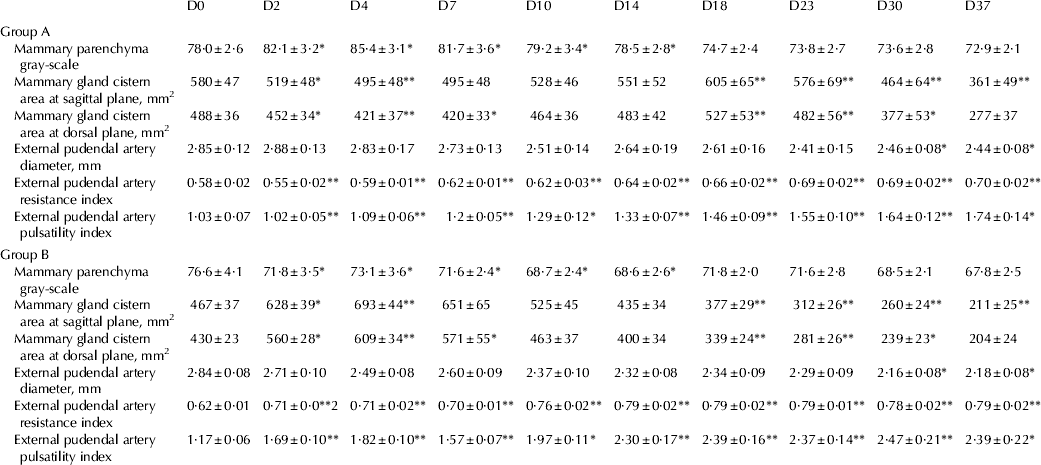
Mean (±sem) gray-scale results in group A were 78·0±2·6 on D0, 78·5±2·8 on D14 and 72·9±2·1 on D37; respective values in group B were 76·6±4·1, 68·6±2·6 and 67·8±2·5 (for differences between groups, P=0·049). In all cases, there was clear evidence that the progressive decrease of gray-scale results throughout the study period was significant in both groups, A or B (P=0·004), as were interactions between time and group (P<0·0005).
Milk clots were imaged in the gland cistern, the earliest on D30 in group A ewes (i.e. 8 d after cessation of milking) and on D7 in group B ewes (i.e. 7 d after cessation of milking). Initially, these appeared as small flakes floating in the gland cistern, later aggregating to form large clots (1–2 cm in diameter), which remained visible ultrasonographically up to 1 month after milk removal had stopped. A temporary increase in cistern area was evident on D2, D4 and D7 in group B animals which was followed by a subsequent decrease thereafter (Fig. 2). Cistern area results did not differ between animals in group A or B (P=0·349 for sagittal plane images, P=0·573 for dorsal plane images). However, differences throughout the study period were significant (P<0·0005), as were interactions between time and group (P<0·0005).
Serial ultrasonographic appearance of gland cistern of ewes during abrupt udder drying-off procedure: (a) D0 (before start of the drying-off procedure), (b) D37. Images taken at an inclined dorsal imaging plane, from the upper part of the intermammary groove towards the teat, which was used as the scanning axis on a MyLab® 30 ultrasonography system with microconvex transducer 3·3 MHz and scanning depth 120 mm.
Diameter of the external pudendal artery differed significantly between the two groups (P=0·007). Mean (±sem) diameters in group A were 2·85±0·12 mm on D0, 2·59±0·19 mm on D14 and 2·44±0·08 mm on D37; respective values in group B were 2·84±0·08 mm, 2·32±0·08 mm and 2·18±0·08 mm. Differences throughout the study period were also significant (P<0·0005), but were interactions between time and group were not (P=0·071).
The spectral display in Doppler mode was observed as a broad band structure. Both RI and PI progressively increased throughout the study period in both groups. There was clear evidence that differences between animals in group A or in group B were significant for both parameters (P<0·0005) (Fig. 3); also, progressive changes in blood flow results throughout the study period were significant in both groups, A or B (P<0·0005), as were interactions between time and group (P<0·0005).
Serial spectral waveforms of the external pudendal artery at the mammary gland of ewes: (a) D2 (2 d after start of the udder drying-off procedure), (b) D37. Images taken and processed on a MyLab® 30 ultrasonography system with linear transducer 10 MHz and scanning depth 60 mm.
Finally, when data at the early stages after cessation of lactation (i.e. D23, D30, D37 in group A and D2, D10, D18 in group B) were used for comparisons between the two groups, there was no evidence that differences in gray-scale results (P=0·425) or cistern area (P=0·537 for sagittal plane images and P=0·163 for dorsal plane images) were significant. Progressive changes throughout the analysis period were significant in cistern area results (P<0·0005), but not in gray-scale (P=0·425). Interactions between time and group were not significant for any of the above parameters (P<0·3). In contrast, differences in RI and PI were significant between the two groups of ewes (P=0·021 for PI and P=0·015 for RI); progressive changes throughout the analysis period were significant in both groups, A or B (P<0·0005), as were interactions between time and group (P<0·015). Detailed results are shown in Table 1.
Quantitative results of ultrasonographic examination of udder in ewes, in which udder drying-off procedure took place progressively (group A) or abruptly (group B); for each parameter examined, mean±sem are presented; results of both mammary glands of ewes are considered together, as no statistical difference (P>0·5) was evident between left and right mammary glands. Results are shown for time (days, D) relative to the start of drying off at D1
* P<0·05, ** P<0·01 between the two groups at respective day for the respective parameter
Discussion
The present study describes, to the best of our knowledge for the first time, the B-mode and Doppler ultrasonographic patterns of the involuting mammary gland of ewes. The technique could be easily applied, with minimal restraint of the animals. Ultrasonographic examination is advantageous in the study of sequential processes, as it affords maintenance of the same experimental subjects with increased welfare, conditions which cannot be met by other techniques (e.g. biopsy sampling).
In B-mode ultrasonographic examination, three fields sufficed to receive a representative image of the entire mammary parenchyma. Images were taken using as reference points mammary anatomical structures (which remained unchanged during involution) rather than dimensions (which changed as the mammary involution progressed). Doppler ultrasonographic examination is useful for measuring mammary blood flow in sheep (Christensen et al. Reference Christensen, Nielsen, Bauer and Hilden1989; Piccione et al. Reference Piccione, Arcigli, Assenza, Percipalle and Caola2004). The ‘Doppler angle’ used in the examination was <60°, as otherwise measurement errors could occur, leading to overestimation of blood flow velocity (Arning, Reference Arning and Arning2002); best velocity accuracy occurs at 45–60° (Ginther, Reference Ginther2007). It was easy to distinguish arteries from veins by means of Doppler ultrasonographic examination, as in the latter vessels the spectral waveform was seen to be flat and broad with no appearance of cardiac cycles.
Changes in ultrasonographic appearance of the mammary parenchyma reflect to some extent histological changes, for example, 30 d after cessation of milk removal, mammary epithelial cells have been found to be greatly reduced in size and cuboidal in shape (Tatarczuch et al. Reference Tatarczuch, Philip and Lee1997). Changes in gray-scale appearance (progressively decreasing echogenicity) of ultrasonographic images of mammary parenchyma during involution could be attributed to decreasing numbers of mammary epithelial cells and, consequently, to the increasing proportion of adipose tissue in the mammary gland. Transfer of milk fat from the alveoli to the cistern slows down with increased time of milk removal, leaving a backup of milk fat in the mammary alveoli (McKusick et al. Reference McKusick, Thomas, Berger and Marnet2002; Castillo et al. Reference Castillo, Such, Caja, Salama, Albanell and Casals2008) which, during involution, is removed by leucocytes (Lee & Lascelles, Reference Lee and Lascelles1969; Tatarczuch et al. Reference Tatarczuch, Philip, Bischof and Lee2000). In general, during involution, extensive remodelling of the extracellular matrix of the mammary gland takes place, which results in a volume increase of glandular stroma (Sordillo & Nickerson, Reference Sordillo and Nickerson1988; Hurley, Reference Hurley1989; Capuco et al. Reference Capuco, Akers and Smith1997; Stavros, Reference Stavros, Rumack, Wilson, Charboneau and Levine2011).
Differences in gray-scale (i.e. in echogenicity) between the two groups reflect the differences in tissue remodelling and the speed at which they are taking place during the involution of the mammary gland. Nevertheless, these changes did not appear to lead to differences in the risk of intramammary infection and of mastitis development (Petridis et al. Reference Petridis, Mavrogianni, Gougoulis, Amiridis, Brozos and Fthenakis2012, Reference Petridis, Mavrogianni, Fragkou, Gougoulis, Tzora, Fotou, Skoufos, Amiridis, Brozos and Fthenakis2013).
The lack of differences in results of cistern area at the sagittal and the dorsal plane of ultrasonographic imaging between the two groups indicates that volume of the structure does not appear to be affected by the procedure followed for involution (initiated or progressive). After cessation of milking, an increase in cisternal volume is the result of accumulation of milk therein. Approximately 1 week after cessation of milking, milk therein was observed to have clotted and to have been reabsorbed which led, thereafter, to a decrease in volume of the cistern. If accumulation of milk in the gland cistern is a potential risk factor for development of mastitis, as discussed above, then obviously the similar volume of the gland cistern between ewes of the two groups indicates that risk of mammary infection and mastitis development is similar, independently of the procedure followed for drying-off. This finding can partly explain the lack of difference in mastitis development between the two procedures (Petridis et al. Reference Petridis, Mavrogianni, Gougoulis, Amiridis, Brozos and Fthenakis2012, Reference Petridis, Mavrogianni, Fragkou, Gougoulis, Tzora, Fotou, Skoufos, Amiridis, Brozos and Fthenakis2013).
No significant difference was evident between left and right cistern area (and volume) in animals of both groups. In relation to that, it is noteworthy that Nudda et al. (Reference Nudda, Pulina, Vallebellla, Bencini and Enne2000) and Castillo et al. (Reference Castillo, Such, Caja, Salama, Albanell and Casals2008) have mentioned that potential differences in cistern size between left and right sides can reflect differences in the force applied during milking or suckling. The present results can be explained in view of those of Nudda et al. (Reference Nudda, Pulina, Vallebellla, Bencini and Enne2000), as a lack of force to withdraw milk at drying-off leading to a similar size of the two gland cisterns of the same animal.
Doppler ultrasonographic examination indices (RI and PI) may be used as alternatives to velocity Doppler measurements. They are useful, especially when placement of ‘Doppler angle’ is difficult, as in tortuous vessels, because they are independent of the Doppler angle as ratios of velocity measurements. Increased RI indicates decreased mammary blood perfusion, whilst increased values of PI indicate decreased blood perfusion of distal tissues. In fact, the two indices are highly correlated with each other. RI is more suitable when blood flow in vessels under study persists during diastole and PI when flow is absent during diastole (Ginther, Reference Ginther2007). The significant decrease in blood flow at the external pudendal artery indicates the importance of this vessel for blood circulation in mammary glands.
In the present study, both indices (RI and PI) increased progressively during the involution procedure, in animals of both groups, obviously being the result of decreased blood perfusion as involution advanced. Reduction of blood flow in the mammary gland may be a consequence of reduced blood requirement due to the reduced milk secretory activity rather than of increased intramammary pressure, because of accumulation of milk after cessation of milk removal (Knight et al. Reference Knight, Peaker and Wilde1998). Decrease of the diameter of the external pudendal artery further contributes to the reduced amount of blood transferred to the mammary gland and in that way contributes to the involution of the mammary tissue. Decreasing blood volumes provide weaker signals for Doppler spectrum analysis because of the smaller number of moving erythrocytes (Widder & Goertler, Reference Widder, Goertler, Widder and Goertler2004).
Previous studies have documented the impaired defensive ability of the mammary gland during involution, as a result of decreased concentration of lactoferrin and lactoferrin:citrate ratio, decreased phagocytic ability of leucocytes and reduced amount of immunoglobulins in lacteal secretions (Petridis & Fthenakis, Reference Petridis and Fthenakis2014). The above predispose the mammary gland to new mastitis cases during involution and support recrudescence of subclinical infections, which had occurred during the previous lactation period (Petridis & Fthenakis, Reference Petridis and Fthenakis2014). Reduced blood volumes during the involution process could further contribute to development of mammary disease, as smaller numbers of leucocytes would be transferred to the mammary gland.
The significant differences between the two groups reflect the differences in blood flow into the mammary gland after cessation of lactation. Initiated involution led to a more rapid decrease of blood flow, whilst progressive involution sustained milk production for a longer period. Nevertheless, these changes did not appear to lead to differences in the risk of intramammary infection and of mastitis development (Petridis et al. Reference Petridis, Mavrogianni, Gougoulis, Amiridis, Brozos and Fthenakis2012, Reference Petridis, Mavrogianni, Fragkou, Gougoulis, Tzora, Fotou, Skoufos, Amiridis, Brozos and Fthenakis2013).
The lack of differences in blood flow between the two mammary glands was first recorded by Nielsen et al. (Reference Nielsen, Jakobsen and Jorgensen1990) in goats and is now confirmed by the present results in ewes. This may be potentially used in identifying early stages of inflammation after mammary infection.
The above findings may be used to describe the normal ultrasonographic appearance of the udder of ewes during involution. We used a combination of clinical, bacteriological (teat duct material and milk) and cytological criteria to confirm udder health of the animals. In previous studies, it has been confirmed that bacteria colonise the teat duct (Fragkou et al. Reference Fragkou, Gougoulis, Billinis, Mavrogianni, Bushnell, Cripps, Tzora and Fthenakis2011) and then can cause mammary infection and mastitis, especially if local udder defences become impaired (Mavrogianni et al. Reference Mavrogianni, Fthenakis, Brooks, Papaioannou, Cripps, Taitzoglou, Brellou and Saratsis2005, Reference Mavrogianni, Cripps, Papaioannou, Taitzoglou and Fthenakis2006, Reference Mavrogianni, Cripps and Fthenakis2007; Fragkou et al. Reference Fragkou, Mavrogianni, Cripps, Gougoulis and Fthenakis2007a, Reference Fragkou, Papaioannou, Cripps, Boscos and Fthenakisb).
In this way, reference standards would be available for interpreting abnormal features subsequently. Perhaps ultrasonographic examination of ewes at the end of the first week after cessation of lactation could be of potential help in identifying early cases of ‘dry-period’ mastitis, which develop slowly and with no striking clinical features. That would be especially helpful in cases of selective administration of antibiotics (Petridis & Fthenakis, Reference Petridis and Fthenakis2014) to ewes at the beginning of the dry-period, in which case administration of antibiotics is performed only to animals found to be infected at the end of a lactation period (Petridis & Fthenakis, Reference Petridis and Fthenakis2014). In that procedure, many ewes remain unprotected against new intramammary infections during the dry-period. In animals with abnormal ultrasonographic findings at that stage, perhaps antibiotic administration might be performed at that stage, although further studies would be required to assess the potential value of such a procedure. As lactation would have ceased, injectable administration of antibiotics would be the preferable route in such cases; the efficacy of injectable administration of antibiotics at various time-points during the dry period has been described by Croft et al. (Reference Croft, Duffield, Menzies, Leslie, Bagg and Dick2000).
Increased blood demand in the udder during lactation (Braun & Forster, Reference Braun and Forster2012) puts a tremendous burden on and affects the entire circulatory system of the animal (Gürtler & Schweigert, Reference Gürtler, Schweigert, Engelhardt and Breves2005) which may adversely affect various other systems and functions of the animal. In contrast, the present results indicate reduced blood requirements in the udder during the dry period, hence easing the burden on the circulatory system (Gürtler & Schweigert, Reference Gürtler, Schweigert, Engelhardt and Breves2005). In view of that, one cannot underestimate the significance of correct health management of ewes during dry period (Fthenakis et al. Reference Fthenakis, Arsenos, Brozos, Fragkou, Giadinis, Giannenas, Mavrogianni, Papadopoulos and Valasi2012), which creates improved conditions for preventing subsequent microbial and parasitic infections of the ewes.
Conclusion
Results of B-mode ultrasonographic examination suggested that there were differences in remodelling of the extracellular matrix in relation to the procedure for udder drying-off. Volume of the gland cistern did not appear to be affected by the procedure for udder drying-off. Results of Doppler ultrasonographic examination indicated that blood flow during initiated drying-off was smaller than during a procedure with progressive drying-off. The findings may be used as reference standards for normal ultrasonographic appearance of the udder of ewes during involution.