Introduction
Living organisms such as diatoms, sponges, and grasses naturally build hierarchical biogenic silica structures at mild physiological conditions.[Reference Patwardhan, Mukherjee, Steinitz-Kannan and Clarson1] Nature uses smart and green routes to produce high-tech structures of nano- and micromaterials. Biosilicification, for example, is the most important process converting inorganic silicates into silicon oxide occurring in some living organisms.[Reference Fernandes, Coradin and Aimé2] It starts with the uptake of silicic acid from environments, which is then accumulated as silicate-derived compounds, and finally deposited and aggregated as amorphous hydrated silica.[Reference Müller and Grachev3] Natural SiOx biosynthesis occurs at low temperatures, low pressures, mild reaction conditions, and without any strong reagents.[Reference Vrieling, Gieskes and Beelen4] Diatoms algae are well-known organisms with active SiOx biosynthesis pathway. Diatoms are unicellular photosynthetic microalgae which populate all water environments. They range from 10 to 200 µm in diameter and they produce siliceous silica shells with both micro- and nanopatterning, called frustules.[Reference De Tommasi, Gielis and Rogato5] Frustules have a pill-box-like structure composed of two planar porous valves connected by lateral rings called girdles. Other remarkable features like high surface area, mechanical properties, and simple chemical functionalization of their shells, make diatoms really appealing for applications in photonics, sensing,[Reference Ragni, Cicco, Vona, Leone and Farinola6] optoelectronics,[Reference Vona, Lo Presti, Cicco, Palumbo, Ragni and Farinola7] biomaterial science fields,[Reference Cicco, Vona, Gristina, Sardella, Ragni, Lo Presti and Farinola8] and biomedicine.[Reference Leone, Vona, Lo Presti, Urbano, Cicco, Gristina, Palumbo, Ragni and Farinola9] A variety of functional molecules can be used to decorate biosilica shells by both the in vivo chemical modification approach,[Reference Ragni, Scotognella, Vona, Moretti, Altamura, Ceccone, Mehn, Cicco, Palumbo, Lanzani and Farinola10] based on feeding the living organism with functional molecules tailored to be anchored to the frustule,[Reference Cicco, Vona, De Giglio, Cometa, Mattioli Belmonte, Palumbo, Ragni and Farinola11] and the in vitro chemical modification strategy.[Reference Ragni, Cicco, Vona and Farinola12]
Surface functionalization with PDA is an easily achievable process due to dopamine (DA) ability of forming uniform films under mild conditions as well as its excellent adhesive ability.[Reference Dreyer, Miller, Freeman, Paul and Bielawski13] For the polymerization reaction, catechol groups of DA are oxidized to o-quinone functionalities,[Reference Ye, Zhou and Liu14] which are the main responsible agents of PDA adhesive properties and its easily cross-linking ability.[Reference Yu, Hwang and Deming15] Basically these thin, surface-adherent polydopamine (PDA) layers mimic the surfaces of the natural adhesive proteins.[Reference Lee, Dellatore, Miller and Messersmith16]
PDA/silica hybrids have been recently presented as dual heterostructures with controlled morphologies.[Reference Ho and Ding17] In particular, the production of functional heterostructures such as silica (or polystyrene)/PDA/Metal composites has been reported: in this case, silica (or polystyrene) is the dielectric layer, PDA is the organic semiconductor layer and “Metal” refers to various conductive metals (Au, Cu, Ag), metal oxides, semiconductive polymers, or nanoparticles.[Reference Ambrico, Ambrico, Ligonzo, Cardone, Cicco, D'Ischia and Farinola18] Moreover, Silica/PDA/AgNPs heterostructures have been exploited in catalysis,[Reference Feng, Zhang, Wang, Liao, Xi and Chen19] sensing,[Reference Wang, Zhang, Deng, Sun, Wang, Ma and Song20] anti-microbial membranes,[Reference Yang, Wu, Wang, Cao and Tang21] and opals.[Reference Choi, Rhee, Park, Oh, Hong, Richardson, Guo, Caruso and Yoo22] Moreover, since AgNPs have broad-spectrum antimicrobial activities, low cytotoxicity and anti-inflammatory characteristics, surface immobilization of silver nanoparticles (AgNPs) open the way to biosilica-based materials with antimicrobial ability.[Reference Park, Neigh, Vermeulen, de la Fonteyne, Verharen, Briedé and de Jong23]
Here, we proposed new diatom-based heterostructures consisting of biosilica extracted from culture of living Thalassiosira weissflogii, coated with PDA by oxidative polymerization of DA in aqueous solution, and silver nano-precipitation on the adhesive surface. We obtained also fluorescent biosilica/PDA composites via H2O2-catalyzed DA polymerization without introducing any dye in the silica structure: this fluorescent labeling of heterostructures could provide important insights into their applications as drug carriers. Finally, deep characterizations with contrast phase and confocal microscopy, Fourier transform infrared spectroscopy (FTIR), scanning electron microscopy (SEM), and energy-dispersive x-ray (EDX) analyses were carried out to demonstrate the adhesion of AgNPs onto the PDA-coated biosilica.
Materials and instrumental data
SEM instrument was a FEG SEM, Zeiss (3 keV, 0° tilting function) and the confocal microscopy was a Leica SP8 X (bright field function). UV–vis spectra were recorded on a Shimadzu UV—2401 PC spectrophotometer (acquisition parameters: slit width 1 nm; scan sampling 1 nm; single beam mode). FTIR spectra were recorded using a Vertex 70 V Bruker spectrometer on silicon wafer (acquisition parameters: silicon baseline; 128 scans; Nernst lamp source; spectral range 400–4000 cm−1; spectral res. 0.5 cm−1). DLS measurements were performed using Horiba LB-550 Nanoparticle Size Analyzer (Horiba Jobin Yvonne). Measurements were performed at 25 °C with 100 sampling. Statistical analyses on average size/frustule per sample were performed investigating on bright-field optical images in reflection, using Axiomat microscope, Zeiss (German).
Experimental details
Diatoms cultures and biosilica extraction
T. weissflogii diatom [culture collection of algae and protozoa (CCAP) strain 1085/10] was grown in a sterile f/2 Guillard enriched by seawater medium in polystyrene flasks (250 mL) in a vertical bioreactor. Sea water was sterilized in autoclave and filtered twice (4–7 µm ø). The salinity rate was around 4%. After Guillard stocks addition (Na2SiO3·9H2O, trace metals, and a vitamin mix), the medium was buffered with NaOH (2 N) till pH value of 7.8. In the first 4 days of subculture glucose was added (0.55 mg/L) for enhancing cell viability. Moreover, in order to avoid bacterial contamination, a low amount of kanamycin (0.5 mg/L) was added.[Reference Cicco, Vona, De Giglio, Cometa, Mattioli Belmonte, Palumbo, Ragni and Farinola11] Growth was controlled following the standard diatoms method: T = 18 °C ± 2; light : dark cycle:16:8; Pump Photon Flux: 5000–10,000 lux (six neon light tubes); relative humidity: 64%.
Cells were collected by centrifugation (550 g, 20 min) and rinsed with doubly distilled water (total volume of 200 mL), and organic matter was removed by acid treatment and oxidation with H2SO4 (five drops, 98% w/w) at 80 °C for 30 min; a further oxidation step with hydrogen peroxide (200 mL, 30% w/w) at 90 °C for 4 h was performed to ensure the exposure of hydroxyl functions on SiO2 surfaces. This treatment was followed by repeated washing steps and soft centrifugations (550 g, 10 min).[Reference Cicco, Vona, De Giglio, Cometa, Mattioli Belmonte, Palumbo, Ragni and Farinola11]
Biosilica coating with PDA
To produce PDA-coated biosilica, NH2-functionalized frustules were produced following a reported procedure with the aim of a better PDA adhesion on surface.[Reference Cicco, Vona, De Giglio, Cometa, Mattioli Belmonte, Palumbo, Ragni and Farinola11] Briefly, frustules were suspended in 500 µL of ethanol and 3 µL of (3-aminopropyl)triethoxysilane (APTES) (d: 0.946 g/mL) were added: reaction was performed at room temperature for 1 h. Amino-functionalized frustules were washed in Tris–HCl buffer solution (10 mM, pH 7.4) for three times and PDA coating was performed as previously described. After the surface functionalization, NH2-frustules (10 mg) were suspended in 2 mL of Tris–HCl buffer solution (10 mM) and DA hydrochloride (2 mg) was added. DA polymerization was performed at pH 8.5. Reaction was carried out for 3 h at room temperature; then PDA-coated frustules were collected with centrifugation and washed with bidistilled water.[Reference Choi, Rhee, Park, Oh, Hong, Richardson, Guo, Caruso and Yoo22]
AgNPs formation on PDA-coated biosilica
The PDA-coated frustules were stirred in aqueous AgNO3 solution (0.1 M, 2 mL). The reaction was carried out for 24 h at room temperature. The Ag-PDA-coated frustules were washed with deionized water.[Reference Choi, Rhee, Park, Oh, Hong, Richardson, Guo, Caruso and Yoo22]
Fluorescent PDA-coated frustules
For the synthesis of fluorescent PDA-coated frustules, the “soft” cleaning procedure was performed as described in Supporting Information section. After the first DA polymerization, PDA-coated frustules (F-PDA) were collected and rinsed with bidistilled water. Then, F-PDA (5 mg) were resuspended in 5 mL of Tris–HCl buffer (pH 7.5) with 2.5 mL of H2O2 was added to the mixture and allowed to react for 10 h, together with DA hydrochloride till the concentration of 0.2 mg/mL, as prescribed previously.[Reference Chen, Yan, Müllner, van Koeverden, Noi, Zhu and Caruso24] The obtained fluorescent structures were washed with Milli-Q water three times through centrifugation/redispersion cycles.
Results and discussion
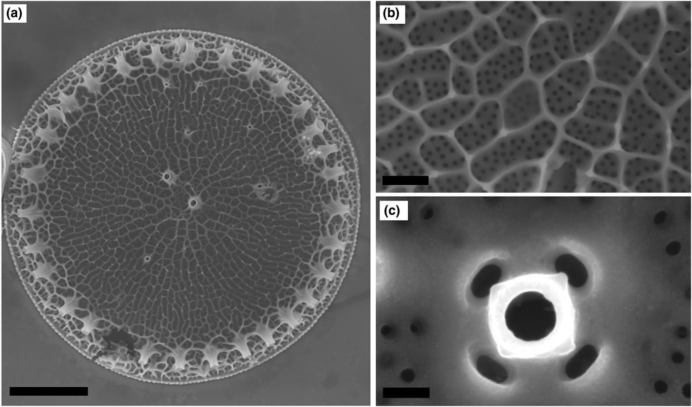
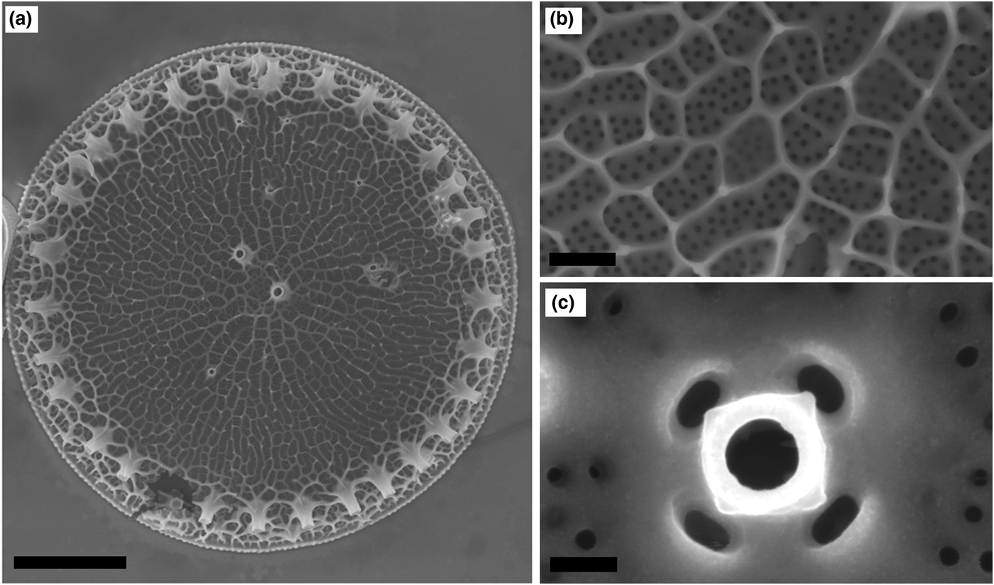
The centric Thalassosiria weissflogii diatom was easily grown in cultures in our laboratories, without expensive equipment or complicated media affording pure discrete nano-porous microcapsules with an average size of 10–15 µm. In vivo, the siliceous parts of diatoms are coated with organic matter (proteins and carbohydrates) that protects them in the aqueous environment. After easy removal of the protoplasm and of the outer organic layer by acidic/oxidative cleaning, the resulting nanostructured biosilica shells have been considered for many applications including photonics, molecular separation, and biosensing, and also as drug-delivery microsystems (Fig. 1).[Reference Ragni, Cicco, Vona, Leone and Farinola6]
SEM images of cleaned frustules of Thalassiosira weissflogii diatom after acid/oxidative treatment: (a) whole valve view (marker: 2 µm), (b) nanostructure view (marker: 200 nm), (c) diatom ocelle (marker: 100 nm).
The fabrication of multifunctional coatings through mussel-inspired chemistry has attracted considerable attention for its promising and exciting applications. PDA is an emerging soft matter that forms adherent PDA thin films by self-polymerization in water, mimicking the composition of the natural adhesive proteins.
PDA-based materials have been studied for biomedical applications for interfacing with cells, drug delivery and biosensing. More recently, PDA-based materials have been investigated for energy and environmental applications.[Reference Yanlan, Kelong and Lehui25]
In this work, we propose a bioinspired silica-based material from diatoms, as an alternative to silicon oxide, whose natural nanostructured surface can be easily functionalized with several organic molecules.
PDA coating of frustules surface was performed in Tris–HCl buffer solution (10 mM) at pH 8.5 at room temperature. During the reaction time (3 h), the transparent aqueous suspension of biosilica tended towards blackish (see Supplementary Information, Fig. S1). By a preliminary analysis via contrast phase microscopy, biosilica shells appeared still integer and transparent, with no coating of PDA, and the presence of black amorphous aggregates around the frustules in the reaction medium was detected. The polymerization reaction was then carried out at different pH (F PDA 9.5 and 10.5) with same results: PDA aggregation occurred but dramatically not on the frustule surface (see Fig. S2, Supplementary Information).
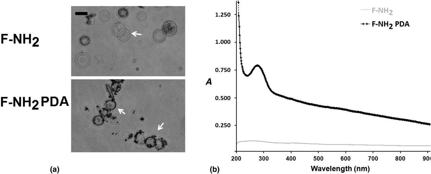
Since DA polymerization reaction occurs quickly at high pH levels, it was conceivable that the intrinsic acidity of silica shell surfaces (due to hydroxyl groups) prevented from the adhesion of PDA films. So we performed the amino-functionalization of silica shells with APTES in order to give a homogeneous intrinsic basicity to the frustules surface. Silica shells with the amino moieties (F-NH2) underwent to PDA coating at pH 8.5. Finally, DA polymerization occurred onto F-NH2 affording black “coin-like” frustules as shown by contrast phase microscopy in Fig. 2(a). The percentage yield of F-NH2 PDA over the total amount of initial shells ranges around 92%. All samples were observed after washing in bidistilled water.
Contrast Phase microscopy images of (a) frustules after APTES functionalization (F-NH2, top), circular and transparent valves are visible, and after polydopamine coating (F-NH2 PDA, bottom), a black coating on the valves appeared; (b) UV–visible spectra comparison F-NH2, F-NH2 PDA, and PDA NPs (A, absorption as y-axis). Marker: 10 µm.
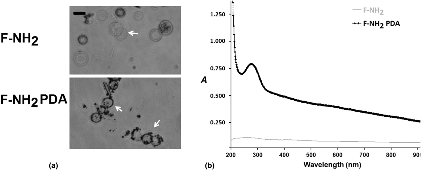
It can be noted that F-NH2 PDA exhibited a PDA accumulation onto biosilica edges, as shown in Fig. (2a).
These F-NH2 PDA hybrids were then characterized by UV–visible spectroscopy (Fig. 2b): the spectra of F-NH2, F-NH2 PDA) were compared.
The UV–vis spectra of F-NH2 PDA showed the broadband absorption typical of PDA due the oxidation of the catechol groups into quinone and the subsequent intramolecular cyclization,[Reference Wei, Zhang, Li, Li and Zhao26] while no UV strong absorption signal was observed for F-NH2.
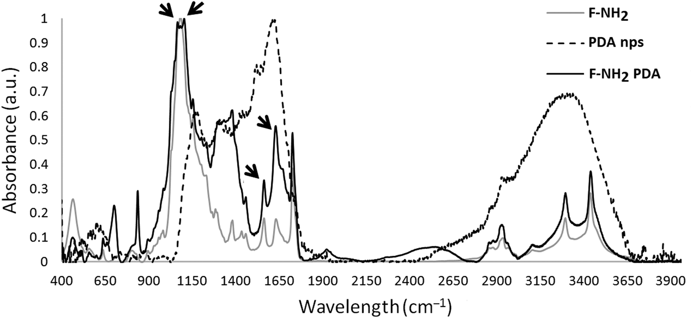
In Fig. 3, the FTIR spectra for the F-NH2, F-NH2 PDA, and PDA NPs are showed. PDA NPs were synthesized following a reported procedure (see Supplementary Information, Fig. S3), characterized by DLS measurements (see Supplementary Information, Fig. S4) and used as standard.
FTIR spectra comparison between F-NH2, F-NH2 PDA, and PDA NPs. Spectra have been normalized to 1101 cm−1 band value. Marks underline typical signals after PDA functionalization of F-NH2 samples.
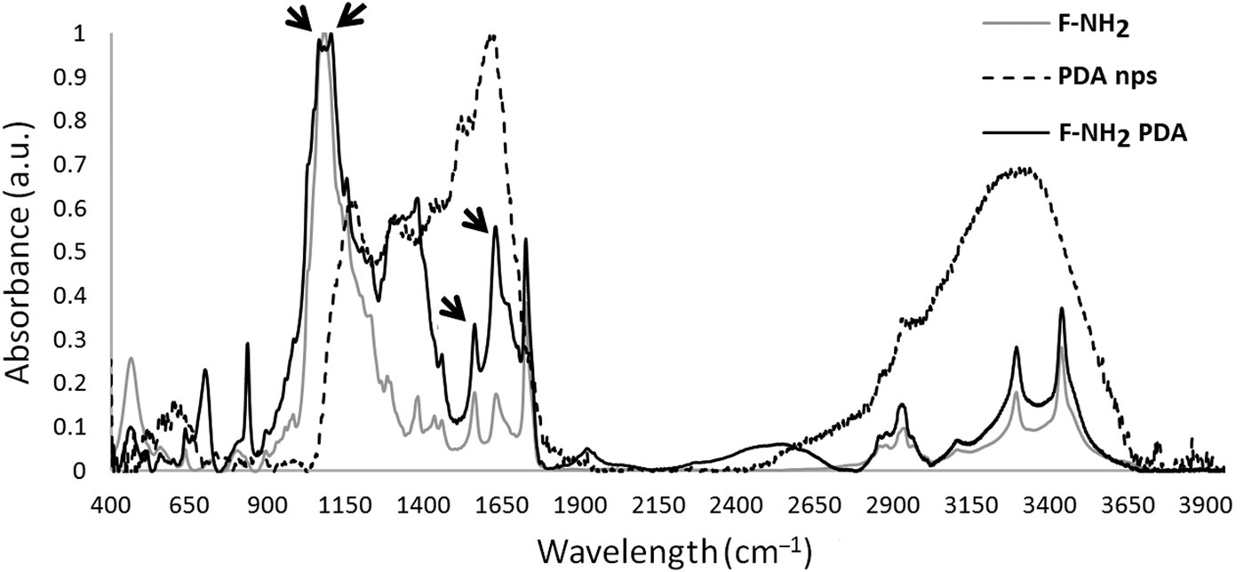
In the FTIR spectra of F-NH2 and F-NH2 PDA, the peaks at 3330 and 3470 cm−1 corresponding to the stretching vibrations of O–H and N–H groups respectively broadened in the FTIR spectrum of PDA NPs. The vibrations at 2973 and 2885 cm−1 belong to the asymmetric and symmetric stretching, respectively, of the methyl group of the carbon chain of alkoxysilane molecules.
The asymmetric Si–O–Si stretching at 1060 cm−1 is detected in the FTIR spectra of F-NH2 and F-NH2 PDA together with the Si–O–C vibration at 1100 cm−1 that confirmed that the diatom frustules were functionalized.
In the lower frequency regions, the detected band at 944 cm−1 is assigned to Si–O stretching of Si–OH groups and usually is connected with amorphous silica framework or marine-produced biosilica. The appeared band at 794 cm−1 is attributed to symmetric stretching vibrations of Si–O–Si bonds. This band is characteristic for amorphous silica such as opal-A.[Reference Adamo, Ghisoli and Caucia27] The FTIR bands at 552 and 443 cm−1 are recognized as relating to Si–O bending vibrations of siloxane groups. Generally, the four bands appearing at 1061, 944, 794, and 443 cm−1 may be considered as FTIR-spectra features of diatom biosilica structure.
Characteristic broad PDA signals were detected in the range 1630–1510 cm−1 assigned to the stretching vibration of aromatic ring, the bending vibration of N–H, the N–H shearing vibration of the amide group, and the C–O vibration.[Reference Zangmeister, Todd. Morris and Tarlov28]
The PDA-coating of biosilica microcapsules provides new opportunities for their application in biology and medicine. In addition to excellent biocompatibility, a fluorescent labeling of the PDA-coated frustules could provide important insights into their applications as drug carriers. In fact, for a drug delivery system to be effective, it must meet requirements such as highly flexibility, a tunable surface chemistry by an easy chemical modification, a high drug loading with a potential “smart” multidrug delivery, and finally the system can readily be made bio-imageable. For example, the covalent attachment of fluorophores for optical microscopy studies makes it possible to study the cell specificity and the biodistribution of the particles both under in vitro and in vivo conditions. In the case of diatom frustules which have been already demonstrated as suitable systems for drug loading/delivery,[Reference Cicco, Vona, De Giglio, Cometa, Mattioli Belmonte, Palumbo, Ragni and Farinola11] fluorescence engineering could allow to obtain traceable biosilica particles, without the use of toxic fluorescent dyes which can in general interfere with the loading/delivery process of the drugs.
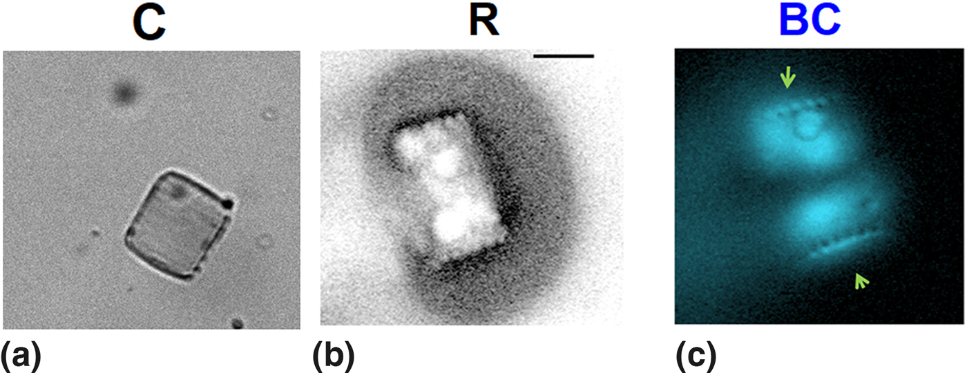
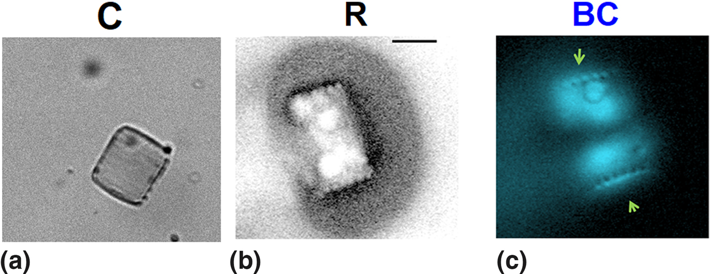
Following a recent study in which the oxidative treatment of DA in the presence of hydrogen peroxide (H2O2) yielded fluorescent precipitates,[Reference Chen, Yan, Müllner, van Koeverden, Noi, Zhu and Caruso24] after the first DA polymerization carried out on the frustules, a second polymerization of DA in the presence of H2O2 was performed in order to obtain fluorescent PDA-coated frustules. Figure 4(a) shows the box-like morphology of frustules (obtained by a “soft” cleaning procedure, see Supplementary Information, Fig. S5) with the first coating of PDA. In Fig. 4(b), the reflection microscopy image demonstrate that the use of H2O2 as an additional reactant for the DA polymerization does not negatively affect the box-like silica structure; finally, the fluorescence microscopy image [Fig. 4(c)] shows that this second chemical treatment yielded fluorescent heterostructures.
(a) Confocal microscopy of biosilica structures obtained via soft cleaning after first PDA coating. Marker: 10 µm; (b, c) Reflection microscopy images: (R) in bright field, PDA-coated biosilica after the second PDA-coating; (BC) blue channel (exc. 400 nm, DAPI filter set) images in fluorescence after the second PDA-coating. Marker [for (a–c) subfigures]: 5 µm.
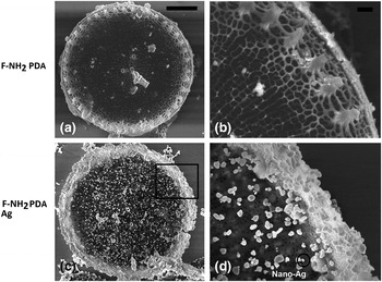
Surface immobilization of AgNPs is a popular method to endow materials with antimicrobial ability. Ag NPs have been reported to display antimicrobial activities with low citotoxicity and anti-inflammatory characteristics.[Reference Park, Neigh, Vermeulen, de la Fonteyne, Verharen, Briedé and de Jong23] Taking advantage from metal ion chelating ability and redox activity of DA, a direct reduction of Ag+ on the DA -coated biosilica surface to form Ag NPs via a one-step reaction was investigated. Biosilica/PDA/silver heterostructures prepared were characterized by SEM (Fig. 5). EDX was used to deeply investigate the nano-precipitated/adhered Ag NPS on the PDA-coated biosilica surface (Table I).[Reference Choi, Rhee, Park, Oh, Hong, Richardson, Guo, Caruso and Yoo22]
SEM images of: (a) frustule valve after APTES functionalization and PDA coating; (b) focus on the outer part of the same valve, with the hierarchical nanotexturization; (c) AgNPs adherent to the frustules surface; (d) focus on the edge zone, with a large density of AgNPs. Marker: (a, b), 2.5 µm; (c, d), 200 nm.

EDX analyses: atomic percentages of Si and Ag elements in all the biosilica structure: F (bare frustules), F-NH2, F-NH2 PDA, F-NH2 PDA Ag.

SEM analyses revealed that Ag nanoparticles covered about all the frustule structure, heading more the edge of the valve. These results are coherent with the DA polymerization on F-NH2, as previously reported, which was more significant on the frustule edges than on the center of the valve.
Finally, EDX analysis was carried out to determine the atomic element percentage in all the biosilica composites synthesized. The presence of silver on biosilica was confirmed by EDX investigation on F-NH2 PDA Ag, where Ag w/w % is 2.385 ± 1.1, with a Ag/Si ratio of 0.037 ± 0.017 (see Table I). As expected, silver was absent into bare biosilica frustules. The EDX signal for the nitrogen confirmed its predictable increased value passing from amino-functionalized frustules (F-NH2) to the PDA-coated ones, also considering the natural presence of nitrogen into the bare frustules (polyamine in the silica network, see Table I). The size of Ag nanoparticles was mainly in the range 96 ± 14 nm, value as determined by Image J test. Our AgNPs size resulted compatible with Ag nanoparticles obtained via traditional physicochemical[Reference Natsuki, Natsuki and Hashimoto29] and natural[Reference Srikar, Giri, Pal, Mishra and Upadhyay30] processes.
To evaluate if detrimental effects on the nano-texturization had been caused by PDA chemical modification and Ag nanoprecipitation, the architecture parameters of the frustules were investigated. From optical microscopy and SEM images, statistical analyses of the number of pores/area and the size of shells showed that no significant differences neither in average sizes of frustules valves (without and with PDA, without and with Ag nanoparticles) nor in average number of pores/500 nm × 500 nm of area (A) in girdles were found (see Supplementary Information, Fig. S6). These data confirm that two consecutive processes of biosilica functionalizations were not detrimental for the micro- and nano-structures of the diatom frustules.
Conclusion
Bioinspired structures from diatoms represent innovative nanostructured multifunctional materials for several applications ranging from nanomedicine to opto-electronics and photonics. To change both the bulk and the surface composition of diatom frustules, many biotechnological methods based on in vivo chemical or genetic modification have been reported.
Here we have reported an efficient protocol for decorating the biosilica shells of the T. weissflogii diatom with AgNPs exploiting the adhesion properties of PDA. After the extraction from the living cultures and a cleaning procedure with an acidic/oxidative treatment, the frustules were coated with a PDA thin film by a self-polymerization of DA in aqueous environment for increasing the adhesion properties of the biosilica surface. With the aim to make these materials even more appealing for applications in bio-nanomedicine, a fluorescent labeling of the PDA-coated frustules was performed by a second polymerization of DA in the presence of hydrogen peroxide (H2O2) yielding fluorescent PDA-coated frustules, without the requirement of an organic dye.
As a last step, AgNPs were precipitated onto the biosilica surface leading to silica platforms with applications as antimicrobial or photonic heterostructures.
Our study represents a further step in the use of diatom microalgae as a low-cost, environmental-friendly source of bioinspired materials whose chemical surface composition can be easily modified, preserving their main morphological features, envisaging future applications in bioelectronics and nanomedicine.
Supplementary material
The supplementary material for this article can be found at https://doi.org/10.1557/mrc.2018.103.
Acknowledgments
For the design of the experiments and some important advices about interpretation of the analyses data, we thank Dr. F. Palumbo (CNR NANOTECH) and Dr. E. Altamura (Università degli Studi di Bari).