Introduction
Transgenic animals have important applications in the pharmaceutical industry, human medicine and agriculture production (Wilmut et al., Reference Wilmut, Archibald, Harris, McClenaghan, Simons, Whitelaw and Clark1990; Stice et al., Reference Stice, Robl, Ponce de Leon, Jerry, Golueke, Cibelli and Kane1998; Bondioli et al., Reference Bondioli, Ramsoondar, Williams, Costa and Fodor2001; Golovan et al., Reference Golovan, Meidinger, Ajakaiye, Cottrill, Wiederkehr, Barney, Plante, Pollard, Fan, Hayes, Laursen, Hjorth, Hacker, Phillips and Forsberg2001; Salamone et al., Reference Salamone, Barañao, Santos, Bussmann, Artuso, Werning, Prync, Carbonetto, Dabsys, Munar, Salaberry, Berra, Berra, Fernandez, Papouchado, Foti, Judewicz, Mujica, Muñoz, Alvarez, Gonzalez, Zimmermann, Criscuolo and Melo2006). To date, several methods have been used for the generation of genetically modified animals; however, these techniques still remain inefficient. Recently, a new strategy to generate in vitro fertilization (IVF) transgenic bovine embryos was developed (Vichera et al., Reference Vichera, Moro and Salamone2010). This efficient technique consists of intracytoplasmic injection of oocytes and IVF zygotes with liposomes that contain exogenous DNA. Contrast to pronuclear microinjection in farm animals, this method avoids the centrifugation of zygotes and also the insertion of the pipette into the pronucleus (Chauhan et al., Reference Chauhan, Nadir, Bailey, Pryor, Butler, Notter, Velander and Gwazdauskas1999; Murakami et al., Reference Murakami, Fahrudin, Varisanga and Suzuki1999). Thus, it is technically easier, and also increases the number of zygotes that can be injected in the same time period.
Previous reports on pronuclear microinjection or cytoplasmic injection have proposed that these procedures cause spontaneous breaks in chromosomes, possibly exacerbated by the micromanipulation practice (Brinster et al., Reference Brinster, Chen, Trumbauer, Yagle and Palmiter1985; Yamauchi et al., Reference Yamauchi, Doe, Ajduk and Ward2007). However, it has also been demonstrated that the early developmental potential of embryos with slight DNA fragmentation is not different from embryos without DNA fragmentation (Alikani et al., Reference Alikani, Calderon, Tomkin, Garrisi, Kokot and Cohen2000; Mateusen et al., Reference Mateusen, Van Soom, Maes, Donnay, Duchateau and Lequarre2005), although it can lead to a large proportion of pregnancy losses.
Assessment of embryonic cells with fragmented DNA has been carried out using the terminal deoxynucleotidyl transferase (TdT) nick-end labelling (TUNEL) assay. This method has been used to detect apoptotic cells in cleaved embryos, morulae, and blastocysts from different species (Jurisicova et al., Reference Jurisicova, Varmuza and Casper1996; Pampfer et al., Reference Pampfer, Vanderheyden, McCracken, Vesela and De Hertogh1997; Byrne et al., Reference Byrne, Southgate, Brison and Leese1999; Matwee et al., Reference Matwee, Betts and King2000; Paula-Lopes & Hansen, Reference Paula-Lopes and Hansen2002). However, apoptosis may not always be related to extensive DNA degradation (Cohen et al., Reference Cohen, Sun, Snowden, Dinsdale and Skilleter1992), as this occurs only as a relatively late event in the apoptotic process (Collins et al., Reference Collins, Schandi, Young, Vesely and Willingham1997).
This study was conducted to evaluate the quality of IVF embryos after intracytoplasmic injection of bovine zygotes either with pCX–EGFP–liposome complexes or pBCKIP2.8–liposome complexes (plasmids that codified human insulin gene). We examined embryo development after both treatments, and the expression levels of enhanced green fluorescent protein (EGFP) when this plasmid was injected. In addition, we used the TUNEL assay to evaluate the timing of the onset of DNA fragmentation in bovine blastocyst that express or not EGFP, at different days of in vitro culture (days 6, 7 and 8).
Materials and methods
Research protocols followed the guidelines stated in the Guide for the Care and Use of Agricultural Animals in Agricultural Research and Teaching. The Comision Nacional de Biotecnologia Argentina (CONABIA) approved the animal confinement conditions for recipient cows.
Reagents
Except otherwise indicated, all chemicals were obtained from Sigma Chemical Company (St. Louis, MO, USA).
Oocyte collection and in vitro maturation
Ovaries were collected at a slaughterhouse and transported to the laboratory. Cumulus–oocyte complexes (COCs) were aspirated from follicles with a diameter of 2 to 8 mm and collected into Dulbecco's phosphate-buffered saline (DPBS; 14287-072; Gibco, Grand Island, NY, USA) that contained 10% fetal bovine serum (FBS; 10499-044; Gibco) and 2% antibiotic–antimycotic (ATB; 15240-096; Gibco). Follicular oocytes covered by at least three layers of granulosa cells were selected for in vitro maturation. The maturation medium was bicarbonate-buffered TCM-199 (31100-035; Gibco) and contained 2 mM glutamine (G8540), 10% fetal bovine serum (FBS), 2 μg/ml follicle-stimulating hormone (NIH-FSH-P1, Follitropin™, Bioniche, Belleville, ON, Canada), 0.3 mM sodium pyruvate (P2256), 100 μM cysteamine (M9768) and 2% v/v ATB. Oocytes were incubated in 100-μl droplets of medium covered with mineral oil (M8410) in 32 mm Petri dishes. in vitro maturation conditions were 6.5% CO2 in humidified air at 39°C for 22 h. After maturation, oocytes were vortexed for 2 min in hyaluronidase (H4272) (1 mg/ml DPBS), to remove cumulus cells, and washed three times in HEPES-buffered Tyrodes medium containing albumin, lactate and pyruvate (TALP-H; Bavister & Yanagimachi, Reference Bavister and Yanagimachi1977).
in vitro fertilization (IVF)
Bovine frozen semen was thawed in a 37°C water bath for 30 s. Spermatozoa were centrifuged twice (490 g/5 min at 37°C) and resuspended in Brackett–Oliphant medium (BO, Brackett & Oliphant, Reference Brackett and Oliphant1975) supplemented with 5 mM caffeine (C4144) and 20 IU/ml heparin (H3149). Spermatozoa were diluted to half concentration with BO that contained 10 mg/ml fatty acid-free bovine serum albumin (BSA; A6003), resulting in a final sperm concentration of 12 × 106 – 15 × 106/ml. Spermatozoa were coincubated with COCs for 5 h at 39°C in a humidified atmosphere of 5% CO2 in air. The presumptive zygotes were then washed three times in TALP-H and cultures were continued as described as follows.
DNA construction
The plasmids used were pCX–EGFP (kindly provided by Dr Masaru Okabe, Osaka University, Japan) that contain an enhanced green fluorescent protein gene (egfp) under the chimerical cytomegalovirus–IE–chicken β-actin enhancer promoter control (Ikawa et al., Reference Ikawa, Kominamik, Yoshimura, Tanaka, Nishimune and Okabe1995), and pBCKIP2.8 that codified the human insulin gene under the β-casein promoter control. Closed plasmids were used in this work, as we have reported previously the successful production of a transgenic cow using a circular plasmid construction (Salamone et al., Reference Salamone, Barañao, Santos, Bussmann, Artuso, Werning, Prync, Carbonetto, Dabsys, Munar, Salaberry, Berra, Berra, Fernandez, Papouchado, Foti, Judewicz, Mujica, Muñoz, Alvarez, Gonzalez, Zimmermann, Criscuolo and Melo2006).
Liposome–DNA coincubation
For the injection experiments, 1 μl of 4 μg/ml of DNA in combination with 3 μl of commercial liposome (Fugene; Boehringer-Manheim, Germany) were coincubated for 15 min. The DNA–liposome mixture was diluted to half concentration with 10% polyvinylpyrrolidone (PVP, 99219; Irvine Scientific, Santa Ana, CA, USA), reaching a final DNA concentration of 0.5 μg/ml.
Intracytoplasmic injection of DNA–liposome complexes
After 16 h of IVF, presumptive zygotes were injected with approximately 2 pl of DNA–liposome/PVP mixture, using an injection capillary (0.7 μm in diameter) attached to a micromanipulator (Medical Systems, Great Neck, NY, USA) mounted on a Nikon Eclipse E-300 microscope (Nikon, Melville, NY, USA).
in vitro culture
Fertilized oocytes were cultured in 100-μl droplets of synthetic oviductal fluid (SOF) (Tervit et al., Reference Tervit, Whittingham and Rowson1972; modified by Holm et al., Reference Holm, Booth, Schmidt, Greve and Callesen1999) and supplemented with 2.5% v/v FBS, in a humidified atmosphere of 5% O2, 5% CO2 and 90% N2 in air at 39°C. Cleavage was evaluated on day 2, and the number of blastocysts on days 6, 7 and 8 of culture.
Determination of EGFP fluorescence in IVF embryos
During in vitro culture, embryos were exposed briefly to blue light using an excitation filter at 488 nm and an emission filter at 530 nm to determine the expression of the egfp gene at different stages of development. Embryos were analyzed on days 3 (cleaved) and 6–8 (blastocysts), after IVF.
TUNEL and confocal microscopy
DNA fragmentation was evaluated using the DeadEnd™ Fluorometric TUNEL System (Promega G3250, Madison, WI, USA). Embryos were fixed in 4% paraformaldehyde in DPBS and washed in BSA solution (1 mg BSA/ml DPBS). After washing, embryos were permeabilized with 0.5% Triton X-100 in DPBS for 15 min at room temperature and washed again in BSA solution. Extensive DNA fragmentation was induced in positive controls by incubation in 50 U/ml DNase (RQ1; Promega; Madison, WI, USA) prior to the TUNEL reaction, and negative controls were generated by omitting terminal transferase from the reaction. After three washes in BSA solution, positive controls and samples were incubated in fluorescein-dUTP and TdT for 2 h at 39°C in the dark. The nuclei were then counterstained with 0.5% propidium iodide (PI) for 30 min at room temperature. Embryos were then washed in BSA solution and mounted on a glass slide in 70% v/v glycerol under a coverslip. Embryos were analyzed on a Nikon Confocal C.1 scanning laser microscope. An excitation wavelength of 488 nm was selected for detection of fluorescein-12-dUTP and a 544 nm wavelength to excite PI. Images of serial optical sections were recorded every 1.5–2 μm vertical step along the Z-axis of each embryo. Three-dimensional images were constructed using software EZ-C1 3.9. Total cell numbers and DNA-fragmented nuclei were counted.
Embryo transfer
Bovine blastocysts were transferred individually to recipient cows on days 6, 7 and 8 of embryo culture after IVF. Each blastocyst was washed several times in TL-HEPES and loaded into a 0.25 ml straw. Fresh embryos were transported to the farm at 35°C within 3 h. Embryos were transferred non-surgically to the uterine horn ipsilateral to the ovary bearing the corpus luteum. Each recipient received one blastocyst. Pregnancies were detected by ultrasonography (7.5 MHz lineal transducer; Aloka Co, Tokyo, Japan) at 23 days after embryo transfer.
DNA extraction and polymerase chain reaction (PCR) analysis
Bovine genomic DNA was isolated from blood and ear tissue according to the manufacturer instructions in the extraction kit (Wizard Genomic DNA Purification Kit, Promega, Madison, WI, USA). Different dilutions 1/10, 1/100, 1/1000 were used as templates in the PCR method for detecting the presence of the transgene (insulin). The PCR reaction contained: 10× PCR buffer, 50 mM MgCl2; 3 mM IP Fwd and Rev; 10 mM dNTPs; and Taq polymerase 500 U/ml. Thermocycling conditions were as follows: one cycle of initial denaturation at 94°C for 3 min, followed by 40 amplification cycles each including denaturation (94°C for 30 s), annealing (60°C for 30 s), and extension (72°C for 30 s). Cycle 40 contained an additional extension at 72°C for 3 min.
The primers used were: Reverse IP GGAAGCATGGAGTCTTGGAC; Forward IP GGGAGGTGAAGGTTTTCA. The positive control was the plasmid pBCKIP2.8, which amplified 700-bp insulin fragments. Sterile water was used instead of DNA as a negative control. The PCR products (10 μl) were analyzed on a 1.2% agarose gel stained with ethidium bromide. The amplified DNA bands were visualized under ultraviolet (UV) light.
Statistical analysis
in vitro embryo development, fluorescence expression and differences between replicates were compared by Fisher's exact test. Differences in total cell number and fragmented nuclei were analyzed using a difference of proportions test. For all statistical analyses, the SAS program was used (SAS Institute Inc. SAS/STAT, 1989). Differences were considered to be significant at a P-value < 0.05.
Experimental design
In the first experiment, IVF presumptive zygotes were injected (16 h post fertilization) with two different plasmid–liposome complexes. The plasmids used were the pCX–EGFP that codifies the egfp gene and the pBCKIP2.8 that codifies the human insulin gene. Embryo development was evaluated in both groups. In the first group we also determined the pattern of EGFP expression on cleaved embryos and blastocysts (days 3 and 7 post fertilization, respectively). The control groups consisted of IVF embryos that were injected with liposomes free of plasmid (IVF-L) and IVF embryos that were not injected. In the second experiment, the blastocysts obtained from all the groups (using pCX–EGFP), expressing and not EGFP, were analyzed by TUNEL assay at days 6, 7 and 8 of in vitro culture, to determine the presence of DNA fragmentation. For TUNEL assay, the following control samples were analyzed: a positive control consisting of a blastocyst with extensive DNA fragmentation induced by incubation in 50 U/ ml DNase, and a negative control generated by omitting terminal transferase from the TUNEL reaction. Finally, IVF bovine blastocysts produced by intracytoplasmic injection of pBCKIP2.8–liposome complexes were transferred to recipient cows after 6, 7 and 8 days of in vitro culture.
Results
Experiment 1. Embryo development and EGFP expression after intracytoplasmic injection of DNA–liposome complexes that contained egfp or insulin into IVF bovine zygotes
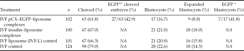
In this experiment we evaluated the development of IVF bovine embryos injected with pCX–EGFP–liposome complexes or insulin–liposome complexes (Table 1). No statistical differences among treatments were seen in cleavage rates either for blastocysts or expanded blastocysts. However, the IVF zygotes injected with the reporter gene egfp showed a tendency towards decreased expanded blastocyst rates with respect to the other groups (IVF insulin, IVF-L and IVF control). In addition, high percentages of EGFP+ embryos were seen on days 3 and 7 of culture (Fig. 1).
Embryo development and EGFP expression of IVF bovine embryos injected with DNA–liposome complexes

†Expression of enhanced green fluorescent protein (EGFP).
Within a column, means do not differ. Fisher's exact test, P < 0.05.
IVF, in vitro fertilization.
Bovine blastocyst with EGFP expression produced by in vitro fertilization (IVF) and intracytoplasmic injection of pCX–EGFP–liposome complexes. The fluorescence was evaluated under blue light (488 nm; original magnification ×200). A colour version of this figure is available in the online version of this paper.
Experiment 2. DNA fragmentation in IVF bovine blastocysts (day 6, day 7 and day 8) evaluated by TUNEL assay
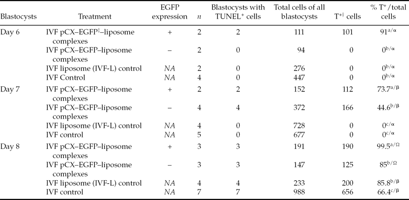
Blastocysts obtained after the injection of pCX–EGFP–liposome complexes, that express or not the transgene, and control groups were evaluated by TUNEL assay to determine the presence of fragmented nuclei, at days 6, 7 and 8 of embryo culture (Bd6, Bd7 and Bd8; Fig. 2). Total cell numbers and T+ cells were counted separately in all blastocysts. The blastocysts incubated with DNase (positive control) showed TUNEL labelling in almost all nuclei. In contrast, none of the nuclei was labelled in the embryos incubated without TdT (negative control, data not shown). There was no evidence of DNA fragmentation either in IVF and IVF-L control groups, at days 6 and 7 of in vitro culture. However, T+ cells were detected in Bd8 in both control groups (85.8 and 66.4%, respectively P < 0.05). On the other hand, all EGFP+ blastocysts showed T+ nuclei at day 6, 7 and 8. Blastocysts that were injected with pCX–EGFP–liposome complexes and did not express the transgene showed statistically lower fragmented nuclei with respect to embryos with EGFP expression, and no positive TUNEL labelling was seen in Bd6. In Table 2 the percentages of T+ cells with respect to total cell numbers for all the groups are detailed.
Evaluation of DNA-fragmented nuclei in IVF bovine blastocysts (day 6, day 7 and day 8) that express or not EGFP
†TUNEL positive.
‡Plasmid that codifies the enhanced green fluorescent protein (EGFP).
IVF, in vitro fertilization.
a,b,cValues from different treatments within the same blastocyst day with different superscripts in a column are significantly different. Difference of proportions test P < 0.05.
α,β,ΩValues from the same treatment among the different blastocyst days with different superscripts in a column are significantly different. Difference of proportions test P < 0.05.
Day 6, day 7 and day 8 blastocysts produced by in vitro fertilization (IVF) and intracytoplasmic injection of pCX–EGFP–liposome complexes, analyzed by TUNEL assay to determine the presence of DNA fragmentation. TUNEL-positive cells are labelled with fluorescein-12-dUTP (green) and nuclei are counterstained with propidium iodide (red). EGFP+, with EGFP expression; EGFP−, without EGFP expression. Original magnification ×200. A colour version of this figure is available in the online version of this paper.
Experiment 3. Embryo transfer, pregnancies and birth of IVF calves
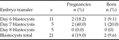
In order to determine the influence of in vitro culture length on pregnancy rates and birth, we compared the transfer of day 6, day 7 and day 8 IVF blastocysts which were injected previously with insulin–liposome complexes. Two of 11 cows (18.2%) that received Bd6 became pregnant while two of five (40%) cows became pregnant from the transfer of Bd7. None of the recipients receiving Bd8 was found to be pregnant. No differences were observed in pregnancy rates among the groups. For all treatments, each recipient received one blastocyst and pregnancies were detected by ultrasonography at 23 days after embryo transfer. Of the four pregnant recipients, two pregnancies developed to term, one had been derived from Bd6 transfer (9.1%) and the other from a Bd7 transfer (20%; Table 3). PCR analysis of blood and tissue samples determined that none of the calves was transgenic for the insulin gene.
Embryo transfer of IVF bovine blastocyst injected with insulin–liposome complexes
Within a column, means do not differ. Fisher's exact test, P < 0.05.
Discussion
In this work, we evaluated the quality and viability of IVF bovine embryos, after intracytoplasmic injection of DNA–liposome complexes. The exogenous DNA consisted of two different plasmids that were used separately: pCX–EGFP, that codifies the reporter gene egfp and pBCKIP2.8 that codifies the human insulin gene. The utilization of the first plasmid allowed transgene expression determinations. We chose the second plasmid in order to produce a cow that expressed insulin in its milk, thereby potentially increasing the amount of hormone available to consumers and decreasing costs for the pharmaceutical industry.
The results confirmed that a successful transfection using an easy technique can be obtained with this new method. Moreover, in agreement with our previous report (Vichera et al., Reference Vichera, Moro and Salamone2010), we demonstrated that EGFP+ embryos can develop to the blastocyst stage, equally as well as controls. A similar result was obtained with the plasmid that contained the insulin gene. Thus, this work has shown that it is possible to use constructs with biomedical or agricultural interest, without any decrease in embryonic development following their injection into IVF zygotes.
Previous reports have demonstrated that various transgenic methods can yield transgenic offspring, but they all suffer from various drawbacks and their efficiency is far from optimal (Jaenisch, Reference Jaenisch1976; Hammer et al., Reference Hammer, Pursel, Rexroad, Wall, Bolt, Ebert, Palmiter and Brinster1985; Lavitrano et al., Reference Lavitrano, Camaioni, Fazio, Dolci, Farace and Spadafora1989; Krimpenfort et al., Reference Krimpenfort, Rademakers, Eyestone, Van derSchans, Van denBroek, Kooiman, Kootwijk, Platenburg, Pieper and Strijker1991; Schnieke et al., Reference Schnieke, Kind, Ritchie, Mycock, Scott, Ritchie, Wilmut, Colman and Campbell1997; Chan et al., Reference Chan, Homan, Ballou, Burns and Bremel1998; Cibelli et al., Reference Cibelli, Stice, Gluekey, Kane, Jerry, Blackwell, Ponce de Leo¢n and Robl1998; Perry et al., Reference Perry, Wakayama, Kishikawa, Kasai, Okabe, Toyoda and Yanagimachi1999). Transgene integration is low and pre and post implantation embryo losses are large (Gandolfi et al., Reference Gandolfi, Terqui, Modina, Brevini, Ajmone-Marsan, Foulon-Gauze and Courot1996; Eyestone, Reference Eyestone1999; Rideout et al., Reference Rideout, Eggan and Jaenisch2001). Most of the work in this field has focused on improving transgene transmission to offspring, while little attention has been paid to enhancing embryo survival after embryo transfer (Wall, Reference Wall2001). Embryo loss may result from DNA ruptures that could directly trigger the apoptosis mechanism. Previous reports have proposed that spontaneous breaks occur in chromosomes, although the microinjection technique can possibly exacerbate them, increasing the number of sites for DNA integration (Brinster et al., Reference Brinster, Chen, Trumbauer, Yagle and Palmiter1985; Yamauchi et al., Reference Yamauchi, Doe, Ajduk and Ward2007). For example, a relationship between chromosome breakage and transgene expression was observed using the ICSI-mediated transgenesis technique (Szczygiel et al., Reference Szczygiel, Moisyadi and Ward2003). For these reasons, it is necessary to establish conditions conducive to maintaining the right balance between integration, transgene expression and preservation of DNA integrity in embryos that will be transferred, in order to avoid pregnancy losses.
In our work, we evaluated the incidence of DNA fragmentation after transgene injection, liposome injection and transgene expression (egfp reporter gene) of day 6, day 7 and day 8 IVF blastocysts, using the TUNEL assay. Fragmented nuclei were noted in control blastocysts cultured for 8 days, but no TUNEL labelling was detected in Bd6 or Bd7. These results contrast with those of Matwee et al. (Reference Matwee, Betts and King2000), who proposed that all bovine blastocysts produced in vitro had apoptotic cells. One of the control groups consisted of zygotes injected with liposomes free of DNA. The development of this group was similar to that obtained for IVF controls and, as previously mentioned, fragmented nuclei were not detected in Bd6 and Bd7. From these observations we conclude that liposomes are not harmful to the embryo when they are used as vectors for exogenous DNA.
Blastocysts obtained from zygotes injected with pCX–EGFP–liposome complexes were also evaluated by TUNEL assay, distinguishing those EGFP+ from those EGFP−. We noticed that all EGFP+ blastocysts showed T+ labelling, whereas blastocysts without transgene expression showed lower DNA fragmentation. These observations, together with the results of embryonic development, suggest that transgene expression affects DNA integrity, but not blastocyst developmental rates. It is important to note that DNA fragmentation is one of the hallmarks of apoptosis, and although apoptosis has certain roles during preimplantation embryo development [such as elimination of abnormal cells or cells with ectopic differentiation patterns (Hardy, Reference Hardy1997; Betts & King, Reference Betts and King2001)], the post implantation developmental potential of embryos can be affected by apoptosis at preimplantation stages.
After having studied embryo development and the incidence of apoptosis, we transferred 21 blastocysts (one per recipient cow) resulting from 6, 7 and 8 days of in vitro culture and injected with insulin–liposome complexes. We achieved four pregnancies from Bd6 and Bd7 but no pregnancies were detected from Bd8 transfers. Despite no statistical differences being seen among groups, there seems to be a correlation between embryos with the greatest percentage of apoptotic cells and the absence of pregnancy. All Bd8 cells showed more than 66% of T+ labelling, even in controls. This finding indicates poor embryo quality, probably accelerated by extensive in vitro culture. In this work we demonstrated that blastocysts with less than 8 days of in vitro culture had a greater potential to develop to term, following transfer. Others have reported similar results after transfer of bovine embryos produced in vivo. They observed a tendency for pregnancy rate to decrease with advancing maturity of the embryo (Coleman et al., Reference Coleman, Dailey, Leffel and Baker1986) and better pregnancy rates after transfer of Bd7 (Niemann et al., Reference Niemann, Lampeter, Sacher and Kruff1982). Our results agree with other studies that support the view that embryo quality is a more accurate predictor of success than the stage of embryonic development (Lindner & Wright, Reference Lindner and Wright1983).
The births we obtained using this strategy did not carry the insulin gene. On the one hand, we confirmed that it is possible to generate viable offspring using this method, but higher numbers of transfers are required to improve transgene integration probabilities. Molecular and cellular studies are needed in order to understand the transgenic mechanism after DNA–liposome injection.
In summary, the present study indicates that IVF bovine embryos can be easily transfected after the injection of DNA–liposome complexes. For DNA fragmentation, it seems that the longer the culture in vitro, the higher the percentage of fragmented nuclei in blastocysts. We also observed that fragmentation increases in blastocysts with transgene expression compared with those carrying the plasmid but without expression. Further research is required to evaluate the pathways of DNA fragmentation and especially to determine the integration efficiency. An optimal balance between transgene expression, transgene integration, preservation of DNA integrity and in vitro culture length before embryo transfer, is an important goal for future research.
Acknowledgements
The authors thank CIALE for providing the biological material. This work was partially supported by PICT CABBIO 2008 N_00145 and UBACYT G808.