Introduction
Schizophrenia has been frequently referred to as a ‘dysconnectivity syndrome’ (Friston and Frith, Reference Friston and Frith1995; Liang et al., Reference Liang, Zhou, Jiang, Liu, Tian, Liu and Hao2006; Stephan et al., Reference Stephan, Friston and Frith2009), since neuroimaging studies have identified a range of abnormalities in the connections among regions of the brain in schizophrenia (Karlsgodt et al., Reference Karlsgodt, Sun and Cannon2010; Whitfield-Gabrieli and Ford, Reference Whitfield-Gabrieli and Ford2012). However, it remains unclear whether the reported connectivity abnormalities observed in psychotic illness are related to the underlying biology of the disease, or if some are attributable to secondary effects of having the illness, including effects of its treatment (Stephan et al., Reference Stephan, Magnotta, White, Arndt, Flaum, O'leary and Andreasen2001; Schmidt et al., Reference Schmidt, Smieskova, Aston, Simon, Allen, Fusar-Poli, McGuire, Riecher-Rössler, Stephan and Borgwardt2013; Sarpal et al., Reference Sarpal, Robinson, Lencz, Argyelan, Ikuta, Karlsgodt, Gallego, Kane, Szeszko and Malhotra2015). This unresolved question has contributed to a recent increased interest in earlier, more broadly-defined ‘psychosis spectrum’ states (Calkins et al., Reference Calkins, Moore, Merikangas, Burstein, Satterthwaite, Bilker, Ruparel, Chiavacci, Wolf, Mentch, Qiu, Connolly, Sleiman, Hakonarson, Gur and Gur2014), including ‘psychotic experiences’ (PEs) (Linscott and van Os, Reference Linscott and van Os2013), that are unaffected by these confounds.
PEs are subclinical expressions of psychotic symptoms that are reported commonly (5–35% prevalence) in the general population (Poulton et al., Reference Poulton, Caspi, Moffitt, Cannon, Murray and Harrington2000; van Os et al., Reference van Os, Linscott, Myin-Germeys, Delespaul and Krabbendam2009; Kelleher et al., Reference Kelleher, Connor, Clarke, Devlin, Harley and Cannon2012; Calkins et al., Reference Calkins, Moore, Merikangas, Burstein, Satterthwaite, Bilker, Ruparel, Chiavacci, Wolf, Mentch, Qiu, Connolly, Sleiman, Hakonarson, Gur and Gur2014). Although the majority of individuals reporting PEs do not develop clinical psychosis (Dominguez et al., Reference Dominguez, Wichers, Lieb, Wittchen and van Os2011; Linscott and van Os, Reference Linscott and van Os2013), several lines of evidence suggest that PEs and clinical psychosis represent distinct stages of a symptomatic and biological continuum of severity. Severe and persistent PEs are associated with a greater than 10-fold increase in risk for the development of clinical psychosis (Poulton et al., Reference Poulton, Caspi, Moffitt, Cannon, Murray and Harrington2000; Dominguez et al., Reference Dominguez, Wichers, Lieb, Wittchen and van Os2011), and similar to clinical psychosis, PEs are familial and heritable (Kelleher and Cannon, Reference Kelleher and Cannon2011). Many of the same epidemiological risk factors that increase risk for schizophrenia (e.g. cannabis use, childhood adversity) also increase risk for PEs (Cougnard et al., Reference Cougnard, Marcelis, Myin-Germeys, De Graaf, Vollebergh, Krabbendam, Lieb, Wittchen, Henquet, Spauwen and Van Os2007; Kelleher and Cannon, Reference Kelleher and Cannon2011; Linscott and van Os, Reference Linscott and van Os2013). Thus, shared etiological mechanisms may underlie these common associations.
In addition, recent neuroimaging studies have provided evidence for biological overlap between PEs and psychosis, identifying changes in brain structure (Kelleher and Cannon, Reference Kelleher and Cannon2011) and function (Fusar-Poli et al., Reference Fusar-Poli, McGuire and Borgwardt2012; Satterthwaite et al., Reference Satterthwaite, Vandekar, Wolf, Bassett, Ruparel, Shehzad, Craddock, Shinohara, Moore, Gennatas, Jackson, Roalf, Milham, Calkins, Hakonarson, Gur and Gur2015) in individuals with PEs that are similar to those observed in patients with psychotic disorders. However, few studies thus far have investigated the neurobiological basis of the clinically apparent heterogeneity of PEs. Like clinical psychosis, PEs comprise a range of seemingly distinct symptoms, such as delusional beliefs and hallucinatory experiences (Yung et al., Reference Yung, Nelson, Baker, Buckby, Baksheev and Cosgrave2009; Capra et al., Reference Capra, Kavanagh, Hides and Scott2013; Ronald et al., Reference Ronald, Sieradzka, Cardno, Haworth, McGuire and Freeman2014). Of the subclinical psychotic phenomena, paranoia is the most heritable (Zavos et al., Reference Zavos, Freeman, Haworth, McGuire, Plomin, Cardno and Ronald2014) and prevalent (Wigman et al., Reference Wigman, Vollebergh, Raaijmakers, Iedema, van Dorsselaer, Ormel, Verhulst and van Os2011) symptom, with a 10–15% prevalence within the general population (Freeman, Reference Freeman2007). Suspiciousness (one manifestation of paranoia) is associated with an increased risk for developing clinical psychosis in at-risk populations (Tarbox et al., Reference Tarbox, Addington, Cadenhead, Cannon, Cornblatt, Perkins, Seidman, Tsuang, Walker, Heinssen, McGlashan and Woods2013; Cannon et al., Reference Cannon, Yu, Addington, Bearden, Cadenhead, Cornblatt, Heinssen, Jeffries, Mathalon, McGlashan, Perkins, Seidman, Tsuang, Walker, Woods and Kattan2016), suggesting that a phenomenological continuum of severity, spanning suspiciousness and paranoia to frank persecutory delusions, may be associated with a common underlying mechanism.
Paranoia or persecutory ideation has been proposed to involve misattributions of threat to non-threatening events, objects, or experiences (Freeman et al., Reference Freeman, Garety, Kuipers, Fowler and Bebbington2002) and is associated with elevated levels of negative affect (Bentall et al., Reference Bentall, Rowse, Shryane, Kinderman, Howard, Blackwood, Moore and Corcoran2009). Because of these findings, prior neuroimaging studies of paranoia have focused on the amygdala, due to its well-established role in fear, threat perception, and negative affect (LeDoux, Reference LeDoux2003; Phelps, Reference Phelps2006; Delgado et al., Reference Delgado, Nearing, Ledoux and Phelps2008; Coombs et al., Reference Coombs, Loggia, Greve and Holt2014; Cullen et al., Reference Cullen, Westlund, Klimes-Dougan, Mueller, Houri, Eberly and Lim2014). For example, one recent study found significantly elevated amygdala perfusion in paranoid compared to non-paranoid patients with schizophrenia (Pinkham et al., Reference Pinkham, Liu, Lu, Kriegsman, Simpson and Tamminga2015). Changes in the connectivity of the amygdala have been observed previously in schizophrenia (Anticevic et al., Reference Anticevic, Tang, Cho, Repovs, Cole, Savic, Wang, Krystal and Xu2014; Ford et al., Reference Ford, Palzes, Roach, Potkin, van Erp, Turner, Mueller, Calhoun, Voyvodic, Belger, Bustillo, Vaidya, Preda, McEwen and Mathalon2015); however, it is not known whether these findings were related to the presence of paranoia. Recent evidence for symptom-specific changes in brain function in psychotic illness (Menon et al., Reference Menon, Anagnoson, Mathalon, Glover and Pfefferbaum2001; Shaffer et al., Reference Shaffer, Peterson, McMahon, Bizzell, Calhoun, van Erp, Ford, Lauriello, Lim and Manoach2015) highlights the importance of investigating the specific neural correlates of individual psychotic symptoms, which are associated with different antecedents (Cannon et al., Reference Cannon, Mednick and Parnas1990), levels of risk (Yung et al., Reference Yung, Phillips, Yuen, Francey, McFarlane, Hallgren and McGorry2003; Demjaha et al., Reference Demjaha, Valmaggia, Stahl, Byrne and McGuire2010; Ruhrmann et al., Reference Ruhrmann, Schultze-Lutter, Salokangas, Heinimaa, Linszen, Dingemans, Birchwood, Patterson, Juckel and Heinz2010), and outcomes (Ho et al., Reference Ho, Nopoulos, Flaum, Arndt and Andreasen1998; Marshall et al., Reference Marshall, Lewis, Lockwood, Drake, Jones and Croudace2005).
Thus, in the current study, we sought to determine whether amygdala connectivity in non-help-seeking youth is altered in association with delusional thinking, particularly persecutory beliefs.
Methods and materials
Study design and participants
Subjects were recruited from a larger pool of participants of a study of psychopathology in college students (Farabaugh et al., Reference Farabaugh, Bitran, Nyer, Holt, Pedrelli, Shyu, Hollon, Zisook, Baer, Busse, Petersen, Pender, Tucker and Fava2012; Nyer et al., Reference Nyer, Farabaugh, Fehling, Soskin, Holt, Papakostas, Pedrelli, Fava, Pisoni, Vitolo and Mischoulon2013), in which college students were assessed at four Boston-area universities during on-campus, in-person mental health screenings. In these screenings, two questionnaires [the Beck Depression Inventory (BDI) (Beck et al., Reference Beck, Ward, Mendelson, Mock and Erbaugh1961) and the 21-item Peters et al. Delusions Inventory (PDI) (Peters et al., Reference Peters, Joseph, Day and Garety2004)] were used to identify young adults with a range of severity of depressive and/or psychotic-like symptoms, in order to characterize young adults currently enrolled in college who are at risk for depression, psychosis, and other types of psychopathology. Students with elevated scores [BDI total score >5, representing approximately the top half of the distribution for this population (Farabaugh et al., Reference Farabaugh, Bitran, Nyer, Holt, Pedrelli, Shyu, Hollon, Zisook, Baer, Busse, Petersen, Pender, Tucker and Fava2012), and/or PDI total score >7, scores that are typical of individuals with clinical psychosis (Preti et al., Reference Preti, Rocchi, Sisti, Mura, Manca, Siddi, Petretto and Masala2007b)], or students who endorsed the suicidal ideation item of the BDI (BDI item #9 > 0), as well as a small number of low-scoring students (BDI total score = 0, included to increase variance in the measures), were invited to participate in: (1) a brief clinical assessment (to identify those who were in immediate need of treatment) (Farabaugh et al., Reference Farabaugh, Bitran, Nyer, Holt, Pedrelli, Shyu, Hollon, Zisook, Baer, Busse, Petersen, Pender, Tucker and Fava2012), (2) a baseline neuroimaging session, and (3) longitudinal follow-up assessments, conducted via self-report questionnaires administered online. Levels of anxiety and hallucinatory experiences [measured using the State-Trait Anxiety Inventory (Spielberger et al., Reference Spielberger, Gorsuch and Lushene1970) and the Launay–Slade Hallucinations Scale-Revised (Bentall and Slade, Reference Bentall and Slade1985), respectively] were also assessed, among other symptoms (Farabaugh et al., Reference Farabaugh, Bitran, Nyer, Holt, Pedrelli, Shyu, Hollon, Zisook, Baer, Busse, Petersen, Pender, Tucker and Fava2012). Written informed consent was obtained from all subjects in accordance with the guidelines of the Partners Institutional Review Board (IRB), and those completing the neuroimaging session provided additional written informed consent in accordance with the guidelines of the Harvard University IRB. Subjects with neurological disorders, a history of substance abuse or dependence, serious medical illness, or contraindications to magnetic resonance imaging (MRI) scanning were excluded from the neuroimaging session.
One hundred and thirty-one subjects completed the neuroimaging portion of the study; the analyses reported here included the 122 subjects (mean age = 19.5 ± 1.3 years; 87 females; 47.5% Caucasian, 30.3% African-American, 7.4% Asian, 8.2% Other; 12.3% Hispanic) whose data passed quality control procedures (see online Supplementary Methods S1). In this sample, 9.8% reported taking psychotropic medications (antidepressants, anxiolytics, and stimulants; no antipsychotic medications). Of the 122 subjects, 56 completed an online follow-up assessment that was conducted 1 year following the baseline assessment.
Analyses
The 21-item PDI (Peters et al., Reference Peters, Joseph, Day and Garety2004), a validated self-report scale widely used to assess unusual thinking within the general population (Verdoux et al., Reference Verdoux, Maurice-Tison, Gay, Van Os, Salamon and Bourgeois1998a, Reference Verdoux, Van Os, Maurice-Tison, Gay, Salamon and Bourgeois1998b; Scott et al., Reference Scott, Welham, Martin, Bor, Najman, O'Callaghan, Williams, Aird and McGrath2008, Reference Scott, Martin, Welham, Bor, Najman, O'Callaghan, Williams, Aird and McGrath2009; Varghese et al., Reference Varghese, Scott and McGrath2008), was used to measure subclinical delusions and persecutory thinking. The PDI measures the total number of delusional beliefs endorsed by the participant (PDI total score) as well as the levels of distress, preoccupation, and conviction associated with these beliefs (the distress, preoccupation, and conviction subscales). Given that: (1) the overall total number and degree of persistence of PEs are established risk factors for the development of clinical psychosis (Poulton et al., Reference Poulton, Caspi, Moffitt, Cannon, Murray and Harrington2000; Dominguez et al., Reference Dominguez, Wichers, Lieb, Wittchen and van Os2011; Kaymaz et al., Reference Kaymaz, Drukker, Lieb, Wittchen, Werbeloff, Weiser, Lataster and van Os2012; van Os and Linscott, Reference van Os and Linscott2012) and (2) higher distress levels associated with delusional beliefs have been linked to both clinical severity (Freeman, Reference Freeman2006; Sisti et al., Reference Sisti, Rocchi, Siddi, Mura, Manca, Preti and Petretto2012) and persecutory ideation (Cutting, Reference Cutting1997; Yung et al., Reference Yung, Nelson, Baker, Buckby, Baksheev and Cosgrave2009; Wigman et al., Reference Wigman, Vollebergh, Raaijmakers, Iedema, van Dorsselaer, Ormel, Verhulst and van Os2011), we examined the contribution of each of these factors, as well as the presence of persecutory beliefs, to amygdala connectivity variation in this cohort. We adjusted the distress subscale score for the total number of items endorsed on the PDI by dividing each subject's distress subscale score by their total score. We measured persecutory beliefs using the ‘persecutory factor’ of the PDI [defined in a prior factor analysis (Peters et al., Reference Peters, Joseph and Garety1999; Peters et al., Reference Peters, Joseph, Day and Garety2004)]. Also, to assess the specificity of our findings with the persecutory factor, we examined two other PDI factors (‘religiosity’ and ‘thought disturbances’) in control analyses (see online Supplementary Methods S2 for additional details).
To test our hypotheses regarding changes in amygdala connectivity associated with subclinical delusions and persecutory thinking, we conducted two sets of categorical analyses (between-group comparisons). First, we compared participants with markedly elevated scores on the PDI [the high (H) group, n = 43, PDI total score >7] to those with low scores [the low (L) group, n = 44, PDI total score <5]. These PDI scores were chosen based on prior evidence showing that PDI scores above 7 are typical of patients with clinical psychosis (Preti et al., Reference Preti, Rocchi, Sisti, Mura, Manca, Siddi, Petretto and Masala2007b) and PDI scores below 5 are typical of healthy subjects (van Os et al., Reference van Os, Verdoux, Maurice-Tison, Gay, Liraud, Salamon and Bourgeois1999; Kim et al., Reference Kim, Chang, Hwang, Yi, Cho and Jung2013). In a second set of analyses, we compared subjects who had at least moderately elevated PDI total scores both at baseline and at 1-year follow-up [the persistently high (PH) group, n = 22, PDI total scores ⩾5 at both time points] to those with persistently low (PL) PDI total scores [the PL group, n = 17, PDI total scores <5 at both time points]. In addition, to determine whether our findings in the between-group comparisons would also be observed using a dimensional approach, we conducted whole brain regressions in the full sample (n = 122), in order to examine the dimensional relationship between amygdala connectivity and (1) PDI total score and (2) PDI distress score. Demographic information about these groups of participants is provided in Table 1; also see online Supplementary Fig. S1 for a schematic summary of the design of the analyses and online Supplementary Table S1 for details on subjects’ current psychotropic medication use.
Characteristics of the High (H) and Low (L), and Persistently High (PH) and Persistently Low (PL), groups

As expected, the H and L groups showed significant differences (p < 0.05) in measures of total number of delusional beliefs and the degree of distress associated with their delusional beliefs, as well as in levels of symptoms of depression, anxiety, and hallucinatory experiences, with significantly higher values in the H compared to the L group. The PH and PL groups showed significant differences (p < 0.05) in measures of total number of delusional beliefs and levels of hallucinatory experiences, but not in other measures. The findings of the analyses remained significant when controlling for these between-group differences in depression, anxiety, and hallucinatory experiences (see the text). There were no significant differences in gender, mean age, relative head motion, cannabis use in the past month, race, or ethnicity between the comparison groups. Race and ethnicity for each group were as follows: for the H group, 39.5% Caucasian, 34.9% African-American, 4.7% Asian, 11.6% Other, and 16.3% Hispanic; for the L group, 54.5% Caucasian, 25.0% African-American, 9.1% Asian, 4.6% Other, and 9.1% Hispanic; for the PH group, 36.4% Caucasian, 50.0% African-American, 0.0% Asian, 9.1% Other, and 4.6% Hispanic; and for the PL group, 52.9% Caucasian, 29.4% African-American, 5.9% Asian, 0.0% Other, and 11.8% Hispanic. F, female, M, male, PDI, Peters et al. Delusions Inventory; BDI, Beck Depression Inventory; STAI, State-Trait Anxiety Inventory (state); LSHS-R, Launay–Slade Hallucinations Scale-Revised;
* Significant between-group difference (independent t test, p < 0.05).
Also, because previous studies have observed correlations between delusional severity and measures of symptoms of depression, anxiety, and hallucinatory experiences (Verdoux et al., Reference Verdoux, van Os, Maurice-Tison, Gay, Salamon and Bourgeois1999; Preti et al., Reference Preti, Bonventre, Ledda, Petretto and Masala2007a; Cella et al., Reference Cella, Cooper, Dymond and Reed2008; Scott et al., Reference Scott, Welham, Martin, Bor, Najman, O'Callaghan, Williams, Aird and McGrath2008; Varghese et al., Reference Varghese, Scott and McGrath2008), we repeated the analyses while covarying for these symptoms, to determine the specificity of our findings to delusional beliefs. Lastly, we measured the contribution of the participants who endorsed persecutory beliefs (+P) and those who did not (−P), using a regions-of-interest (ROI) approach.
MRI data acquisition
All MRI data were collected on a 3T Siemens Tim Trio scanner using a 12-channel head coil at the Harvard Center for Brain Science. The scans collected during the scan session (Holmes et al., Reference Holmes, Hollinshead, O'Keefe, Petrov, Fariello, Wald, Fischl, Rosen, Mair, Roffman, Smoller and Buckner2015) included one T1 anatomical scan and two 6.2-min resting-state scans. See online Supplementary Methods S1 for scan parameters and quality control procedures.
Seed-based functional connectivity: voxel-wise and ROI-based analyses
A seed-based functional connectivity analysis was conducted using a bilateral amygdala seed, using standard preprocessing techniques for resting-state functional connectivity analyses (Buckner et al., Reference Buckner, Sepulcre, Talukdar, Krienen, Liu, Hedden, Andrews-Hanna, Sperling and Johnson2009; Van Dijk et al., Reference Van Dijk, Hedden, Venkataraman, Evans, Lazar and Buckner2009; Yeo et al., Reference Yeo, Krienen, Sepulcre, Sabuncu, Lashkari, Hollinshead, Roffman, Smoller, Zöllei, Polimeni, Fischl, Liu and Buckner2011; Van Dijk et al., Reference Van Dijk, Sabuncu and Buckner2012). The seed was defined by thresholding the Harvard-Oxford Atlas amygdala ROI within FSL (https://fsl.fmrib.ox.ac.uk/fsl) at 50%; each voxel included in the seed had a greater than or equal to 50% chance of falling within the amygdala. Pearson correlations between the averaged time series across all voxels within the amygdala seed and the time series corresponding to each voxel across the brain were computed and group-level functional connectivity maps were constructed. The primary voxel-wise analyses were performed at a voxel-wise p-value threshold of p < 0.01; clusters which met a cluster-level false discovery rate (FDR)-corrected level of significance across the whole brain (p < 0.05) were considered significant. FDR-corrected p-values are reported in all tables. The average amygdala connectivity map for all subjects (n = 122) showed the expected pattern of amygdala connectivity (Roy et al., Reference Roy, Shehzad, Margulies, Kelly, Uddin, Gotimer, Biswal, Castellanos and Milham2009) (online Supplementary Fig. S2).
An ROI analysis was conducted to investigate the contribution of the data of individuals with persecutory beliefs to the findings of the categorical analyses. The average signal at each time point of the two averaged resting-state scans was extracted from the bilateral amygdala ROI and the Jülich Atlas V1 ROI. The Pearson's correlation between the two time courses within each subject was then computed, followed by a Fisher's r-to-z transformation. The z-scores for each subject were then used as dependent variables in subsequent analysis of variance (ANOVA) and multiple regression analyses. See online Supplementary Methods S1 for additional details.
Results
Amygdala connectivity in youth with delusional beliefs: relationship to number, persistence, and associated distress
Categorical analyses
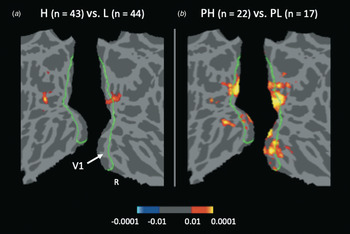
Youth with high levels of delusional beliefs (the H group, n = 43) exhibited significantly greater connectivity between the amygdala and visual cortex compared to those with low levels of delusional beliefs (the L group, n = 44) [Fig. 1a, Table 2a; MNI (Montreal Neurological Institute) coordinates of peak in extrastriate visual cortex (x, y, z) = −34, −80, 10, z = 3.62, p = 0.034, 325 voxels; MNI coordinates of peak in striate cortex (x, y, z) = 20, −88, 10, z = 3.31, p = 0.034, 345 voxels; see online Supplementary Fig. S3 for the effects of global signal regression]. No other clusters met whole-brain-corrected level of significance.
Amygdala-visual cortex connectivity is elevated in youth with high levels of delusional beliefs. Flattened voxel-wise maps of parietal and occipital cortex showing clusters that met FDR whole-brain correction in the comparisons of amygdala connectivity of the H v. L groups (a); and PH v. PL groups (b). The border of V1 is shown in green. The p-values of the clusters showing significant between-group differences in these maps [H > L in (a); PH > PL in (b); the reverse contrasts, L > H and PL > PH, showed no significant differences] are represented by warm colors. All significant clusters are shown.

Results of between-group comparisons of amygdala connectivity and regressions of amygdala connectivity v. total number of delusional beliefs and associated distress

Peaks that survived FDR whole-brain correction. (a) and (b) describe findings of the comparisons between the H and L groups, and the PH and the PL groups, respectively. There were no areas that showed greater connectivity in the L compared to the H group, or the PL compared to the PH group. (c) and (d) describe the dimensional relationships found between amygdala connectivity and: (1) the number of subclinical delusions (PDI total score) and (2) the degree of associated distress (PDI distress score), respectively. For (c), there were no significant negative correlations. The proportion of the significant cluster that was located in striate v. extrastriate cortex and other regions is listed in the column furthest to the right (‘Distribution of effects’).
Next, the magnitude of amygdala connectivity of youth who exhibited persistently high levels of subclinical delusional thinking (the PH group, n = 22) was compared to those with persistently low levels (the PL group, n = 17). Compared to the PL group, the PH group exhibited significantly elevated amygdala connectivity with visual cortex (Fig. 1b, Table 2b; MNI coordinates of peak in striate cortex (x, y, z) = 16, −90, 4, z = 4.20, p < 0.001, 2390 voxels). No other clusters met whole-brain-corrected level of significance.
Regression analyses
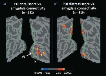
A whole-brain regression in the full sample (n = 122) was conducted to determine whether a continuous relationship was present between amygdala-visual cortex connectivity and (1) levels of delusional beliefs (PDI total score) and/or (2) the degree of distress associated with these beliefs (PDI distress score, corrected for the total number of beliefs). Both outcome measures (total number of beliefs and associated distress) were significantly positively correlated with the strength of functional connectivity between the amygdala and a widely distributed area of visual cortex, including both striate and extrastriate areas (Fig. 2, Table 2c and d).
The number of delusional beliefs and degree of associated distress correlate with amygdala-visual cortex connectivity. Flattened voxel-wise maps of parietal and occipital cortex showing clusters that met FDR whole-brain correction following whole-brain regression of amygdala connectivity with PDI total score (a) or PDI distress score (b) as regressors. These maps reveal similar patterns of correlations between these measures and amygdala-visual cortex connectivity [in (a), MNI coordinates of peak in temporal occipital fusiform cortex (x, y, z) = 36, −50, −10, z = 3.84, p = 0.001, 681 voxels; in (b), MNI coordinates of peak in extrastriate cortex (x, y, z) = 10, −54, 2, z = 3.60, p < 0.001, 795 voxels]. The border of V1 is shown in green. The p-values of the clusters are represented by warm (positive correlations) and cool (negative correlations) colors. Two clusters (negative correlations) in (b) are not shown; see Table 2d for details.

Specificity of findings
In the full sample (n = 122), levels of symptoms of depression, anxiety, and hallucinatory experiences were found to be significantly correlated with levels of subclinical delusional thinking (r = 0.38, p < 0.001; r = 0.31, p = 0.001; r = 0.64, p < 0.001, respectively), as expected (Cella et al., Reference Cella, Cooper, Dymond and Reed2008; Varghese et al., Reference Varghese, Scott and McGrath2008). Given these relationships, we repeated the categorical analyses above with levels of depression, anxiety, and hallucinatory experiences included as covariates; the findings remained significant [p < 0.05, FDR whole-brain-corrected; MNI coordinates of peak of the H v. L group comparison (x, y, z) = −24, −84, −2, z = 4.69, p = 0.020, 1523 voxels; MNI coordinates of peak of the PH v. PL group comparison (x, y, z) = −28, −78, 2, z = 5.83, p = 0.003, 1756 voxels]. Additionally, findings in all primary comparisons remained significant when excluding those currently taking psychotropic medication (p < 0.01, FDR whole-brain-corrected). Moreover, lowering the significance thresholds used in the analyses did not alter the pattern of findings, which remained primarily limited to visual cortex (online Supplementary Fig. S4).
Next, to explore whether the origins of the amygdala-visual cortex pathway affected by delusional thinking could be further localized within the amygdala, we created a 3-mm spherical seed centered on the strongest peak in the primary analyses [MNI (x, y, z) = 16, −90, 4; see Table 2] and repeated the categorical analyses above using this visual cortex seed. Significant differences in visual cortex-amygdala connectivity were observed in both analyses (p < 0.01, FDR whole-brain-corrected), with significant clusters found within the basolateral nucleus of the amygdala (online Supplementary Fig. S5), consistent with the known topography of the projection of the amygdala to the visual cortex in non-human primates (Amaral et al., Reference Amaral, Behniea and Kelly2003).
Lastly, in light of the close anatomical proximity and functional relationship between the amygdala and hippocampus, we repeated the two categorical analyses using a bilateral hippocampus (rather than amygdala) seed. The analyses using the hippocampal ROI revealed a similar but weaker pattern of results compared to those observed using the amygdala ROI, suggesting some specificity of our results to the amygdala (online Supplementary Fig. S6).
Persecutory beliefs
In the full sample (n = 122), the subjects who endorsed persecutory beliefs (n = 28) had a significantly greater overall number of delusional beliefs (t (120) = 5.00, p < 0.001) and associated distress (t (114) = 2.65, p = 0.009), and also showed a higher rate of persistence of high levels of delusional beliefs [χ2 (1, n = 56) = 10.06, p = 0.002], compared to those without persecutory beliefs (n = 94). Thus, based on these data and our a priori hypothesis, additional analyses were conducted to determine the specific contribution of persecutory ideation to the above findings.
First, an anatomical ROI analysis was conducted. Amygdala-V1 connectivity values were extracted for all subjects and mean amygdala-V1 connectivity values were compared among the following groups: (1) members of the high group with persecutory beliefs (H + P, n = 19), (2) members of the high group without persecutory beliefs (H − P, n = 24), and (3) the members of the low group (L, n = 43; excluding one L subject who endorsed persecutory beliefs). These groups were well-matched on all demographic and symptom variables (see online Supplementary Table S2), with the exception of a small but significant difference in age between the H + P and H − P groups; however, all reported effects (see below) remained significant after controlling for age.
A one-way ANOVA revealed a significant effect (F (2,83) = 3.80, p = 0.026) that arose from greater amygdala-V1 connectivity of the H + P group compared to both the H − P (p = 0.043) and L (p = 0.006) groups, with no difference observed between the H − P and L groups (p = 0.760) (Fig. 3a). Similar findings were observed for the PH + P, PH − P, and PL groups in the cohort with 1-year follow-up data (F (2,36) = 7.42, p = 0.002; Fig. 3b).
Amygdala-V1 connectivity is elevated in individuals with, compared to those without, persecutory beliefs. In an ROI analysis, average amygdala-V1 connectivity values (z scores) were extracted. The mean values are plotted here for the following groups: members of the H group with persecutory beliefs (H + P, n = 19), the H group without persecutory beliefs (H − P, n = 24), and the L group (L, n = 43) in (a); and members of the PH group with persecutory beliefs (PH + P, n = 10), the PH group without persecutory beliefs (PH − P, n = 12), and the PL group (PL, n = 17) in (b). In (a), post-hoc t tests showed that amygdala-V1 connectivity of the H + P group was significantly greater in magnitude than that of both the H − P and L groups (p = 0.043 and p = 0.006, respectively), while there was no significant difference between the H − P and L groups (p = 0.760) in amygdala-V1 connectivity. Similarly, the PH + P group showed significantly greater amygdala-V1 connectivity compared to both the PH − P and PL groups (p = 0.045 and p = 0.001, respectively), while the difference between the PH − P and PL groups did not reach significance (p = 0.099). Note that the majority of the extracted z-scores are negative in value because of the effects of the global mean regression (see online Supplementary Fig. S3). Error bars represent standard errors of the mean.

To determine the relative contribution of the number, associated distress, and persecutory nature of participants’ delusional beliefs to the above findings, a multiple regression analysis was then conducted. A statistically significant effect of persecutory thinking on amygdala-V1 connectivity (β = 0.29, p = 0.032, n = 86) was found; no other factors were significant in the model. An analysis of the sample with 1-year follow-up data yielded similar results; persecutory thinking was again the only significant factor (β = 0.40, p = 0.027, n = 39). Lastly, to explore whether the findings were specific to persecutory thinking, these two regression analyses were repeated including two other PDI factors [religiosity and thought disturbances (Peters et al., Reference Peters, Joseph and Garety1999; Peters et al., Reference Peters, Joseph, Day and Garety2004)] as additional regressors; persecutory thinking remained the only significant predictor (p = 0.034 and p = 0.025, respectively).
Discussion
Summary of main findings
In this study, young adults with high levels of delusional beliefs showed greater functional connectivity between the amygdala and visual cortex than young adults with low levels of delusional beliefs. This pattern of results was even stronger in those with delusional beliefs that persisted over the course of 1 year. Subsequent analyses revealed that this effect was primarily driven by participants who endorsed persecutory beliefs, suggesting that excessive functional coupling between the amygdala and visual cortex may represent a vulnerability marker or a consequence of persecutory thinking.
Relationship to prior work
Prior studies of amygdala responses or connectivity in psychosis have demonstrated somewhat inconsistent findings; some of these discrepancies may be attributable to differences in experimental paradigms, stage of illness effects, or treatment effects. For example, studies have detected significant elevations (Kosaka et al., Reference Kosaka, Omori, Murata, Iidaka, Yamada, Okada, Takahashi, Sadato, Itoh, Yonekura and Wada2002; Holt et al., Reference Holt, Kunkel, Weiss, Goff, Wright, Shin, Rauch, Hootnick and Heckers2006), no differences (Anticevic et al., Reference Anticevic, Repovs and Barch2011), or significant reductions (Schneider et al., Reference Schneider, Weiss, Kessler, Salloum, Posse, Grodd and Muller-Gartner1998; Gur et al., Reference Gur, McGrath, Chan, Schroeder, Turner, Turetsky, Kohler, Alsop, Maldjian, Ragland and Gur2002; Anticevic et al., Reference Anticevic, Van Snellenberg, Cohen, Repovs, Dowd and Barch2012) in task-elicited responses of the amygdala in schizophrenia patients compared to demographically-matched healthy controls. Reductions in amygdala responses have also been reported in paranoid compared to non-paranoid schizophrenia patients (Williams et al., Reference Williams, Das, Harris, Liddell, Brammer, Olivieri, Skerrett, Phillips, David, Peduto and Gordon2004; Russell et al., Reference Russell, Reynaud, Kucharska-Pietura, Ecker, Benson, Zelaya, Giampietro, Brammer, David and Phillips2007). However, studies conducted in first-degree relatives of schizophrenia patients (Rasetti et al., Reference Rasetti, Mattay, Wiedholz, Kolachana, Hariri, Callicott, Meyer-Lindenberg and Weinberger2009; Cao et al., Reference Cao, Bertolino, Walter, Schneider, Schafer, Taurisano, Blasi, Haddad, Grimm, Otto, Dixson, Erk, Mohnke, Heinz, Romanczuk-Seiferth, Muhleisen, Mattheisen, Witt, Cichon, Noethen, Rietschel, Tost and Meyer-Lindenberg2016) have suggested that the reported reductions in amygdala responses in schizophrenia may represent a consequence of antipsychotic treatment. Consistent with this interpretation is a recent finding of significantly elevated amygdala responses to negatively-valenced faces in unmedicated, non-help-seeking adolescents with subclinical psychotic symptoms, in comparison to typical adolescents (Wolf et al., Reference Wolf, Satterthwaite, Calkins, Ruparel, Elliott, Hopson, Jackson, Prabhakaran, Bilker, Hakonarson, Gur and Gur2015). Reports of increased task-elicited amygdala responses in schizophrenia (Kosaka et al., Reference Kosaka, Omori, Murata, Iidaka, Yamada, Okada, Takahashi, Sadato, Itoh, Yonekura and Wada2002; Holt et al., Reference Holt, Kunkel, Weiss, Goff, Wright, Shin, Rauch, Hootnick and Heckers2006) and increased baseline amygdala cerebral blood flow in schizophrenia patients compared to healthy controls (Taylor et al., Reference Taylor, Phan, Britton and Liberzon2005) and in paranoid compared to non-paranoid schizophrenia patients (Pinkham et al., Reference Pinkham, Liu, Lu, Kriegsman, Simpson and Tamminga2015) also suggest that the amygdala is overactive during psychotic states. In addition, two resting-state functional connectivity studies of patients with schizophrenia have observed overconnectivity of the amygdala with downstream projection sites, such as the brainstem (Anticevic et al., Reference Anticevic, Tang, Cho, Repovs, Cole, Savic, Wang, Krystal and Xu2014) and visual cortex (Ford et al., Reference Ford, Palzes, Roach, Potkin, van Erp, Turner, Mueller, Calhoun, Voyvodic, Belger, Bustillo, Vaidya, Preda, McEwen and Mathalon2015). Consistent with these prior findings and the continuum model of psychosis, the current study shows that amygdala overconnectivity is present in non-help-seeking youth with subthreshold persecutory beliefs.
Potential functional implications of findings
The increase in connectivity observed in the delusional compared to the non-delusional youth in the current study resulted from a statistically significant difference between the magnitude of negative correlations between the amygdala and visual cortex in each group. Although the source of negative correlations (or ‘anti-correlations’) may vary across studies (Buckner et al., Reference Buckner, Andrews-Hanna and Schacter2008; Fox et al., Reference Fox, Zhang, Snyder and Raichle2009; Murphy et al., Reference Murphy, Birn, Handwerker, Jones and Bandettini2009), here the negative correlations in our data were attributable to the regression of the global mean during preprocessing. Thus, these results suggest that resting blood oxygenation level dependent activity of the amygdala and visual cortex is weakly positively correlated in humans, which is consistent with the established presence of excitatory projections from the amygdala to the visual cortex (Amaral et al., Reference Amaral, Behniea and Kelly2003). Work in non-human primates has shown that fibers originating primarily in the basal nucleus of the amygdala project to all levels of the ventral visual stream [the object processing, or ‘what,’ pathway of the visual system (Ungerleider and Haxby, Reference Ungerleider and Haxby1994)], terminating in synaptic connections (Tigges et al., Reference Tigges, Walker and Tigges1983; Iwai and Yukie, Reference Iwai and Yukie1987; Amaral et al., Reference Amaral, Behniea and Kelly2003). Referred to as the ‘temporal-occipital amygdalo-cortical pathway,’ these projections are distributed continuously across both striate and extrastriate cortices in a primarily unidirectional, nonreciprocal manner, with a point-to-point, non-diffuse organization (Freese and Amaral, Reference Freese and Amaral2005). For example, projections from dorsal portions of the basal nucleus of the amygdala terminate in the posterior visual cortical areas, i.e. striate cortex/V1, whereas neurons in the ventral portion of the basal nucleus of the amygdala terminate in more anterior visual cortical areas (Amaral et al., Reference Amaral, Behniea and Kelly2003). Our data suggest that the dorsal basal nucleus-V1 pathway is altered in youth with elevated delusional thinking. Follow-up studies conducted using high field strength MRI in independent samples will be necessary to confirm these observations.
Functional neuroimaging work in humans has suggested that these feedback-like projections to visual cortex from the amygdala modulate lower-level visual processing. One study showed that both healthy controls and patients with hippocampal lesions (but an intact amygdala) exhibit greater activation of visual cortex in response to fearful faces compared to neutral faces, whereas patients with amygdala lesions fail to show this response (Vuilleumier et al., Reference Vuilleumier, Richardson, Armony, Driver and Dolan2004). These findings are consistent with single-unit recordings conducted in non-human primates that suggest that top-down processes bias early perception (Lee, Reference Lee2002). Therefore, projections from the amygdala to the primary visual cortex may influence sensory processing, particularly in the context of environmental threats.
Abnormal sensory-limbic coupling in psychosis
Prior evidence for increased amygdala-visual cortex connectivity in schizophrenia patients with visual hallucinations suggests that coupling between perceptual and affective processing is altered in psychotic illness (Ford et al., Reference Ford, Palzes, Roach, Potkin, van Erp, Turner, Mueller, Calhoun, Voyvodic, Belger, Bustillo, Vaidya, Preda, McEwen and Mathalon2015). These findings are generally consistent with evidence for abnormalities in early sensory processing in schizophrenia (Javitt, Reference Javitt2009), as well as with the ‘aberrant salience’ model of psychosis (Kapur, Reference Kapur2003). This model suggests that perceptual processing is disrupted by changes in dopaminergic neurotransmission during pre-illness or subthreshold psychotic states, leading to misattributions of motivational salience to neutral or irrelevant stimuli in the environment. Abnormal function of the amygdala-visual cortical pathway in psychosis may contribute to this putative cycle of misattribution of threat to incoming sensory signals (Das et al., Reference Das, Kemp, Flynn, Harris, Liddell, Whitford, Peduto, Gordon and Williams2007). It has been proposed that delusional thinking may represent a consequence of these misattributions, reflecting a compensatory effort to make sense of inappropriately labeled perceptual experiences (Maher, Reference Maher2005).
Limitations and future directions
In this study, we enrolled a large sample of young adults, the majority (90.2%) of whom were medication-naïve, yet displayed a wide range of subthreshold psychotic symptoms. Although all subjects initially underwent a brief, in-person assessment by a clinician, we relied primarily on self-report measures, in order to reduce subject burden and facilitate recruitment. Also, we did not formally assess whether our participants had, or later developed, clinical psychosis. However, the unmedicated status of the majority of our subjects and their concurrent enrollment in college suggest that the symptoms endorsed by this cohort were generally not severe enough to merit clinical intervention or cause substantial functional impairment and were likely predominantly subthreshold in nature. An additional limitation was that persecutory ideation was measured by responses to only two items on a self-report scale; although paranoia is somewhat categorical in nature, future investigations could also assess paranoia in a dimensional manner.
Lastly, it is unclear whether the identified changes in amygdala-visual cortex connectivity represent a vulnerability to or a consequence (which could be either maladaptive or compensatory) of persecutory ideation. Studies that perturb this neural system may shed further light on the role of this pathway in persecutory thinking. For example, pharmacological interventions with compounds such as psilocybin that are known to inhibit threat-induced increases in amygdala-striate cortex connectivity (Kraehenmann et al., Reference Kraehenmann, Schmidt, Friston, Preller, Seifritz and Vollenweider2016), or psychosocial interventions that decrease threat attributions, could be tested for potential effects on this circuitry and paranoia.
Conclusions
The demonstration of a robust association, using both categorical and dimensional analyses between a quantitative imaging phenotype (amygdala-visual cortex functional connectivity) and a symptom (delusional beliefs and persecutory ideation) that has been linked to increased risk for clinical psychosis (Cannon et al., Reference Cannon, Yu, Addington, Bearden, Cadenhead, Cornblatt, Heinssen, Jeffries, Mathalon, McGlashan, Perkins, Seidman, Tsuang, Walker, Woods and Kattan2016) and functional disability (Yung et al., Reference Yung, Nelson, Baker, Buckby, Baksheev and Cosgrave2009; Pinkham et al., Reference Pinkham, Harvey and Penn2016) may represent a novel biomarker of psychosis. Following further validation, such a marker could serve as a target for interventions that could slow or interrupt progression of psychosis-related changes in this pathway, or be used to identify individuals who merit further monitoring.
Supplementary material
The supplementary material for this article can be found at https://doi.org/10.1017/S0033291718004221.
Author ORCIDs
Stephanie N. DeCross, 0000-0001-8993-4413.
Financial support
This work was supported by internal funds of the Massachusetts General Hospital Department of Psychiatry, the National Institutes of Health (R01MH095904 to DJH; 1S10RR023043; 1S10RR023401), and the National Science Foundation Graduate Research Fellowship (DGE1144152/DGE1745303 to SND).
Acknowledgements
We are grateful for the essential support of Tammy Moran, Marissa Hollingshead, and other staff of the Harvard University Center for Brain Science during data acquisition.
Conflict of interest
None of the authors have any competing interests related to the work described. Dr. Holt has received research support from Janssen Scientific Affairs, EnVivo Pharmaceuticals and the Stanley Medical and Research Insitute. Dr. Fava's disclosures are listed here: https://mghcme.org/faculty/faculty-detail/maurizio_fava).
Ethical standards
The authors assert that all procedures contributing to this work comply with the ethical standards of the relevant national and institutional committees on human experimentation and with the Helsinki Declaration of 1975, as revised in 2008.