Introduction
Vascular abnormalities of the temporal bone, such as aberrant internal carotid artery, persistent stapedial artery, dehiscent carotid artery canal and dehiscent high jugular bulb, are very important, especially in patients scheduled for middle-ear surgery.Reference Lin, Wang, Liu and Chen 1 High jugular bulb is the most common vascular abnormality of the petrous part of the temporal bone, with an incidence of 3.5–34 per cent reported in the literature.Reference Ball, Elloy, Vaidhyanath and Pau 2 – Reference Atilla, Akpek, Uslu, Ilgit and Işik 4 It is defined as the apex of the jugular foramen being above the inferior bony annulus of the tympanic membrane.Reference Overton and Ritter 5 It is more common on the right side, and bilateral high jugular bulb is rare.Reference Ball, Elloy, Vaidhyanath and Pau 2 , Reference Wadin, Thomander and Wilbrand 6
High jugular bulb is often discovered as an asymptomatic, incidental finding.Reference Lin, Wang, Liu and Chen 1 Tinnitus, hearing loss, vestibular disorders and aural fullness can be seen in patients with a high jugular bulb.Reference Woo, Wie, Park, Kong, Lee and Goh 7 , Reference Weiss, Zahtz, Goldofsky, Parnes and Shikowitz 8 Dehiscent high jugular bulb, defined as superior and lateral extension of the jugular bulb to the middle-ear cavity, is more commonly seen in association with a high jugular bulb. There may be a defect in the bone surrounding the jugular bulb, covered by thin mucosa.Reference Atmaca, Elmali and Kucuk 9 The incidence of dehiscent high jugular bulb is 1–7 per cent. The symptoms of dehiscent high jugular bulb are vertigo, syncope, tinnitus and hearing loss.Reference Atilla, Akpek, Uslu, Ilgit and Işik 4 , Reference Hourani, Carey and Yousem 10 , Reference Tomura, Sashi, Kobayashi, Hirano, Hashimoto and Watarai 11
Hearing loss associated with high jugular bulb is uncommon. However, sensorineural hearing loss (SNHL) or mixed-type hearing loss can be seen in high jugular bulb patients, accompanied by compression of the vestibular aqueduct or internal acoustic canal.Reference Weiss, Zahtz, Goldofsky, Parnes and Shikowitz 8 Hearing loss associated with dehiscent high jugular bulb has been reported in the literature.Reference El-Begermy and Rabie 12
This article aims to inform on the incidence of high jugular bulb and dehiscent high jugular bulb, and on the symptoms related to these vascular anomalies.
Materials and methods
This study was approved by the Clinical Trials Ethics Committee of the Samsun Training and Research Hospital.
Temporal bone computed tomography (CT) scanning of 3285 patients with various ear symptoms, including vertigo, tinnitus, hearing disturbances and pre-syncope, was performed between January 2012 and May 2014, and the scans were retrospectively reviewed. The temporal bone CT scanning was performed by one radiologist (ATS) using a Toshiba 120 kV 16-slice scanner (Alexion 16 system; Toshiba Medical Systems, Tokyo, Japan); the images were acquired in axial planes with 1 mm thickness. Coronal plane images were reconstructed from raw images.
The exclusion criteria were: age under 15 years, congenital anomalies in the middle or inner ear, tumour or inflammatory mass surrounding the jugular bulb, and history of previous ear surgery.
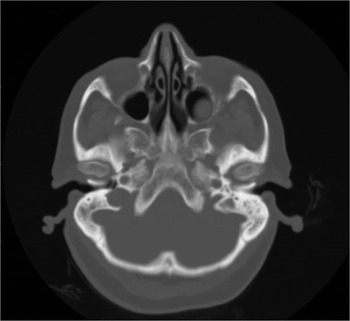
The position of the jugular bulb and jugular dehiscence in the middle-ear cavity were evaluated by temporal bone CT. We defined high jugular bulb as the apex of the jugular foramen above the inferior bony annulus of the tympanic membrane on axial temporal bone CT images (Figure 1). A total of 730 patients met the criteria.
High jugular bulb was defined as the apex of the jugular foramen above the inferior bony annulus of the tympanic membrane, as seen on this axial computed tomography image of the temporal bone.

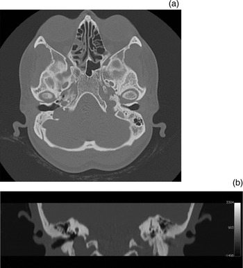
Dehiscent high jugular bulb was defined as any defect of the bony structures covering the jugular foramen in the tympanic cavity, or protrusion of soft tissue from the jugular foramen into the tympanic cavity, on axial temporal bone CT images (Figure 2a, b). All images were evaluated using bone and soft-tissue window settings for the dehiscence. Coronal images were examined if necessary. Twenty-six of 730 patients had a dehiscent high jugular bulb.
Axial (a) and coronal (b) computed tomography images showing bony dehiscence of the high jugular bulb.

The medical records of patients with a dehiscent high jugular bulb were evaluated for patient complaints and audiometry findings. Patients who did not undergo audiometry were called by telephone and invited to the hospital for pure tone audiometry testing.
Pure tone audiometry and speech audiometry were performed using an Interacoustics™ AC40 clinical audiometer. TDH-39 standard headphones were used for air conduction thresholds and speech tests, and a RadioEar™ B-71 bone vibrator was used for bone conduction. Air thresholds were established for frequencies of 250, 500, 1000, 2000, 4000 and 8000 Hz, and bone conduction thresholds were established for 500, 1000, 2000 and 4000 Hz. The pure tone average was calculated as the average (in decibels) of the thresholds for pure tones at 500, 1000 and 2000 Hz.
Statistical analysis
The SPSS statistical software package (version 21 for Windows; SPSS, Chicago, Illinois, USA) was used to perform all statistical calculations. Significant differences between qualitative variables were examined by Fisher's exact test or Pearson's chi-square test. Average age differences between females and males were analysed by the independent samples t-test. A value of p < 0.05 was considered significant in all statistical analyses. All data were expressed as means ± standard deviations.
Results
A total of 3285 patients were evaluated for high jugular bulb between January 2012 and May 2014 by temporal bone CT. A high jugular bulb was detected in 730 patients, for an incidence of 22 per cent. The study group consisted of 471 females (64.5 per cent) and 259 males (35.5 per cent). Mean age was 42.6 ± 17 years for females and 46.5 ± 18.4 years for males.
High jugular bulb was right-sided in 510 patients (69.9 per cent) and left-sided in 220 (30.1 per cent); this finding was statistically significant (p < 0.01). Of the females, 314 (66.6 per cent) had a high jugular bulb on the right side and 157 (33.3 per cent) had it on the left. Of the males, 196 (75.6 per cent) had a high jugular bulb on the right side and 63 (24.3 per cent) had it on the left. Thus, high jugular bulb was more prominent on the right side both for females and males; this finding was statistically significant (p < 0.01).
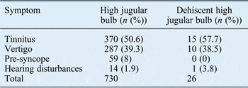
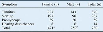
Symptoms of the patients with high jugular bulb and dehiscent high jugular bulb are shown in Table I. Table II shows the symptoms of high jugular bulb patients according to gender distribution.
Symptoms of patients with high jugular bulb and dehiscent high jugular bulb

Symptoms of patients with high jugular bulb according to gender

*64.5 per cent; †35.5 per cent
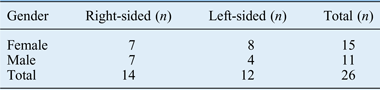
One-sided temporal bone defects compatible with dehiscence were detected in 26 (15 females, 11 males) of the 730 patients. The overall incidence of dehiscent high jugular bulb was 3.5 per cent. The distribution of dehiscent high jugular bulb according to gender is shown in Table III. The mean age of patients with dehiscent high jugular bulb was 31 ± 21 years for the females and 45 ± 17 years for the males.
Dehiscent high jugular bulb sidedness

Fifteen of the 26 dehiscent high jugular bulb patients (57.7 per cent) had complaints of tinnitus, 10 (38.5 per cent) had vertigo and 1 (3.8 per cent) had hearing loss. There were no statistically significant associations between dehiscent high jugular bulb and vertigo, tinnitus or hearing disturbances.
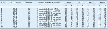
Pure tone audiometry was suggested to the dehiscent high jugular bulb patients for evaluation of hearing loss. Sixteen patients did not come to the hospital, for a variety of reasons, and pure tone audiometry was performed on only 10 patients. The audiometric findings of these patients are shown in Table IV.
Audiometric findings of dehiscent high jugular bulb patients

Pure tone average (PTA) values are shown in decibels. ‘PTA1’ = 250 Hz; ‘PTA2’ = (500 + 1000 + 2000 Hz)/3; ‘PTA3’ = (4000 + 8000)/2. *Of dehiscent high jugular bulb. Pt no. = patient number; y = years; L = left; R = right; F = female; SNHL = sensorineural hearing loss; CHL = conductive hearing loss; M = male; HL = hearing loss
There were no clinical or radiological factors to explain the hearing loss except for dehiscent high jugular bulb. Close proximity between the tympanic membrane and dehiscent high jugular bulb was found in four of the seven patients with conductive hearing loss (two on the right side, two on the left). The audiometric evaluation of these patients revealed an air–bone gap, closing from low frequency to high frequency. There was no contact with the tympanic membrane or obstruction in the round window niche in the other three patients.
Discussion
The incidence and definition of high jugular bulb vary widely among studies. Overton and Ritter examined 257 temporal bone specimens, and described high jugular bulb as a jugular bulb above the level of the inferior bony annulus.Reference Overton and Ritter 5 They reported a high jugular bulb incidence of 6 per cent. Wadin et al. evaluated 245 temporal bone specimens, and defined high jugular bulb as a jugular bulb above the level of the lower border of the round window; they reported an incidence of 24 per cent.Reference Wadin, Thomander and Wilbrand 6 Atilla et al. retrospectively evaluated high-resolution CT scans of 700 temporal bones, and defined high jugular bulb as a jugular bulb above the basal turn of the cochlea. They reported a high jugular bulb incidence of 20 per cent.Reference Atilla, Akpek, Uslu, Ilgit and Işik 4 Woo et al. defined high jugular bulb as the apex of the jugular foramen above the inferior bony annulus of the tympanic membrane on axial temporal bone CT images, and reported an incidence of 9.5 per cent.Reference Woo, Wie, Park, Kong, Lee and Goh 7 In our study, we defined high jugular bulb as the apex of the jugular foramen above the inferior bony annulus of the tympanic membrane, as per Woo et al., and 730 patients in our study met this criteria. The incidence of high jugular bulb was 22 per cent in our study, in accordance with the literature.
A jugular foramen apex above the inferior bony annulus of the tympanic membrane may easily be injured during transtympanic interventional procedures.Reference Woo, Wie, Park, Kong, Lee and Goh 7 It is therefore important to establish the presence of a high jugular bulb in all patients, especially in those who undergo transtympanic interventional procedures.
Atilla et al. reported that 142 (20.3 per cent) of 700 temporal bones had a high jugular bulb; 94 (26.9 per cent) were on the right side, 48 (13.7 per cent) were on the left and 14 (4 per cent) were bilateral.Reference Atilla, Akpek, Uslu, Ilgit and Işik 4 High jugular bulbs are usually seen on the right side due to wide dural sinuses and jugular vein in 75 per cent of individuals.Reference Weiss, Zahtz, Goldofsky, Parnes and Shikowitz 8 In our study, 510 (69.9 per cent) of the 730 high jugular bulbs were on the right side and 220 (30.1 per cent) were on the left side on axial temporal bone CT images. We found that a statistically significantly greater number of high jugular bulbs were on the right side compared to the left side (p < 0.01).
Normally, the dome of the jugular bulb is covered by a layer of bone that forms the floor of the hypotympanum. When elevated, the jugular bulb can occupy the middle-ear cavity. The high jugular bulb is normally located in the posterior-inferior part of the tympanic membrane. Rarely, the high jugular bulb extends into the middle-ear cavity with no bony covering, resulting in a dehiscent high jugular bulb. In addition, a dehiscent high jugular bulb shows more extension anteriorly than expected.Reference Weiss, Zahtz, Goldofsky, Parnes and Shikowitz 8
Atilla et al. reported 27 (3.9 per cent) dehiscent high jugular bulbs; 20 (74 per cent) were on the right side, 7 (26 per cent) were on the left side and 2 (7 per cent) were bilateral.Reference Atilla, Akpek, Uslu, Ilgit and Işik 4 In our study, we found 26 dehiscent high jugular bulbs, with an incidence of 3.5 per cent. Fourteen (53.8 per cent) of these were on the right side and 12 (46.2 per cent) were on the left. Dehiscent high jugular bulbs were found more frequently on the right side in our study, consistent with the literature. However, there was no statistically significant difference between the number of dehiscent high jugular bulbs on the left and right sides (p = 0.070).
Dehiscent high jugular bulb can be accompanied by vascular abnormalities of the temporal bone. Lin et al. reported aberrant internal carotid artery accompanying dehiscent high jugular bulb.Reference Lin, Wang, Liu and Chen 1 Serious bleeding can occur in patients with an aberrant internal carotid artery accompanying a dehiscent high jugular bulb during middle-ear surgery.Reference Lin, Wang, Liu and Chen 1 Wang et al. reported a statistically significant association between carotid canal dehiscence and the presence of a high jugular bulb.Reference Wang, Shi, Liu, Wang, Huang and Chen 13 They found that high jugular bulb was associated with a thinner carotid canal wall. Therefore, accompanying anomalies should be evaluated in patients with a dehiscent high jugular bulb.
High jugular bulb is usually asymptomatic and often detected incidentally during otological examination. It can have variable symptoms related to the extension of the jugular bulb and the presence of dehiscence. The most common symptom is tinnitus. Also, vertigo, hearing disturbances and pre-syncope can occur with high jugular bulb.Reference El-Begermy and Rabie 12 , Reference Cama, Inches, Muzzi, Sadushi, Santarelli and De Colle 14 In our study, 368 of 730 patients (50.4 per cent) had tinnitus, 285 (39 per cent) had vertigo, 57 (7.8 per cent) had pre-syncope and 14 (1.9 per cent) had hearing disturbances. The most common symptom in our study was tinnitus, consistent with literature. However, this finding was not statistically significant. Audiometry testing revealed no hearing loss in 14 patients with complaints of hearing disturbances.
Symptoms of dehiscent high jugular bulb include vertigo, tinnitus, pre-syncope and hearing disturbances. Friedmann et al. reported inner-ear dehiscence associated with tinnitus, vertigo and hearing loss.Reference Friedmann, Eubig, Winata, Pramanik, Merchant and Lalwani 15 In our study, 15 of the 26 dehiscent high jugular bulb patients had complaints of tinnitus, 10 had vertigo and 1 had hearing loss. All of the patients were radiologically normal except for the dehiscent high jugular bulb. El Begermy and Rabie reported seven patients with dehiscent high jugular bulb, five of whom had normal hearing levels on pure tone audiometry.Reference El-Begermy and Rabie 12 One of the other patients in that study had mild bilateral high frequency SNHL, and the last patient had moderate bilateral conductive hearing loss.Reference El-Begermy and Rabie 12 Lin et al. reported on a 24-year-old male patient with pulsatile tinnitus and mild conductive hearing loss due to a dehiscent high jugular bulb.Reference Lin, Wang, Liu and Chen 1 A 52-year-old female with moderate right-sided SNHL and a 42-year-old male with left-sided conductive hearing loss associated with dehiscent high jugular bulb were described by Ball et al.Reference Ball, Elloy, Vaidhyanath and Pau 2
In our study, we evaluated the audiograms of 10 of the 26 dehiscent high jugular bulb patients; 7 of these patients had conductive hearing loss, 2 had SNHL and 1 had mixed hearing loss on pure tone audiometry. Different frequencies and types of hearing loss can be detected by simultaneous audiometric tests in patients with dehiscent high jugular bulb who have no complaints of hearing disturbances. We suggest that the lack of such complaints may be because of unilateral hearing loss, mild to moderate hearing loss and good adaptation associated with slow progression of hearing loss over a prolonged period. There was mild contact with the eardrum in four of the seven patients with conductive hearing loss, and the audiometric evaluation of these patients revealed an air–bone gap, closing from low frequency to high frequency. Therefore, patients with an air–bone gap should be evaluated for dehiscence by temporal bone CT before any interventional procedures are performed.
Patients with a dehiscent high jugular bulb do not always complain of hearing loss. Most cases are asymptomatic and are frequently detected incidentally. Patients whose dehiscent high jugular bulb is detected on temporal bone CT can undergo audiometric examination to assess for hearing loss.
-
• High jugular bulb, often an asymptomatic, incidental finding, is the most common vascular abnormality of the petrous part of the temporal bone
-
• Tinnitus, hearing loss, vestibular disorders and aural fullness can be seen in high jugular bulb patients
-
• Dehiscent high jugular bulb is more commonly seen in association with high jugular bulb
-
• Dehiscent high jugular bulb symptoms include vertigo, syncope, tinnitus and hearing loss
-
• This study aimed to inform on high jugular bulb and dehiscent high jugular bulb incidence, and the related symptoms
-
• Various degrees of hearing loss were detected in dehiscent high jugular bulb patients, despite no hearing disturbance complaints
Compared with previous studies, our study has the largest number of high jugular bulb cases. Only having the audiometry results for 10 dehiscent high jugular bulb patients was a limitation of our study. However, the presence of various degrees of hearing loss ipsilateral with the dehiscence, and conductive hearing loss in patients with dehiscence causing mild contact with the eardrum, were striking findings.
Conclusion
In our study, the incidences of high jugular bulb and dehiscent high jugular bulb were 22 per cent (730 out of 3285) and 3.5 per cent (26 out of 730), respectively. High jugular bulb and dehiscent high jugular bulb were observed more frequently in females and on the right side. The most common presenting symptom in patients with a high jugular bulb or dehiscent high jugular bulb was tinnitus. We found varying degrees of hearing loss on pure tone audiometry in patients with dehiscent high jugular bulb, although they had not complained of prominent hearing loss.