INTRODUCTION
A regular feature of NG2 cells throughout grey and white matter in the developing and adult brain is to detect neurotransmitter release from neighbouring axons (Bergles et al., Reference Bergles, Roberts, Somogyi and Jahr2000; Chittajallu et al., Reference Chittajallu, Aguirre and Gallo2004; Lin and Bergles, Reference Lin and Bergles2004; Lin et al., Reference Lin, Huck, Roberts, Macklin, Somogyi and Bergles2005; Kukley et al., Reference Kukley, Capetillo-Zarate and Dietrich2007, Reference Kukley, Kiladze, Tognatta, Hans, Swandulla and Schramm2008; Ziskin et al., Reference Ziskin, Nishiyama, Rubio, Fukaya and Bergles2007; Karadottir et al., Reference Karadottir, Hamilton, Bakiri and Attwell2008). Mechanistically, the release of neurotransmitter onto NG2 cells is identical to synaptic transmitter release and involves action potential induced calcium entry and rapid fusion of transmitter-filled small vesicles (Kukley et al., Reference Kukley, Capetillo-Zarate and Dietrich2007). The membrane surface of NG2 cells is in very close contact with such release sites and allows the glial cell to detect released transmitter with an exquisite sensitivity and makes NG2 cells the only non-neuronal cells which respond to the release of single vesicles with a rapid and significant change in membrane conductance (Bergles et al., Reference Bergles, Roberts, Somogyi and Jahr2000; Lin and Bergles, Reference Lin and Bergles2004; Lin et al., Reference Lin, Huck, Roberts, Macklin, Somogyi and Bergles2005; Kukley et al., Reference Kukley, Capetillo-Zarate and Dietrich2007, Reference Kukley, Kiladze, Tognatta, Hans, Swandulla and Schramm2008; Ziskin et al., Reference Ziskin, Nishiyama, Rubio, Fukaya and Bergles2007). This dedicated cell-to-cell signalling, which we refer to as synaptic signalling for its mechanistical equivalence to neuro-neuronal synapses, provides a high spatial and temporal resolution which potentially allows NG2 cells to track and to respond to the pattern of activity of individual neurons (Gallo et al., Reference Gallo, Mangin, Kukley and Dietrich2008).
Glutamate is the most prevalent transmitter which is released onto NG2 cells and synaptic currents mediated by ionotropic glutamate receptors are present in all NG2 cells throughout all brain regions which have been tested so far (Bergles et al., Reference Bergles, Roberts, Somogyi and Jahr2000; Lin and Bergles, Reference Lin and Bergles2004; Lin et al., Reference Lin, Huck, Roberts, Macklin, Somogyi and Bergles2005; Kukley et al., Reference Kukley, Capetillo-Zarate and Dietrich2007, Reference Kukley, Kiladze, Tognatta, Hans, Swandulla and Schramm2008; Ziskin et al., Reference Ziskin, Nishiyama, Rubio, Fukaya and Bergles2007; Mangin et al., Reference Mangin, Kunze, Chittajallu and Gallo2008) but see Chittajallu et al. (Reference Chittajallu, Aguirre and Gallo2004) and Karadottir et al. (Reference Karadottir, Hamilton, Bakiri and Attwell2008). There are three major subtypes of ionotropic glutamate receptors: NMDA receptors, AMPA receptors and kainate receptors (Bettler and Mulle, Reference Bettler and Mulle1995; Paoletti and Neyton, Reference Paoletti and Neyton2007; Jane et al., Reference Jane, Lodge and Collingridge2009). Previous work in various brain regions has shown that NMDA receptors do not contribute to the fast synaptic currents generated by vesicular glutamate release because these synaptic responses are entirely blocked by antagonists specific for AMPA/kainate receptors (Bergles et al., Reference Bergles, Roberts, Somogyi and Jahr2000; Chittajallu et al., Reference Chittajallu, Aguirre and Gallo2004; Lin et al., Reference Lin, Huck, Roberts, Macklin, Somogyi and Bergles2005; Kukley et al., Reference Kukley, Capetillo-Zarate and Dietrich2007; Ziskin et al., Reference Ziskin, Nishiyama, Rubio, Fukaya and Bergles2007). Furthermore, NMDA receptors are expressed at very low levels such that even saturating doses of NMDA only produce barely detectable currents at the physiological membrane potential of NG2 cells (Ziskin et al., Reference Ziskin, Nishiyama, Rubio, Fukaya and Bergles2007). In the cerebellar molecular layer, synaptic currents in NG2 cells are exclusively mediated by AMPA receptors as revealed by their sensitivity to an AMPA receptor-specific antagonist (Lin et al., Reference Lin, Huck, Roberts, Macklin, Somogyi and Bergles2005). However, it is currently unknown whether NG2 cells express any functional kainate receptors and whether these receptors participate in neuroglial signalling in other brain regions.
In hippocampal neurons there is a coarse cell type-specific function of AMPA (GluR1-4) and kainate receptors (GluR5-7, KA-1 and KA-2) (Frerking and Nicoll, Reference Frerking and Nicoll2000; Huettner, Reference Huettner2003). Synaptic transmission onto excitatory principal neurons is predominantly mediated by AMPA receptors. Kainate receptors are also expressed by certain principal neurons but they contribute little to the total EPSCs (Castillo et al., Reference Castillo, Malenka and Nicoll1997; Vignes and Collingridge, Reference Vignes and Collingridge1997). In contrast, there is a prominent role for kainate receptors in mediating direct glutamatergic synaptic excitation of hippocampal GABAergic interneurons (Cossart et al., Reference Cossart, Epsztein, Tyzio, Becq, Hirsch and Ben-Ari2002). Interneuronal AMPA receptors, on the other hand, are comparably less involved in synaptic transmission and are at certain synaptic inputs specialised to detect glutamate spillover (Cossart et al., Reference Cossart, Epsztein, Tyzio, Becq, Hirsch and Ben-Ari2002). Because of their slow kinetics kainate receptor-mediated EPSCs in hippocampal interneurons have been proposed to be critical for the recruitment of interneurons and their coordination of neuronal oscillations (Frerking and Ohliger-Frerking, Reference Frerking and Ohliger-Frerking2002).
To further explore the functional connectivity of NG2 cells in the hippocampal circuitry, we tested for the presence of functional kainate receptors and what are the implications for hippocampal NG2 cells in the CA1 region when interneuronal kainate receptors are activated.
METHODS
Slice preparation
Horizontal hippocampal slices (300 µm) were prepared with a vibrating blade microtome (Leica) from 7 to 13 days old C57black mice and maintained in an interface chamber perfused with artificial cerebrospinal fluid (ACSF) containing (in mM): NaCl 124, KCl 3, NaH2PO4 1.25, MgCl2 2, CaCl2 2, NaHCO3 26, glucose 10, osmolality 300 mOsm/kg, pH 7.4, gassed with a 95% O2 and 5% CO2 mixture. Individual slices were transferred to a submerged recording chamber mounted on the stage of an upright microscope (Nikon E600FN), and continuously superfused (2 ml/min) with carbogenated ACSF. Drugs were added to the superfusion solution.
Voltage-clamp recordings
Cells were selected using infrared differential interference contrast video microscopy. During our previous work we established how to identify NG2 cells in un-stained life slices of mouse hippocampus (Kukley et al., Reference Kukley, Kiladze, Tognatta, Hans, Swandulla and Schramm2008). In this study, we used the same methodology in the stratum radiatum of the CA1 region and verified in two cells following patch clamp recordings and filling with Lucifer yellow that they are immunoreactive for NG2. Whole-cell voltage-clamp experiments were performed in standard ACSF. Patch pipettes were pulled from borosilicate glass on a vertical puller (Model PP-830, Narishige, Japan) and had resistance of 4.5–5.0 MΩ when filled with internal solution containing (in mM): potassium gluconate 125, Na2-ATP 2, MgCl2 2, EGTA 0.5, HEPES 10, KCl 20, NaCl 3, pH 7.3 and osmolality 280–290 mOsm/kg. To minimise noise and to improve the stability of the baseline some recordings were performed with a Cs-gluconate-based solution containing (in mM): Cs-gluconate 100, MgCl2 4, Na2-ATP 4, EGTA 0.5, HEPES 10, CsCl 30, pH 7.3 and osmolality 280–290 mOsm/kg. The fluorescent tracer Lucifer yellow (0.1%) was included into the pipette solution. Cells were voltage clamped at −80 mV with an EPC-7 amplifier (HEKA, Germany) and the holding potential (V h) was corrected for a liquid junction potential of −10 mV prior to seal formation. Evoked synaptic responses were elicited with an isolated pulse stimulator (A-M Systems, Science Products GmbH, Hofheim, Germany) using a mono-polar glass electrode (resistance 1 MΩ, filled with ACSF) placed at a distance of ~50–150 µm from the recorded cell as described (Kukley et al., Reference Kukley, Capetillo-Zarate and Dietrich2007). Single or paired (40 ms inter-pulse interval) rectangular pulses of 100–200 µs duration were applied every 10–15 s. The responses were low-pass filtered at 1–2 kHz, digitised with a sampling frequency of 20 kHz (ITC-16, HEKA Instruments Inc., USA) and analysed using Igor Pro software (WaveMetrics, Lake Oswego, USA).
Analysis
Changes in the holding current during bath application of drugs when keeping NG2 cells at −80 mV in voltage-clamp mode were measured as difference currents. For this we subtracted from the steady holding current measured during the end of the application period the holding current measured during a stable period immediately before drug application.
Spontaneous IPSCs were analysed by a sliding template algorithm provided by Neuromatic (V1.71) for Igor Pro (V5) with a threshold of −2.5. A template was created which closely matched the rise and decay times of the recorded currents (IPSCs) and was used for all cells. All automatically detected events were visually inspected and discarded in case of doubt. In each cell, for each condition, an average was calculated across all events. Amplitudes were measured as a difference between baseline immediately before an event and the peak of the current. The 20–80% rise time was determined as an interval between time points where the amplitude of the current reached 20 and 80% of its peak value determined as described above, respectively. Decay time constant was determined from an exponential fit to the mean current.
Evoked IPSCs: To improve the accuracy of amplitude measurements we aimed at removing any residual stimulus artefact currents by subtracting failure trials. For this failures were manually selected through all recorded sweeps and averaged. To estimate the amplitude of evoked IPSCs under control conditions, 10–15 successive sweeps recorded shortly before kainate applications were averaged and failures were subtracted from this average. To estimate the amplitude of evoked IPSCs during kainate, 10–15 successive sweeps recorded 4–6 min after beginning of kainate applications were averaged and failures were subtracted from this average.
RESULTS
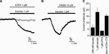
We obtained whole-cell patch clamp recordings from NG2 cells in hippocampal slices from mice 1 to 2 weeks after birth as described previously (Kukley et al., Reference Kukley, Kiladze, Tognatta, Hans, Swandulla and Schramm2008). Cells were held in voltage-clamp mode near their physiological membrane potential at −80 mV. We pharmacologically isolated kainate receptors by blocking NMDA receptors (25 µM APV or 50 µM MK801) and AMPA receptors (30 µM GYKI 53655). To test for the presence of functional kainate receptors we bath applied a low concentration of the non-specific AMPA/kainate receptor agonist kainate (250 nM) and monitored the holding current. At this concentration and under our conditions kainate will only partially activate kainate receptors but bind to all subtypes of kainate receptors (GluR5-7, KA-1 and KA-2) and not to AMPA receptors (Jane et al., Reference Jane, Lodge and Collingridge2009). As soon as kainate reached the recorded cell in the slice we observed a persistent inward current of 6.4±2.4 pA indicating that indeed functional kainate receptors are present in NG2 cells (Fig. 1C, n = 6). At a higher concentration (1 µM) kainate induced a significantly larger inward current (18±3.5 pA, n = 6) which is still very likely to be exclusively mediated by kainate receptors because AMPA receptors are potently blocked by GYKI 53655 (Fig. 1A,C) (Jane et al., Reference Jane, Lodge and Collingridge2009). As expected, the current induced by 1 µM kainate was almost completely reversed by the AMPA/kainate receptor antagonist CNQX which shows that kainate is not acting at a different receptor system (Fig. 1B). To test which kainate receptor subtypes are involved we applied ATPA, an agonist specific for GluR5 containing kainate receptors (Jane et al., Reference Jane, Lodge and Collingridge2009). 1 µM ATPA did not produce a detectable inward current in NG2 cells indicating that they do not express the GluR5 subunit to a significant degree (Fig. 1A,C).
NG2 cells express functional kainate receptors which lack the GluR5 subunit. (A) Continuous holding current recordings of NG2 cells patch clamped in the CA1 area of the hippocampus. Cells were held at −80 mV and AMPA receptors were blocked pharmacologically. Application of kainate (indicated by the black bar) induces a clear inward current consistent with the opening of a mixed potassium and sodium conductance. In contrast, ATPA, which potently and specifically activates GluR5 containing kainate receptors, does not induce a discernible inward current. (B) As expected for the involvement of kainate receptors the current induced by kainate is strongly reversed by the AMPA/kainate receptor antagonist CNQX. (C) Summary bar graph of the changes in holding current recorded under various conditions. Note that even 250 nM kainate causes a small but detectable change in the holding current. The kainate receptor-mediated inward current is not dependent on action potential generation and persists in TTX (right most bar).

To verify that the inward currents observed in NG2 cells are not secondary due to a potential excitatory effect of kainate on neighbouring neurons which may in turn release transmitters onto NG2 cells, we repeated the experiments in 0.5 µM TTX. TTX prevents the generation of action potentials that are required for activity-dependent release of transmitter. Despite the presence of TTX, kainate receptor activation produced an inward current of comparable amplitude (15±1.1 pA, n = 3, Fig. 1C) substantiating the conclusion that kainate and ATPA directly act at receptors on the membrane surface of NG2 cells.
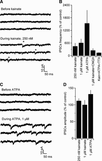
During the above-mentioned experiments we noted that kainate not only induced a persistent inward current but that it also caused the frequent appearance of relatively small and transient inward currents which closely resemble GABAergic synaptic currents previously described in hippocampal NG2 cells (Lin and Bergles, Reference Lin and Bergles2004; Kukley et al., Reference Kukley, Kiladze, Tognatta, Hans, Swandulla and Schramm2008). Under control conditions, i.e. before drug application, these currents were very infrequent (0.08±0.02 Hz, n = 35, Fig. 2A) and displayed mean amplitudes, rise times and decay time constants of 3.4±0.2 pA, 1.0±0.1 ms (20–80%) and 21±1.3 ms, respectively. We and others have previously shown that these currents are entirely blocked by GABA-A receptor antagonists (Lin and Bergles, Reference Lin and Bergles2004; Kukley et al., Reference Kukley, Kiladze, Tognatta, Hans, Swandulla and Schramm2008) and therefore refer to them as IPSCs. Upon application of 250 nM kainate (in the presence of 30 µM GYKI 53655) there was a dramatic, more than four-fold, increase in the frequency of these IPSCs (Fig. 2A) while their amplitudes remained unchanged (Fig. 2D). At 1 µM, kainate produced an even greater increase in the frequency of spontaneous IPSCs consistent with the view that the lower concentration of kainate does not fully activate kainate receptors. The AMPA/kainate receptor antagonist CNQX (10 µM) almost completely reversed the effect of kainate (1 µM) and brought the frequency of IPSCs nearly back to control values (Fig. 2B).
We next tested whether the GluR5-specific agonist ATPA mimics the effect of kainate. While, as described above, 1 µM ATPA did not activate kainate receptors on NG2 cells, it produced a massive increase in spontaneous IPSCs in NG2 cells which strongly exceeds the increase induced by 1 µM kainate (by 1537±622%, n = 17, Fig. 2B,C). As was the case for kainate, ATPA only changed the frequency but the amplitude of spontaneous IPSCs was largely unchanged (Fig. 2D). It is known that hippocampal GABAergic interneurons highly express functional GluR5 containing kainate receptors (Cossart et al., Reference Cossart, Esclapez, Hirsch, Bernard and Ben-Ari1998; Wondolowski and Frerking, Reference Wondolowski and Frerking2009). Therefore, we asked whether kainate and ATPA act by depolarising and firing GABAergic interneurons which project onto NG2 cells and which in turn may cause an increase in the frequency of IPSCs in NG2 cells by action potential-driven vesicular release of GABA. In that scenario the increase in frequency of IPSCs in NG2 cells by kainate receptor activation should be prevented if action potential firing is inhibited. Indeed, in the presence of 0.5 µM TTX, a combination of kainate and ATPA (both at 1 µM) did not induce spontaneous IPSCs (Fig. 2B).
Activation of GluR5 on the somatodendritic membrane of interneurons provokes a strong increase in the frequency of spontaneous IPSCs in NG2 cells. (A) Continuous whole-cell voltage-clamp recording of an NG2 cell held at −80 mV in the presence of AMPA and NMDA receptor antagonists. Before kainate application there are hardly any spontaneous synaptic currents. However, briefly after application of a low concentration of kainate fast rising transient inward currents are observed (appearing as downward deflections, some larger ones are marked by asterisks). (B) Summary bar graph showing the percentage increase in IPSCs frequency under various conditions. Note that there is a concentration-dependent increase in the frequency of IPSCs by kainate which is strongly exceeded by 1 µM ATPA. Further, CNQX and TTX largely prevent the increase in spontaneous currents (right most 2 bar graphs) showing that this increase depends on action potential firing and on activation of kainate-type glutamate receptors. For each bar the frequency during drug application was normalised (and expressed as per cent) on the frequency under control conditions in the same cell. (C) As in (A) but for another NG2 cell onto which 1 µM ATPA was applied. As above, before application no spontaneous currents are discernible but during application of the GluR5-specific agonist there was a pronounced increase in the frequency of IPSCs which are clearly seen as transient downward deflections. (D) Summary bar graph showing that during drug application the mean amplitude of the IPSCs remains largely constant. There is an approximately 30% increase in the IPSC amplitude under 1 µM ATPA which reaches the level of statistical significance (P < 0.05). However, this apparent increase has to be treated with some some care as the low frequency of currents before drug application makes a reliable statistical assessment difficult.

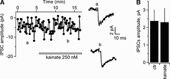
The data, however, does not allow us to say whether there are also presynaptic kainate receptors located on those GABAergic synaptic terminals contacting NG2 cells which possibly directly alter transmitter release. Presynaptic effects of kainate are well known in the hippocampus for both excitatory and inhibitory neuronal synapses (Frerking and Nicoll, Reference Frerking and Nicoll2000; Jane et al., Reference Jane, Lodge and Collingridge2009). Such presynaptic effects on GABAergic terminals contacting NG2 cells, regardless whether inhibitory or facilitating, may be masked by the strong increase in action potential firing caused by kainate/ATPA application which strongly changes the balance between miniature and action potential-dependent vesicle release from control to under drug conditions. To test directly whether there is a presynaptic effect of kainate, we recorded stimulated mono-synaptic IPSCs in NG2 cells. We used a weak and local extracellular stimulation which produced IPSCs with a very low amplitude and frequent release failures (2.4±0.6 pA, n = 4, Fig. 3A). These currents are due to a stimulation of only a few if not only one GABAergic axon and are most likely mono-synaptic because AMPA and NMDA receptors are blocked. Application of kainate (in the presence of 30 µM GYKI 53655) did not alter the amplitude of stimulated IPSCs (2.3±0.8 pA, n = 4, Fig. 3A,B) arguing against a presynaptic effect of kainate at the interneuronal projection onto NG2 cells.
Stimulated mono-synaptic IPSCs in NG2 cells are not altered by kainate receptor activation. (A) Time course of the peak amplitude of stimulated IPSCs recorded in an NG2 cell in the CA1 region. A low stimulation intensity was used which activates at most a handful of GABAergic axons. For this reason the amplitudes are small and are strongly fluctuating. Likewise frequent failures were observed, indicating that only few fibres have been activated. Note that kainate (application time is indicated by the open bar) did not systematically alter the amplitude of the stimulated responses. The inset on the right shows two average responses from time points indicated by the lowercase letters. The example traces are small because they represent averages over successive trials containing responses and failures. (B) Summary bar graph showing the mean amplitudes of IPSCs before (ctr) and during kainate application (250 nM).

DISCUSSION
The main finding of the present study is a dual subtype-specific effect of hippocampal kainate receptor activation on NG2 cells in the CA1 subfield. Kainate receptor activation causes in NG2 cells both a depolarising persistent inward current and a dramatic increase in spontaneous IPSCs. The first effect is due to functional kainate receptors expressed on the membrane surface of NG2 positive OPCs. These kainate receptors are largely free of the GluR5 subunits and are predominantly localised to extra synaptic sites. The second effect is due to a strong synaptic projection of GluR5 expressing GABAergic interneurons onto NG2 cells in the CA1 subfield.
This study is the first to report functional kainate receptors in OPCs in situ. In our eyes the most likely reason why kainate receptors have not been described before simply is that previous studies concentrated on investigating AMPA and not kainate receptors. For example, the pioneering work of the group of Steinhaeuser on ionotropic glutamate receptors on so-called ‘complex glial cells’, which are most likely identical to NG2 cells studied here, did not report the presence of the kainate class of glutamate receptors (Seifert and Steinhauser, Reference Seifert and Steinhauser1995; Seifert et al., Reference Seifert, Rehn, Weber and Steinhauser1997). However, the authors activated glutamate receptors by applying millimolar concentrations of kainate which does not allow to differentiate between AMPA and kainate receptors and thus, in the light of the present study, it is likely that some part of their current was carried by kainate receptors.
The presence of functional kainate receptors in NG2 cells in our study is evidenced by a concentration-dependent inward current induced by low concentrations of kainate (0.25–1 µM) in the presence of a potent AMPA receptor antagonists and by a reversal of this current by a specific AMPA/kainate receptor antagonist. Because this current was also observed when neuronal activity was blocked it is probable that kainate acted directly on receptors in the membrane of NG2 cells. GluR5 containing receptors do not contribute to this current as the effect of kainate was not mimicked by a GluR5-specific agonist. The unavailability of further subtype-specific receptor tools precluded an identification of the kainate receptor subtypes involved. Nevertheless, our data show that when keeping in mind that hippocampal principal neurons also do not express GluR5 (Huettner, Reference Huettner2003; Jane et al., Reference Jane, Lodge and Collingridge2009) and that hippocampal astrocytes completely lack ionotropic glutamate receptors (Zhou and Kimelberg, Reference Zhou and Kimelberg2000; Matthias et al., Reference Matthias, Kirchhoff, Seifert, Huttmann, Matyash and Kettenmann2003), solely GABAergic interneurons are expressing this receptor subtype in the hippocampus.
It could be argued that the inward current in NG2 cells observed upon activation of kainate receptors may not be due to kainate receptors in the membrane of NG2 cells. Instead it may be due to a rise in the extracellular potassium concentration caused by a strong activation of kainate receptors on interneurons which are depolarised and in turn release potassium. While this situation is theoretically possible we believe it is unlikely because the spatial density of interneurons is relatively low and because the input resistance of NG2 cells is quite high. For comparison, CA1 pyramidal cells have an approximately three-fold lower input resistance and should therefore show a three-fold larger inward current upon activation of kainate receptors if bath application of kainate receptor agonists causes an elevation of the extracellular potassium concentration. On the contrary, application of kainate did not induce in inward current in CA1 cells in a previous study (Melyan et al., Reference Melyan, Wheal and Lancaster2002).
It has previously been shown that synaptic glutamate release exclusively activates AMPA but not kainate receptors on NG2 cells (Lin et al., Reference Lin, Huck, Roberts, Macklin, Somogyi and Bergles2005). This means that the kainate receptors we have identified in our study are most likely primarily located extra-synaptically, that is remote from vesicle release sites such that they are not reached by the transmitter content of the vesicles. Therefore, kainate receptors may be specialised to detect changes in the ambient extracellular glutamate concentration. The ambient glutamate concentration is tightly regulated by the glutamate uptake activity of astrocytes but when larger populations of neurons are synchronously active, transport capacity may temporarily be overwhelmed and glutamate can accumulate in the extracellular space and activate extra-synaptic high affinity glutamate receptors such as kainate or metabotropic receptors (Gueler et al., Reference Gueler, Kukley and Dietrich2007; Tzingounis and Wadiche, Reference Tzingounis and Wadiche2007). A further possibility is that kainate receptors on NG2 cells are specialised to detect ambient glutamate which is released from non-synaptic sources. Astrocytes, for example, may be a non-synaptic source of glutamate and have been suggested to release glutamate exocytotically or through pores into the extracellular space (Ye et al., Reference Ye, Wyeth, Baltan-Tekkok and Ransom2003; Takano et al., Reference Takano, Kang, Jaiswal, Simon, Lin and Yu2005; Ni et al., Reference Ni, Malarkey and Parpura2007). In fact, it has been shown that release of glutamate from astrocytes activates kainate receptors on hippocampal interneurons (Liu et al., Reference Liu, Xu, Arcuino, Kang and Nedergaard2004). It is therefore tempting to speculate that kainate receptors on NG2 cells represent a target for astrocyte to NG2 cell signalling.
It may be argued that kainate receptors are not functionally relevant in NG2 cells because the overall current amplitudes observed in this study were relatively small. However, taking into account that 30 µM GYKI 53655 may also partially block kainate receptors (Perrais et al., Reference Perrais, Pinheiro, Jane and Mulle2009) and that 1 µM kainate also does not fully activate all kainate receptor subtypes (Jane et al., Reference Jane, Lodge and Collingridge2009), the total kainate receptor-mediated current in NG2 cells most likely amounts to at least 30 pA. For comparison, this is approximately 30% of the current reported in GluR5 containing hippocampal interneurons (Cossart et al., Reference Cossart, Esclapez, Hirsch, Bernard and Ben-Ari1998). In these interneurons, kainate receptor-driven depolarisation is sufficient to cause a tonic firing of action potentials. Moreover, in comparison to interneurons NG2 cells display at least three-fold higher input resistance of up to 1 GΩ and more (Morin et al., Reference Morin, Beaulieu and Lacaille1998; Kukley et al., Reference Kukley, Capetillo-Zarate and Dietrich2007, Reference Kukley, Kiladze, Tognatta, Hans, Swandulla and Schramm2008). Therefore, the electrical impact of kainate receptor activation on NG2 cells can be considered to be comparable to that on interneurons and can be predicted to depolarise NG2 cells by 30 mV and more. However, as of now the functional consequences of depolarising the membrane potential of NG2 cells are unclear and remain to be revealed by future studies.
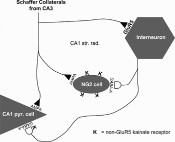
Activation of GluR5 containing kainate receptors that are exclusively found in interneurons in the hippocampus (see Discussion above) leads to a dramatic, 15-fold, increase in the frequency of spontaneous GABA-A receptor-mediated synaptic currents in NG2 cells. This effect is mediated by receptors on the somatodendritic surface of interneurons which depolarise the interneurons and make them tonically fire action potentials. A similar increase in spontaneous IPSCs upon activation of interneuronal GluR5 receptors was observed in CA1 pyramidal neurons (Cossart et al., Reference Cossart, Esclapez, Hirsch, Bernard and Ben-Ari1998) suggesting that most hippocampal interneurons that contact CA1 pyramidal cells also send collaterals to NG2 cells. The GABAergic nerve terminals on CA1 cells, however, seem to differ from those at collaterals which synapse onto NG2 cells. The former contain presynaptic kainate receptors which inhibit the release of GABA (Frerking and Nicoll, Reference Frerking and Nicoll2000; Huettner, Reference Huettner2003; Jane et al., Reference Jane, Lodge and Collingridge2009). This was not observed in our study because 250 nM kainate, which was sufficient to strongly excite interneurons (compare for Fig. 2), did not alter stimulated IPSCs recorded in NG2 cells (compare for Fig. 3). This differential distribution of kainate receptors between nerve terminals on different branches of the same axon contacting either glial cells or CA1 neurons may suggest that GABAergic interneurons are trafficking kainate receptors in a postsynaptic target-specific way (Fig. 4).
Localisation of kainate receptors causing direct and indirect effects on NG2 cells. The scheme integrates findings of the present study and previous observations on the distribution of AMPA and kainate receptors on CA1 pyramidal cells and interneurons (see text for details). Schaffer collaterals represent one of the main glutamatergic inputs to the CA1 region. The collaterals excite CA1 pyramidal cells and NG2 cells by glutamate release onto synaptic AMPA receptors (indicated by AMPA). NG2 cells also express non-GluR5 receptors (denoted by K) but these receptors are not localised to postsynaptic densities and are therefore not activated by glutamate release from Schaffer collaterals but have been activated in this study by application of kainate receptor agonists. Schaffer collaterals also release glutamate onto interneurons. At this synapses the collaterals activate GluR5 receptors (denoted by GluR5). These GluR5 receptors were activated by kainate and ATPA and caused the increase in the frequency of spontaneous IPSCs in NG2 cells in the present study. GABAergic axons of these interneurons form synapses with NG2 cells and CA1 pyramidal cells at which they activate GABA-A receptors. However, the GABAergic terminals contacting the two types of postsynaptic targets seem to differ in presynaptic receptors: GABA release onto the CA1 cells is modulated by presynaptic kainate receptors (as indicated by the K at the interneuron-CA1 pyramidal cell synapses) while such a modulation was not observed for GABAergic input to NG2 cells in the present study.

The parallel innervation of CA1 cells and NG2 positive OPCs implies that these glial cells experience the same pattern of GABAergic synaptic input as do pyramidal cells. It is well established that the rhythmic activity of interneurons is very important to synchronise the hippocampal neural network and to generate neuronal oscillations (Cobb et al., Reference Cobb, Buhl, Halasy, Paulsen and Somogyi1995). These oscillations dramatically change during the postnatal development of the hippocampus (Lahtinen et al., Reference Lahtinen, Palva, Sumanen, Voipio, Kaila and Taira2002; Mohns and Blumberg, Reference Mohns and Blumberg2008). The early neonatal hippocampus primarily only shows very slow network oscillations (minutes) which are, by the end of the first postnatal week, supplemented by gamma frequency activity and which finally cease within the second week and give way for the more adult pattern dominated by theta frequency oscillations. Therefore, the rhythm of occurrence of IPSCs in NG2 cells can be predicted to contain information about the stage of maturation of the brain. Note that CA1 pyramidal cells are inhibited by GABAergic IPSCs and that this inhibition together with an oscillatory propensity is the basis for their synchronisation (Cobb et al., Reference Cobb, Buhl, Halasy, Paulsen and Somogyi1995) but that, in contrast, NG2 cells are excited by the activation of GABA-A receptors. This is due to the higher intracellular chloride concentration of NG2 cells which renders GABAergic currents depolarising (Lin and Bergles, Reference Lin and Bergles2004). Thus, the interneuronal input rhythmically excites NG2 cells according to the ongoing network oscillations.
In summary, NG2 cells use at least three different classes of ionotropic receptors to detect neurotransmitter and the activation of all three classes is converted into a depolarising and excitatory action. Firstly, AMPA receptors are used to detect the release of single glutamate-filled vesicles from individual neighbouring axons. Secondly, GABA-A receptors are used to detect the liberation of GABA-filled synaptic vesicles which are released in synchrony with ongoing network oscillations. Thirdly, extra-synaptic kainate receptors are specialised to detect glutamate which has accumulated in the extracellular space during acute highly synchronous activity of a larger population of excitatory neurons. Currently, it is hardly understood what is the downstream effect of this neurotransmitter input to NG2 cells. However, considering the strong evidence for the modulation of the development of oligodendroglial cells/myelination on neuronal activity (Fields, Reference Fields2005) and the indications that this effect may involve action potential activity in axons (Barres and Raff, Reference Barres and Raff1993; Demerens et al., Reference Demerens, Stankoff, Logak, Anglade, Allinquant and Couraud1996; Zalc and Fields, Reference Zalc and Fields2000) and pharmacological neurotransmitter activation on NG2 cells (Yuan et al., Reference Yuan, Eisen, McBain and Gallo1998), it is tempting to hypothesise that the activation of the three different classes of receptors by synaptic and extracellular transmitters has an influence on the process of differentiation, axon selection or myelination. If so, then AMPA receptors would be ideally suited to guide the selection of individual axons for myelination because they encode the activity of a single neuron. Kainate receptor activation may instruct NG2 cells on the acute activity of an entire bundle of bypassing axons. Finally, differentiation of NG2 cells may be kept in pace with the general maturation of the brain based on the frequency of the permanently ongoing oscillatory activity conveyed by GABAergic synaptic input. However, a series of further studies is needed to explore whether any of these signalling pathways is necessary for or has an effect on the proper development of oligodendroglial cells and on myelination of the brain.
ACKNOWLEDGEMENTS
This study was supported by Deutsche Forschungsgemeinschaft (SFB TR3, DI 853/2, DI 853/3, DI 853/4) and University Clinic Bonn grants (BONFOR). We thank P. Stausberg, S. Buchholz and J. Enders for their excellent technical assistance.