Introduction
The management of cerebrospinal fluid (CSF) leaks used to be the domain of neurosurgery, involving craniotomy and repair. However, this approach leads to higher morbidity as it involves open surgery, craniotomy and retraction of the brain. In 1982, Wigand documented the first case of successful endoscopic CSF leak repair.Reference Wigand1 Currently, the endonasal approach has largely replaced open methods of repair for leaks involving the anterior skull base. However, CSF leaks of the middle temporal fossa are commonly repaired through a lateral craniotomy.Reference de Lara, Ditzel Filho, Prevedello, Carrau, Kasemsiri and Otto2
We demonstrate a case of an expansive CSF leak involving both the middle temporal fossa and the sphenoid due to osteoradionecrosis. An endoscopic endonasal transpterygoidal approach provided a wide surgical corridor to visualise both defects for successful repair. This case illustrates that an endoscopic endonasal approach is a viable alternative to a lateral approach in repairing CSF leak of the middle cranial fossa.
Case report
A 37-year-old male patient presented with a CSF leak involving the left middle cranial fossa and sphenoid sinus. A prior attempt to repair the CSF leak in another hospital using lateral subtemporal craniotomy was not successful.
He had received radiation therapy for nasopharyngeal carcinoma in 2010 and then again in 2011 after a recurrence. Clinical examination confirmed CSF leakage through the nose and throat.
Clinical examination revealed a conscious and alert patient with multiple cranial palsies involving all the left cranial nerves except the IInd and XIth nerves. There was no evidence for nasopharyngeal carcinoma recurrence.
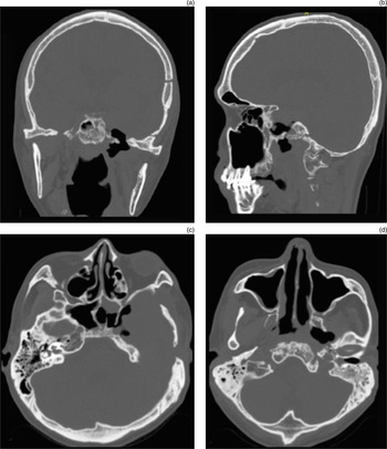
Computed tomography and magnetic resonance imaging revealed extensive osteoradionecrosis of the entire left skull base, with air in the left parapharyngeal space and pneumocranium at the left temporal bone region (Figure 1). There was a ‘Y’-shaped skull base defect, with one limb of the Y extending from the middle cranial fossa floor and the other limb from the sella turcica. Both limbs connected at the region of the pterygoids and then extended inferiorly to the parapharyngeal space.
Coronal (a), sagittal (b) and axial (c and d) pre-operative computed tomography scans, showing extensive osteoradionecrosis of the entire left skull base, involving the greater wing of the sphenoid, clivus, temporal bone, carotid canal and lateral wall of the sphenoid. Imaging also shows left pterygoid muscle necrosis with air tracking into the left parapharyngeal space and a pneumocranium at the left temporal lobe region.

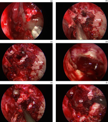
The CSF leak was repaired using a left endoscopic endonasal transpterygoidal approach (Figure 2). The posterior wall of the maxilla was removed after an extended medial maxillectomy to expose the pterygoid plate. Next, the pterygoid plate was removed using a Kerrison punch, and the defect at the temporal lobe was exposed. Removing the lateral wall of the left sphenoid exposed the internal carotid artery, and gave adequate visualisation of both the defect at the temporal lobe and the defect at the sphenoid sinus.
Intra-operative images of the endoscopic endonasal transpterygoidal approach. Customised removal of bone provides a wide surgical corridor that aids visualisation of the skull base defect and allows instrumentation for defect repair. (a) View after extended endoscopic medial maxillectomy. (b) Next, the posterior wall of the maxilla was removed and the pterygoid plate was visualised. (c) After removal of part of the pterygoid plate, the temporal lobe came into view. (d) Endoscopic view of the cerebrospinal fluid leak site at the middle temporal fossa, with exposed temporal lobe. (e) Removal of the lateral wall of the sphenoid exposed the lateral defect of the sphenoid. (f) Final view before defect repair, with exposure of the internal carotid artery. NS = nasal septum; MT = middle turbinate; NLS = nasolacrimal duct; PWM = posterior wall of maxilla; PP = pterygoid plate; TL = temporal lobe; SS = sphenoid sinus; LWS = lateral wall of sphenoid; ICA = internal carotid artery

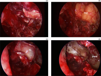
An imaging guidance system (StealthStation S7; Medtronic, Minneapolis, Minnesota, USA) was used intra-operatively. At this stage, adequate exposure for visualisation and instrumentation was provided. The defect was repaired in several layers (Figure 3). First, fascia lata was placed as an inlay. Next, fat was put on top, before another layer of fascia lata was laid over the fat. A middle turbinate flap further stabilised this repair. All layers were stabilised with Tisseel® (tissue glue). The nose was packed using Surgicel (Ethicon, Cincinnati, Ohio, USA) and Gelfoam (Baxter Healthcare, Hayward, California, USA) (haemostatic agents), and Merocel nasal dressings (Medtronic).
Intra-operative images of the defect repair. Adequate visualisation and space for instrumentation created by the endoscopic endonasal transpterygoidal corridor is vital for successful defect repair. (a) First layer of repair was an inlay of fascia lata. (b) Second layer of repair was fat harvested at the same site as the fascia lata. (c) Another layer of fascia lata and a middle turbinate flap were placed. (d) Final view before the nose was packed. FL = fascia lata; FAT = fat; MTF = middle turbinate flap; PWM = posterior wall of maxilla

The post-operative course was uneventful. Intravenous antibiotics (amoxicillin-clavulanate) were given at the time of induction and were continued post-operatively until the patient's nasal packing was removed 5 days after surgery. The patient was instructed not to blow his nose and to avoid strenuous activities for three months. Saline nasal irrigations were started one week after surgery to help clear residual debris and crusting. The patient showed extensive crusting in the first weeks after surgery, and debris and crusting were removed endoscopically on a weekly basis.
At three months' follow up, there were no complaints of any nasal symptoms, and endoscopic endonasal evaluation revealed no crusting in the nasal cavity, with no evidence of CSF leakage.
Discussion
Radiation therapy with or without chemotherapy is the ‘gold standard’ nasopharyngeal carcinoma treatment. Unfortunately, the dose that is required can exceed the radiation dose tolerance of the normal tissue of the skull base. One common side effect of radiation to the skull base is osteoradionecrosis.Reference Birzgalis, Ramsden, Farrington and Small3–Reference Lim, Pang, Low and Tan5
While details of the actual radiation dose the patient received were unavailable, the patient was treated with radiation for the second time a year after the primary treatment due to a recurrence. Higher total radiation dose increases the risk of osteoradionecrosis.Reference Lee, Foo, Chappell, Fowler, Sze and Poon6 In our patient, osteoradionecrosis was further complicated by CSF leakage.
Persistent CSF leaks may lead to bacterial meningitis; therefore, surgical closure should be considered the treatment of choice to prevent ascending meningitis.Reference Bernal-Sprekelsen, Alobid, Mullol, Trobat and Tomas-Barberan7 The goal of surgical closure is repair of the dura defect contributing to the CSF leak.Reference Stone, Castillo, Neelon and Mukherji8 This case report illustrates the successful repair of a large CSF leak of the middle temporal fossa and sphenoid using a novel approach.
We used an endoscopic endonasal transpterygoid approach to facilitate a corridor to the defects that allowed adequate exposure and space for instrumentation. We believe that the first surgical procedure, using a lateral subtemporal craniotomy, failed due to inadequate exposure of both defects. Extensive retraction of the brain is needed to access the lateral sphenoid wall when lateral subtemporal craniotomy is utilised. Moreover, the cavernous sinus further limits the exposure.
• Persistent cerebrospinal fluid leaks can lead to bacterial meningitis
• Surgical closure should be the treatment of choice to prevent ascending meningitis
• Traditionally, middle cranial fossa pathologies have been treated through lateral transpterional or transpetrosal routes
• Endoscopic endonasal approaches provide an alternative technique for skull base disorder treatment
• Unlike lateral approaches, endoscopic endonasal approaches require no manipulation and retraction of neural tissue
Traditionally, middle cranial fossa pathologies have been treated through lateral routes, such as transpterional or transpetrosal approaches.Reference de Lara, Ditzel Filho, Prevedello, Carrau, Kasemsiri and Otto2 However, endoscopic endonasal approaches bring a new perspective to the treatment of skull base disorders and have become a feasible treatment option.Reference de Lara, Ditzel Filho, Prevedello, Carrau, Kasemsiri and Otto2 Unlike lateral approaches, endoscopic endonasal approaches require no manipulation and retraction of neural tissue.Reference Prevedello, Ditzel Filho, Solari, Carrau and Kassam9 Therefore, an endoscopic endonasal approach is an excellent alternative to avoid neural tissue and vascular retraction.Reference Kassam, Prevedello, Carrau, Snyderman, Thomas and Gardner10, Reference Theodosopoulos, Guthikonda, Brescia, Keller and Zimmer11