Fukuyama congenital muscular dystrophy is the second most common cause of muscular dystrophy in Japan. In 1960, Dr Fukuyama originally described the disease and its effects on the human body, including skeletal muscle weakness, vision impairment, and cerebral structural abnormalities.Reference Louhichi, Triki and Quijano-Roy 1 Fukuyama congenital muscular dystrophy is an autosomal recessive disorder linked to chromosome 9q31, which is primarily responsible for the production of a protein called Fukutin.Reference Nakanishi, Sakauchi and Kaneda 2
Fukutin is postulated to assist with the glycoslyation of the muscle membrane protein α-dystroglycan, which helps anchor skeletal muscle to laminin, an extracellular support structure.Reference Hayashi, Ogawa and Tagawa 3 Fukutin gene mutations exist in two forms: homozygous and heterozygous mutations. Homozygous mutations comprise 80% of the defects in Japan. The heterozygous form results in a non-functioning protein equivalent, frequently producing a more severe phenotype.Reference Saito, Osawa and Wang 4 Patients commonly present as toddlers with delayed gross motor development. Although most practitioners are familiar with the skeletal muscle manifestations of congenital muscular disorders, it is not commonly known that these patients can also develop severe cardiomyopathy in the second decade of life. As a result, all patients with congenital muscular dystrophy are recommended to undergo screening echocardiograms every 1–2 years between the ages of 6 and 10 years with yearly screening after 10 years of age or after diagnosis.Reference Bushby, Muntoni and Bourke 5
Current therapies for patients with Fukuyama congenital muscular dystrophy who have a declining ejection fraction are based on studies on Duchenne and Becker muscular dystrophy patients. All three conditions are progressive dystrophinopathies with similar pathology. The focus of current therapies is to reduce cardiac strain and slow the production of cardiac fibrosis with angiotensin converting enzyme inhibitors. β-Blockers are frequently added secondarily with the added benefit of controlling disordered automaticity associated with dilated cardiomyopathy.Reference Frankel and Rosser 6 Arrhythmias, however, are relatively uncommon in patients with Fukuyama congenital muscular dystrophy. In general, the shared electrocardiogram findings between the three diseases include a tall R wave on V1.Reference Nakanishi, Sakauchi and Kaneda 2 Unlike Duchenne and Becker muscular dystrophy, Fukuyama congenital muscular dystrophy does not commonly show a narrow Q wave on leads I, aVL, V5, and V6; however, it is occasionally reported.Reference Nakanishi, Sakauchi and Kaneda 2
Case report
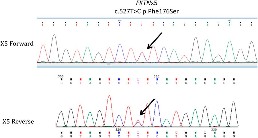
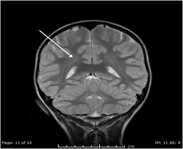
Our patient was a 14-year-old boy who had been relatively hypotonic since birth with mildly delayed gross motor development noted in his limited ability to run and climb stairs compared with his peers. By 4 years of age, the patient exhibited 30% delay in locomotion as well as the Gower sign. He was able to climb and descend stairs but non-reciprocally without a rail. When climbing stairs, he placed his right hand on his right knee for upper body support, and when he descended stairs he turned to the side to descend. Initial blood tests demonstrated elevated levels of creatinine phosphokinase. The patient’s younger brother was the first to present with signs of muscular dystrophy and underwent a muscle biopsy that served to diagnose both siblings as they were both displaying similar gross motor delays. The younger brother’s muscle biopsy revealed reduced levels of α-dystroglycan with microscopic atrophic and hypertrophic fibres as well as scattered areas of necrosis. Immunofluorescent staining of the fibres showed significantly reduced staining of α-dystroglycan, confirming the diagnosis of congenital muscular dystrophy. Our patient’s mother underwent genetic sequencing, which showed that she is a carrier for Fukuyama congenital muscular dystrophy (see Fig 1). As the mother is asymptomatic, it is possible that a second mutation occurred in the germ cells given the phenotypes of both siblings. A brain MRI was performed because of the association of muscular dystrophies with neurocognitive manifestations. The brain MRI showed bright hyperintensity on T2 imaging in the parietal and frontal lobes of unknown significance (see Fig 2). The patient’s paediatric metabolism specialist referred the patient to paediatric cardiology specialists because of the association of congenital muscular dystrophy with cardiomyopathy.
The genetic sequencing performed on the patient’s mother. It demonstrates a heterogenous missence mutation (T>C) in exon 5 on the FKTN gene leading to the substitution of phenylalanine for serine. This mutation, when present in two copies, is expected to be causative for congenital muscular dystrophy. This study has demonstrated that the mother is a carrier for the FKTN gene mutation.

A brain MRI, T2-weight image performed during the initial presentation of the patient’s symptoms at the age of 4 years. The arrow demonstrates an area of hyperintensity, which may be a variant of normal.

Upon first presentation to paediatric cardiology at the age of 4 years, the patient was noted to have a normal sinus rhythm on electrocardiogram as well as a normal ejection fraction on echocardiography. Other paediatric subspecialty groups including ophthalmology, pulmonology, and orthopaedic subspecialty services saw the patient in the outpatient setting for other complications associated with his congenital disorder. Between the age of 4 years and 10 years, he was seen every 2 years, but after 10 years of age he was followed-up every year. His mother reports that he compensated very well and followed the outpatient physical therapy and occupational therapy. He never required any orthotic devices, but his exercise stamina was much less compared with his peers. His mother reported that he had difficulty climbing two flights of stairs at baseline. The family installed a chair lift and obtained a scooter to enhance his mobility.
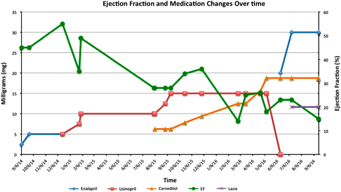
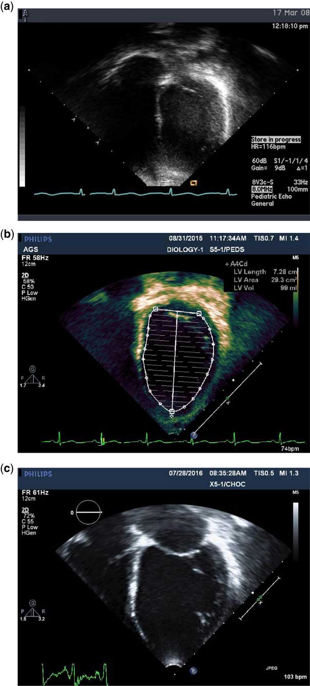
During a routine annual follow-up appointment in September, 2014, a screening echocardiogram showed decreased ejection fraction of 42% in the absence of any reported symptoms. He was started on 2.5 mg of enalapril two times a day. Serial echocardiograms in the outpatient setting every 1–2 months demonstrated a further decrease in the ejection fraction, and several sequential changes were made to his medication regimen (see Fig 3). Our initial strategy was to start with monotherapy from the beginning and progress to dual therapy as a second line. Over time, his echocardiogram – May, 2016 – showed an ejection fraction of 18% with an increased end-diastolic left ventricular filling volume of 153 ml and an indexed volume of 108 ml/m2, which were more than 2 standard deviations above normal. After August, 2015, his symptoms included weakness with walking and difficulty in running, but he denied any chest pain, cyanosis, dizziness, pallor, palpitations, or syncope. Cardiopulmonary stress testing, performed on December, 2015, revealed a moderately reduced exercise capacity with an observed normal chronotropic response up to 77% of maximum heart rate for age. No arrhythmias or ischaemic changes were noted on the electrocardiogram during the stress test. He had been admitted to the cardiovascular intensive care unit every 4–6 months starting from August, 2015 for milrinone maintenance therapy (1 mcg/kg/minute×2–3 days) and medication adjustment. In September, 2016, his ejection fraction decreased to 15%, and his parents reported that he had a much lower exercise tolerance than his previous baseline. Figure 4 demonstrates the progression of his dilated left ventricle from first presentation to immediately before transplantation. As a result, he was placed on the transplant list and waited for approximately 5 weeks before receiving orthotopic heart transplantation. He tolerated the procedure well, and his postoperative course was complicated by seizures and acute kidney injury. He recovered some strength but has not fully reached his previous baseline level of functioning. His postoperative weakness may be a function of diffuse muscle atrophy secondary to reduced activity before transplantation.
The change in ejection fraction over time with the changes/transitions in medications prescribed for treatment. As the ejection fraction decreased, higher doses of angiotensin converting enzyme inhibitors (lisinopril or enalapril) and β-blockers (carvedilol) were added. Lasix was added shortly before the patient received the transplant.

The progression of left ventricular dilatation. (a) The first echocardiogram demonstrating the presenting left ventricular end diastolic volume (LVED volume 13 cm2). (b) A follow-up echocardiogram demonstrating a significant increase in left ventricular filling volume seven years later (LVED volume 29.3 cm2). (c) Last echocardiogram 1 year after the previous echo just before orthotopic heart transplantation (LVED volume 36.1 cm2).

Discussion
This case highlights how quickly cardiac function in patients with congenital muscular dystrophies can deteriorate, making frequent screening a priority. This patient was unusual because of his mild musculoskeletal symptoms, but there are reports of patients with Fukuyama congential muscular dystrophy who have the ability to walk and climb stairs.Reference Saito, Osawa and Wang 4 Our patient is one of the few individuals who have minimal musculoskeletal symptoms, but his heart function rapidly deteriorated over 1 year. As a result, one can argue that Fukuyama has greater similarity to Becker’s muscular dystrophy for its rapid rate of progression of cardiomyopathy. It is postulated that children with Becker muscular dystrophy have an earlier presentation of cardiomyopathy compared to children with Duchenne muscular dystrophy because of the reduced mobility of Duchenne patients. The reduced mobility has a cardioprotective effect by forcing the child to undergo lower cardiac strain.Reference Hoogerwaard, de Voogt and Wilde 7 The lack of symptoms can be misleading to practitioners, and therefore we strongly recommend that these patients be screened every 6 months, rather than yearly, after the age of 10 years to avoid missing this rapid, potentially asymptomatic decline. This recommendation, however, is based on our experience with a single patient, and we understand that an alternative approach may be necessary in clinics that have very high patient volumes. One can consider screening patients yearly and increase the frequency to every 6 months if there is any decrease in ejection fraction, increase in cardiac strain, or increase in left ventricular dilation.
The current literature recommends that treatment be initiated when the ejection fraction is less than 55%; however, recent studies and our experience challenge this practice and argue for earlier treatment.Reference Duboc, Meune and Pierre 8 Duboc et alReference Duboc, Meune and Pierre 8 demonstrated that recent advances in echocardiograms and MRIs can detect earlier occurrences of cardiac peak circumferential strain before a decrease in ejection fraction. They suggest that earlier prophylactic treatment for individuals under the age of 10 years could be beneficial.Reference Duboc, Meune and Pierre 8 We agree with this suggestion based on our own experience demonstrating the rapid decline. It is possible that prophylactic therapy can prolong cardiac function even further; however, further studies will help elucidate this.
Sometimes medication therapy is not enough to halt the progression of the disease, and patients may require heart transplantation. Our patient is the perfect example of an individual who required heart transplantation within 1 year of the initial decline even with therapy. It is possible that if the patient was not compliant, the rate may have been even faster. Other children who are not screened as frequently may have a similar rapid decline that goes unnoticed and may be too late. As a result, we remind practitioners of the fastidiousness of this disease, we recommend that parents be counselled early of the possibility for a heart transplant and its outcomes. Heart transplant outcomes have significantly improved over the years and have been shown to significantly enhance quality measures in patients with dilated cardiomyopathy.Reference Tsirka, Trinkaus and Chen 9 One study by Tsirka et al demonstrated an increase in 5-year survival from 50% without transplant to 80% with a transplant. Only 11/91 children in the study, however, had a dystrophin gene defect, and the subset multivariate analysis found that patients aged>12 years had the highest risk of mortality.Reference Tsirka, Trinkaus and Chen 9 It is possible that the age discrepancy may be confounded by the dystrophin mutations that present during adolescence. Therefore, parents should be counselled with caution that these optimistic statistics may not be truly applicable to their child.
At this time only supportive measures exist for patients with Fukuyama congenital muscular dystrophy, but there are other promising options that may develop soon. For patients with severe heart failure, home milrinone or dobutamine is frequently considered, but have several well-reported side effects. Other medications for home therapy are currently in development including idebenone, which is a benzoquinone with antioxidant properties that works to improve respiratory chain function and muscle cellular energy metabolism.Reference Buyse, Goemans and van den Hauwe 10 Idebenone has been shown to enhance cardiac function in the left ventricular inferolateral wall.Reference Buyse, Goemans and van den Hauwe 10
There are also promising advances in the field of pharmacogenetics. Pharmacogenetics is a type of precision medicine that tailors medical treatment to a patient’s exact genetic defect.Reference Jarmin, Kymalainen, Popplewell and Dickson 11 There are currently three popular methods that target specific genome mutations to correct the underlying disorder. These methods include antisense oligonucleotide-induced exon skipping, homing endonucleases, and recombinant adeno-associated viral vector dystrophin complementary deoxyribonucleic acid transfer.Reference Jarmin, Kymalainen, Popplewell and Dickson 11 There is hope that after studying a patient’s specific genetic defect, these techniques can be used for individualised treatment.
Conclusion
Fukuyama congenital muscular dystrophy is a progressive degenerative muscle disorder associated with cardiomyopathy in the second decade of life. Patients with less skeletal muscle involvement often have greater cardiac muscle involvement secondary to greater cardiac demands from activity. Therefore, we urge practitioners to screen patients with congenital muscular dystrophy with an echocardiogram every 6 months after age 10 to monitor this silent deterioration and start prompt treatment when indicated (current consensus: ejection fraction<55%). This frequency of screening, however, may not be feasible for some practices, so we would urge physicians to start screening patients yearly and increase the frequency to every 6 months if there is any decrease in ejection fraction, increase in cardiac strain, or increase in left ventricular dilation. We also believe that earlier/prophylactic therapy can benefit these patients, but more research will be needed to clarify these findings. The rate of cardiomyopathic progression can be rapid and occasionally resistant despite appropriate, aggressive medical treatment; therefore, parents should be counselled on the possibility of a heart transplant. Promising advances in pharmacogenetics may significantly alter the progression of these diseases.
Acknowledgements
The authors thank the patient’s parents for all their continued support. They were an invaluable source of information. The authors also thank Dr Jose E. Abdenur for his assistance. Audrey Gardner also contributed, and the authors thank her for her assistance in obtaining excellent echocardiographic images.
Financial Support
This research received no specific grant from any funding agency or from commercial or not-for-profit sectors.
Conflicts of Interest
None.
Ethical Standards
The authors assert that all work performed for the purpose of this study abide by national ethical standards.