Introduction
Reproductive efficiency, the ability of cows to become pregnant and produce a healthy live calf, is one the primary factors affecting productivity in the beef or dairy industry. Reproductive efficiency has been shown to be determined by a number of factors, including maternal environment and nutrition, embryo quality, heat stress (Bilodeau-Goeseels & Kastelic, Reference Bilodeau-Goeseels and Kastelic2003; Hansen, Reference Hansen2007) and maternal and/or paternal breeds (Fryer et al., Reference Fryer, Marion and Farmer1958; Fischer et al., Reference Fischer, Bernal, Gutierrez-Robayo and Rutledge2000; Casas et al., Reference Casas, Thallman and Cundiff2011).
Paternal effects on reproductive efficiency have been shown to operate through various mechanism, including influence on fertilization and embryonic survival rates (Giritharan et al., 2007), blastocyst apoptotic index (Jeong et al., Reference Jeong, Cui, Kim, Kim, Kim, Chung and Kim2005; Giritharan et al., 2007) and placental development and characteristics (Constancia et al., Reference Constancia, Hemberger, Hughes, Dean, Ferguson-Smith, Fundele, Stewart, Kelsey, Fowden, Sibley and Reik2002); however, the exact mechanisms are still unclear. There is a growing body of evidence which shows that new sets of epigenetic marks, established during both gametogenesis and in early embryonic development followed by genome-wide reprogramming, could significantly influence gene expression pattern in the embryo and beyond (for review, see Chason et al., Reference Casas, Thallman and Cundiff2011). In addition, some of the epigenetics marks that are established during the preimplantation period are stable during replication, and may affect gene expression at later stages of development (Dean et al., Reference Dean, Bowden, Aitchison, Klose, Moore, Meneses, Reik and Feil1998; Khosla et al., Reference Khosla, Dean, Reik and Feil2001; Shao et al., Reference Shao, Tao, Gao, Xie and Zhao2008).
Previous studies have provided strong evidence for the importance of expression of imprinted genes in embryonic (Rappolee et al., Reference Rappolee, Sturm, Behrendtsen, Schultz, Pedersen and Werb1992; Perecin et al., Reference Perecin, Meo, Yamazaki, Ferreira, Merighe, Meirelles and Garcia2009) and fetal development (Wu et al., 2004; Kwong et al., Reference Kwong, Miller, Ursell, Wild, Wilkins, Osmond, Anthony and Fleming2006). The maternally imprinted insulin-like growth factor II (IGF-II) gene on bovine chromosome 29 is one of the best characterized epigenetically regulated loci (Goodall & Schmutz, Reference Goodall and Schmutz2003; Gebert et al., Reference Gebert, Wrenzycki, Herrmann, Groger, Thiel, Reinhardt, Lehrach, Hajkova, Lucas-Hahn, Carnwath and Niemann2009). Insulin-like growth factors have been shown to play important roles in early embryonic (Rappolee et al., Reference Rappolee, Sturm, Behrendtsen, Schultz, Pedersen and Werb1992; Wang et al., Reference Wang, Feng, Ma, Cang, Li, Yan, Zhou, Wen, Bou and Liu2009), placental, and fetal development (Sibley et al., Reference Sibley, Coan, Ferguson-Smith, Dean, Hughes, Smith, Reik, Burton, Fowden and Constancia2004; Su et al., Reference Su, Yang, Wang, Li, Xiong, Wang, Guo and Zhang2011), probably through combined activation of multiple cell signaling pathways critical to survival, growth, and differentiation (Kim et al., Reference Kim, Song, Gao, Farmer, Satterfield, Burghardt, Wu, Johnson, Spencer and Bazer2008). In addition, the IGF-II gene provides an excellent model for studying RNA involvement in maintenance of nucleosome-retained regions of the sperm chromatin in spite of nearly complete replacement of DNA-bound histones with protamines (Jenkins & Carrell, Reference Jenkins and Carrell2011). It is suggested that retained histones may contain modifications that play a critical role in epigenetic regulation (Jenkins & Carrell, Reference Jenkins and Carrell2012). Accordingly, given that epigenetic patterns govern gene expression (for review see Niemann et al., Reference Niemann, Tian, King and Lee2008), it can be hypothesized that paternal breed effect on reproductive efficiency is mediated through, at least in part, influence on expression levels of IGF-II gene.
The aim this study was to assess the effects of paternal breed on the early embryo development and relative expression of IGF-II in day 8 bovine blastocysts. As a second objective, we examined whether the paternal breed influences blastocyst quality, which is considered the most reliable factor influencing embryo viability, by analysis of blastocyst transcript abundance of selected apoptosis-associated genes such as BCL2-L1 (anti-apoptotic) and BAK1 (pro-apoptotic) (Jeong et al., Reference Jeong, Cui, Kim, Kim, Kim, Chung and Kim2005).
Materials and methods
Chemicals and reagents were obtained from Sigma-Aldrich Co. (St. Louis, MO, USA) unless otherwise stated.
In vitro production (IVP) of embryos
Oocyte collection and in vitro maturation (IVM)
Bovine ovaries, originating mainly from culled Danish dairy cows and heifers, were obtained from a local abattoir, (Herlufmagle Slaughtery, Denmark) and transported to the laboratory within 3 h of retrieval in 0.9% physiological saline solution at 29–32°C in a thermocontainer. The temperature from ovary collection throughout all manipulations outside the incubator was kept within this temperature range. Immature cumulus–oocyte complexes (COCs) were retrieved from antral follicles (2–20 mm in diameter) by use of a vacuum pump (KNF-Neuberger N86 KN.18, VWR International, Copenhagen, Denmark) connected with an 18-gauge needle (18-G × 1.5, 1.2 × 40, Terumo Neolus Luer) and collected into a 50-ml tube containing 140 μl heparin (5000 i.u./ml, Leo Pharma, Ballerup, Denmark). The COCs were retrieved and washed three times in HEPES-buffered TCM-199 wash medium (M 2520, Sigma) supplemented with, 50 μg/ml gentamycin (G 3632, Sigma) and 0.5 mg/ml bovine serum albumin (BS; A4919, Sigma). The COCs were washed once after retrieval in in vitro maturation (IVM) medium before maturation. Groups of 35–40 COCs were placed in 500 μl maturation medium in four-well dishes (Nunc, Roskilde, Denmark) and cultured without oil overlay for 24 h at 38.8 ± 0.2°C in 5% CO2 in ambient humidified air. The IVM medium consisted of Dulbecco's Eagle's Modified Medium (DMEM; D6046, Sigma) supplemented with 5% fetal calf serum (FCS; produced by the Danish Veterinary Institute, Copenhagen, Denmark), 15 IE/ml equine serum gonadotrophin (PMSG)-HCG (Suigonan, VetPharm, Løgstør, Denmark), 0.2 mM pyruvate (P3662, Sigma), 50 ng/ml epidermal growth factor (EGF; E4127, Sigma) and 50 μg/ml gentamicin (G1264, Sigma).
Sperm preparation and in vitro fertilization (IVF)
The COCs were evaluated for cumulus expansion and viscoelasticity and scored from 0–3 on an arbitrary scale and transferred in groups of 40 into four-well dishes containing 400 μl fertilization TALP medium. All experiments were carried out using frozen–thawed semen from proven unrelated high fertile Brown Swiss and Holstein bulls, (both from Viking Genetics, Randers, Denmark). Straws containing frozen semen from the test bulls were thawed at 37°C and washed twice by centrifugation at 328 g for 5 min in non-capacitation medium (NCM: 0.5 mM NaHCO3 (S 4019, Sigma); 113 mM NaCl (S 5886, Sigma); 4.78 mM KCl (P 5405, Sigma); 1.19 mM MgSO4 (M 2643, Sigma); 1.2 mM KH2PO4 (P 5655, Sigma); 21.6 mM sodium lactate (L4263, Sigma); 50 μg/ml gentamicin (G1264, Sigma); pH 7.4). The spermatozoa were counted in a Makler chamber and diluted in Sperm-TALP to give a concentration of 50 × 106 sperm cells/ml. An appropriate volume of this suspension was added to fertilization wells to obtain a final concentration of 2 × 10⁶ spermatozoa/ml. The spermatozoa were co-incubated with matured COCs in TALP medium for 22–24 h without oil overlay at 38.8 ± 0.2°C in 5% CO2 in ambient humidified air. The TALP medium contained 25 mM sodium bicarbonate (S4019, Sigma), 10 mM lactate (L4263, Sigma), 6 mg/ml BSA (A4919, Sigma), 0.25 mM pyruvate (P3662, Sigma), 30 μg/ml heparin (H3149,Sigma), 50 μg/ml gentamicin (G1264, Sigma), 20 μM penicillamine (P4875, Sigma), 10 μM hypotaurine (H1384, Sigma), and 1 μM epinephrine (E4250, Sigma).
Embryo culture after fertilization
After 22–24h of in vitro fertilization (IVF), the inseminated oocytes were denuded of surrounding cumulus cells and excess spermatozoa were removed by vortex agitation. After three washes in HEPES-buffered tissue culture medium (TCM-199, M 2520, Sigma), the zygotes were washed once in culture medium (BA1216 medium, Origio, Måløv, Denmark). The inseminated oocytes were cultured in groups of 35 in 500 μl culture medium (BA1216 medium) with an overlay of oil in low oxygen, 5% O2 and 5% CO2, at 38.8 ± 0.2°C for 168 h. On day 8, embryos from all groups were scored for the rate of blastocyst development (day 0 = IVF). The resulting blastocyst rates were expressed per inseminated oocyte placed into in vitro culture (IVC). Day 8 blastocysts were used for mRNA transcript abundance analysis using reverse-transcription–qualitative polymerase chain reaction (RT-qPCR).
Gene expression analysis
Isolation of total RNA and cDNA synthesis
In vitro-produced day 8 blastocysts were first transferred from the culture medium into TCM-199 and groups of 10 blastocysts were rinsed three times in phosphate-buffered saline (PBS), and finally transferred into 500-μl centrifuge tubes with a minimal amount of PBS. Samples were immediately snap-frozen in liquid nitrogen and stored at –80°C until RNA isolation. Total RNA was isolated from a pool of eight in vitro produced (IVP) blastocysts using a NucleoSpin® RNA XS Kit (Clontech, Mountain View, CA, USA) according to the manufacturer's instructions. To eliminate any contaminating DNA, a DNase digestion step was performed. The quantity and purity (260 nm/280 nm ratio) of total RNA was estimated using a NanoDrop ND-1000 spectrophotometer (Thermo-Scientific, Wilmington, DE, USA). The total RNA of each pool was reverse transcribed into cDNA using RevertAid™ First Strand cDNA Synthesis Kit (Thermo-Scientific) with random hexamer primers according to the manufacturer's instructions. The reverse-transcription reaction was carried out in thermo-cycler using the following temperature conditions: 25°C for 10 min, 42°C for 60 min, 95°C for 5 min, and cooled down to 4°C.
Primer design
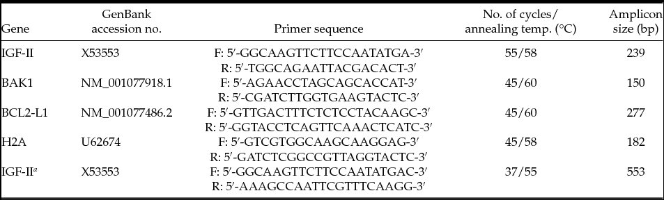
PCR primers except H2A and BCL2-L1 were designed using Primer Premier 5 software (Premier Biosoft International, Palo Alto, CA, USA) based on public transcript and genomic sequence data from cattle, mice and humans (see Table 1). Primer sequences for H2A and BCL2-L1 genes were obtained from previously published data (Robert et al., Reference Robert, McGraw, Massicotte, Pravetoni, Gandolfi and Sirard2002, Hwang et al., Reference Hwang, Park, Moon, Shim, Kim, Yang, Ko, Yang, Cheong and Im2008).
Primer sequences and cycling conditions used in quantitative real-time PCR and for sequencing

aThis primer pair was designed for amplification of the bovine IGF-II DMR located at exon10 for DNA sequencing.
For each gene analyzed, a BLAST search (http://www.ncbi.nlm.nih.gov/BLAST) also was conducted to ensure the specificity of the designed primers and to assure that they were not designed from any homologous regions, coding for other genes. The primer sequences, expected fragment sizes and the sequence references are shown in Table 1.
Reverse-transcription quantitative real-time PCR (RT-qPCR)
Relative expression levels were quantified in real time on a LightCycler® 480 Instrument (Roche Diagnostics, Indianapolis, IN, USA) with LightCycler® 480 SYBR Green I Master mix (Roche Applied Science, Germany) as detection chemistry. The PCR mixture consisted of 2 μl diluted cDNA (10 ng), 0.5 μl forward and reverse primers (10 mM), and 5 μl SYBR Green I master mix, and 2 μl water to a total volume of 10 μl. PCR reactions were performed according to the manufacturer's instructions. First, cDNA was denatured and pre-incubated by heating for 10 s at 95°C. Template was then amplified by 45–55 cycles consisting of denaturation for 10 s at 95°C, annealing for 10 s at 58–60°C, and extension for 20 s at 72°C. At least three replicates were used for each reaction, and a reaction without template served as negative control. To verify RT-PCR product identity, melting point curves were analyzed following amplification.
The relative quantification of gene expression was analyzed by the comparative Cp method (Pfaffl et al., Reference Pfaffl, Horgan and Dempfle2002), using LightCycler 480 software (release 1.5.0 SP4). Quantification was normalized to the endogenous control, H2A (Robert et al., Reference Robert, McGraw, Massicotte, Pravetoni, Gandolfi and Sirard2002). The mathematical model used is based on the PCR efficiencies and the crossing point deviation between targets and reference gene of each sample. The PCR product sizes were confirmed by gel electrophoresis on a standard 2% agarose gel stained with ethidium bromide and visualized by exposure to ultraviolet light.
DNA extraction from sperm and amplification
Genomic DNA was extracted from frozen/thawed sperm using DNeasy Blood and Tissue kit (Qiagen, Hilden, Germany) according to the manufacturer's protocol. After extraction, the quality and quantity of total DNA was determined using a NanoDrop spectrophotometer. Sperm DNA was amplified in the differentially methylated region (DMR) and shore of exon 10 of the bovine IGF-II gene using primers (Table 1). This region included all 27 putative CpG sites between nucleotides 257 and 676 bp of the coding region (Gebert et al., Reference Gebert, Wrenzycki, Herrmann, Groger, Reinhardt, Hajkova, Lucas-Hahn, Carnwath, Lehrach and Niemann2006). Amplification was performed in a 25-μl reaction volume, using Taq PCR master mix (Qiagen), which included 1 μl of sperm DNA, 10 mM each primer, 12.5 μl of Taq PCR master mix and 10.5 μl RNase-free water. The temperature cycles were as follows: 95°C for 5 min, followed by 37 cycles of denaturation at 94°C for 45 s, annealing at 58°C for 45 s, extension at 72°C for 45 s, and a final extension at 72°C for 10 min. Amplified gene-specific PCR products were analyzed on a 2% agarose in 1× TBE buffer gel containing 0.2 mg/ml ethidium bromide (EtBr) which were run for 40 min at 90 V. A 100-bp DNA ladder (Fermentas, Germany) was used as a size marker to determine the size of PCR products.
Sequencing of bovine DMR2 DNA Fragments
PCR products were separated by gel electrophoresis and gel bands were cut out and purified using the Wizard® SV Gel and PCR Clean-up kit (Promega, Fitchburg, WI, USA) according to the manufacturer's protocol to remove primers and dNTPs, and then sequenced to detect possible differences across samples using commercial sequencing service (DNA Technology, Risskov, Denmark).
Statistical analysis
The IVP experiments were repeated four times. Relative quantification of target gene expression levels as fold-differences was based on at least three sets of biological samples and three technical samples. Embryos development data were analyzed with the chi-squared test using logistic regression model. The percentage of cleaved embryos and blastocysts formed are presented as the mean ± standard error of the mean (SEM).
Linear mixed model (Chen et al., Reference Chen, Delongchamp, Tsai, Hsueh, Sistare, Thompson, Desai and Fuscoe2004) followed by multiple pair-wise comparisons using the Student–Newman–Keuls multiple comparisons test was used for analysis of variance components of gene expression data obtained by RT-qPCR. The following mixed model was used was for analysis gene expression data: y ijk = μ + S i + O j + K ij + e ijk, where: y ijk is the gene expression in blastocysts, μ is the overall mean, S i is the fixed effect of the sperm genotype (1, 2), O j is the random effect of the batch of ova from ovary collected in each replicate, K ij is the interaction effect of sperm genotype and batch of oocyte, and e ijk is the random residual error. Pearson's pair-wise correlation coefficient was used to determine the degree of correlation between in vitro embryo development and gene expression in blastocyst stage embryos. Data were statistically analyzed using the computer program JMP 7.0 (SAS Institute Inc., Cary, NC, USA).
Results
In the present study, the embryonic developmental competence and mRNA expression profiles of IGF-II, BAK1 and BCL2-L1 were compared in bovine preimplantation embryos derived from two different paternal genetic backgrounds (breeds).
In four replicates, in total 897 oocytes were utilized for IVP of bovine embryos using sperm from two different bulls’ breeds: Holstein and Swiss Brown. The cleavage and blastocyst rates are presented in Table 2. No statistical differences were found in both cleavage and blastocyst rates between the two sperm breeds derived embryos (P > 0.05) (Table 2).
Cleavage and blastocyst rates following IVP using semen from either Holstein or Brown Swiss bullsa

aFour replicated trials were carried out, and blastocysts were analyzed on day 8.
The blastocyst rate was calculated from the number of inseminated oocytes.
Values with same letters are not statistically different (P > 0.05).
IVP, in vitro production; SEM, standard error of the mean.
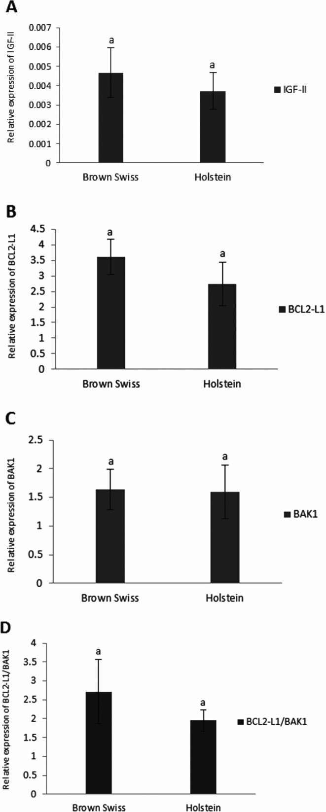
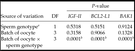
The mRNA expressions of IGF-II, BAK1, and BCL2-L1 genes were determined by RT-qPCR. The genes selected for the analysis have been shown to play decisive roles in the regulation and execution of apoptosis. There was no significant effect of sperm breeds and/or batch of oocytes on relative expression of mRNA of IGF-II (Fig. 1A), BCL2-L1 (Fig. 1B), BAK1 (Fig. 1C) and the ratio of BCL2-L1/BAK1 (Fig. 1D), however relative expression of IGF-II, BCL2-L1 and BAK1 were significantly influenced by the interaction between sperm genotype and four separate batches of oocytes (P < 0.05) (Table 3).
Levels of IGF-II (A), BCL2-L1 (B), BAK1 (C) and ratio of BCL2-L1/BAK1 (D) expression in day 8 bovine blastocysts following in vitro production (IVP) using semen from either Holstein or Brown Swiss bulls (mean ± standard error of the mean (SEM)). Bars with same letters are not statistically different (P > 0.05).
Analysis of variance for IGF-II, BAK1 and BCL2-L1 gene expression in blastocysts derived from two unrelated sperm origins
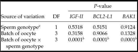
aSperm genotype was considered a fixed effect while all other effects were considered random.
bInteraction between sperm genotype and batch of oocyte significantly influence on gene expression (P < 0.05).
DF = degree of freedom.
Pearson's pair-wise correlation coefficient shows that the blastocyst production rate was positively correlated (r = 0.66, P < 0.1) with IGF-II gene expression levels of the blastocyst. Furthermore, level of IGF-II expression highly correlated with BCL2-L1 (r = 0.72, P < 0.05) and ratios of BCL2-L1/BAK1 (r = 0.78, P < 0.05), however level of IGF-II expression had no correlation with BAK1 expression.
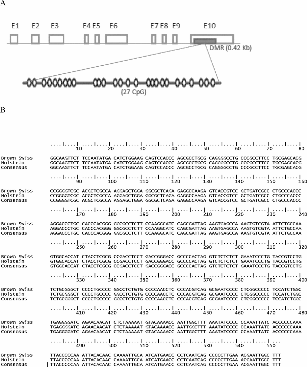
In order to explore breed-specific genetic markers and determination of their potential role in explanations of interaction between sperm breeds and batches of oocytes and/or environment (stochastic extrinsic factors in replicate) on the IGF-II expression pattern, we compared sequences of DMR within the last exon of the bovine IGF-II gene between Holstein and Brown Swiss sperm (Fig. 2A). The resulting DNA sequences were compared with GenBank database entries using a BLAST search. DNA sequence alignment analysis indicated that no sequence differences exist between the analyzed sperm DNA (Fig. 2B).
(A) Molecular structure of the bovine IGF2 gene. Differentially methylated regions (DMRs) in the last exon of bovine IGF-II containing 27 CpG islands is indicated in the blue box. (B) Alignment with the previously published sequence of bovine IGF-II gene (GenBank accession no. X53553) revealed no sequence differences between the analyzed sperm DNA.
Discussion
The preimplantation period of mammalian embryogenesis is notable for many critical events that affect the genome both before and after implantation. During this time, reshuffling of the parental genomes, as well as genome-wide epigenetic reprogramming provide a new set of instructions that guide both early embryonic and life-long growth and development (Chason et al., Reference Chason, Csokmay, Segars, DeCherney and Armant2011).
In the present study, we found that sperm species neither influenced the rate of fertilization as assessed by the cleavage rates (P > 0.0.5), nor blastocyst formation rate (P > 0.0.5) (Table 2). These results are in agreement with those reported in other breeds of cattle in which Holstein oocytes have been fertilized with semen from Nelore or Angus bulls (Barros et al., Reference Barros, Pegorer, Vasconcelos, Eberhardt and Monteiro2006; Eberhardt et al., Reference Eberhardt, Satrapa, Capinzaiki, Trinca and Barros2009). In contrast, several studies reported differences in cleavage and/or blastocyst formation rate between sire breeds; i.e. Nelore oocytes fertilized with semen from Nelore or Angus bulls (Barros et al., Reference Barros, Pegorer, Vasconcelos, Eberhardt and Monteiro2006; Eberhardt et al., Reference Eberhardt, Satrapa, Capinzaiki, Trinca and Barros2009) or Holstein oocytes, fertilized with semen from Holstein or brown Swiss bulls (Lazzari et al., Reference Lazzari, Colleoni, Duchi, Galli, Houghton and Galli2011). The cause of this discrepancy is not clear, but it could be attributed to the inbreeding depression (Lazzari et al., Reference Lazzari, Colleoni, Duchi, Galli, Houghton and Galli2011) or species-specific sperm–egg interactions (Fischer et al., Reference Fischer, Bernal, Gutierrez-Robayo and Rutledge2000; Zi et al., Reference Zi, Yin, Chen, Liang, Zhang and Guo2009). Using norm-finder software, we found that the mRNA levels of bovine histone H2A are more constant across the replicate than bovine B-actin (ACTB) and ubiquitin B (UBB) (data not shown). Our data are in agreement with reports showing that mRNA levels of bovine histone H2A are more constant across the preimplantation period than other endogenous housekeeping genes (Robert et al., Reference Robert, McGraw, Massicotte, Pravetoni, Gandolfi and Sirard2002; Lonergan et al., Reference Lonergan, Rizos, Gutierrez-Adan, Moreira, Pintado, Fuente and Boland2003a; Vigneault et al., Reference Vigneault, Gilbert, Sirard and Robert2007).
The experimental results and statistical analysis show that neither the sperm breed, nor the oocyte population (batch of ova) affected IGF-II, BAK1 and BCL2-L1 expression (P > 0.05). In contrast, their interaction significantly influence on the level of IGF-II, BAK1 and BCL2-L1 expression in blastocysts (P < 0.05) (Table 3). Overall, the lack of significant effects of sperm breed and the presence of significant interactions tend to imply that the IGF-II, BCL2-L1and BAK1 expression can be modulated by heritable and non-heritable factors. Precise interpretation of gene expression results is difficult given the probable differences in oocyte populations (breed, age, quality) and/or stochastic extrinsic factors connected with the experimental procedure (environment factors). However, numerous studies have demonstrated that variable culture conditions [due to maturation (Warzych et al., Reference Warzych, Wrenzycki, Peippo and Lechniak2007) and/or culture media composition (Natale et al., Reference Natale, Sousa, Westhusin and Watson2001), incubation temperatures (Jin et al., Reference Jin, Lee, Choi, Kim, Cui and Kim2007; Yadav et al., Reference Yadav, Singh, Singh, Saini, Palta, Manik, Singla, Upadhyay and Chauhan2013), oxygen concentration (Harvey, Reference Harvey2007) and media osmolality (Hwang et al., Reference Hwang, Park, Moon, Shim, Kim, Yang, Ko, Yang, Cheong and Im2008)] can lead to alteration in expression levels of developmentally important genes in in vitro developed embryos. Along with these reports, Feinberg & Irizarry have recently developed an evolutionary model in which DNA mutations that do not change the mean phenotype could modify the variability of phenotype; and this could be mediated epigenetically through tissue-specific differentially methylated regions (T-DMRs) (Feinberg & Irizarry, Reference Feinberg and Irizarry2010). In consistence with their evolutionary model, a set of orthogonal contrasts within replicates among the transcript level of the endogenous IGF-II in blastocysts derived from cattle oocytes fertilized either with Brown Swiss or Holstein spermatozoa, shows significant differences in levels of IGF-II expression (p < 0.05); Thus for investigation of this likelihood we explored the sequence variation at a recently identified intragenic DMR within the last exon (exon10) of the bovine IGF-II gene (Gebert et al., Reference Gebert, Wrenzycki, Herrmann, Groger, Reinhardt, Hajkova, Lucas-Hahn, Carnwath, Lehrach and Niemann2006, Reference Gebert, Wrenzycki, Herrmann, Groger, Thiel, Reinhardt, Lehrach, Hajkova, Lucas-Hahn, Carnwath and Niemann2009). The sequencing result revealed that no differences existed in this DMR of IGF-II between the analyzed sperm samples, which may imply that these regions are conserved in evolution (Gebert et al., Reference Gebert, Wrenzycki, Herrmann, Groger, Reinhardt, Hajkova, Lucas-Hahn, Carnwath, Lehrach and Niemann2006; Han et al., Reference Han, Lee and Szabo2008a). In addition, the effects of certain combinations of parental alleles (dominance, over dominance and epistatic components) on propensity to blastomere cytofragmentation and/or embryo development which are well documented in the cow and mouse, may partially explain these interactions (Han et al., Reference Han, Mtango, Patel, Sapienza and Latham2008b; Zi et al., Reference Zi, Yin, Chen, Liang, Zhang and Guo2009).
The present study has shown for the first time a strong correlation between BCL2-L1/BAK1 ratios and endogenous IGF2 expression at the bovine blastocyst stage. The ratio of BCL2-L1 to BAK1 has been shown to be a critical determinant of cell fate (Willis et al., Reference Willis, Chen, Dewson, Wei, Naik, Fletcher, Adams and Huang2005) such that elevated BCL2-L1 favors extended survival of cells and increasing levels of BAK1 expression accelerate cell death (Jeong et al., Reference Jeong, Cui, Kim, Kim, Kim, Chung and Kim2005; Hwang et al., Reference Hwang, Park, Moon, Shim, Kim, Yang, Ko, Yang, Cheong and Im2008). Typically, BCL2-L1 transcripts are more abundantly expressed in in vivo rather than in vitro produced embryos and/or morphologically grade 1 than grade 2 blastocyst (which are generally accepted to differ in their developmental potential) (Jeong et al., Reference Jeong, Cui, Kim, Kim, Kim, Chung and Kim2005; Gyu-Jin Rho et al., Reference Gyu-Jin, Balasubramanian, Dong-Sik, Woo-Jin, Sang-Rae, Jung-Gon, Mohana kumar and Sang-Young2007; Lloyd et al., Reference Lloyd, Romar, Matas, Gutierrez-Adan, Holt and Coy2009). These correlations tend to imply that the level of endogenous IGF-II may act as an anti-apoptotic factor by enhanced expression of the anti-apoptotic gene BCL2-L1 and influence the ratio of anti-apoptotic to pro-apoptotic related gene expression. This observation supported previous findings that during preimplantation development in vitro, IGF-II reduce apoptosis and increase embryo viability in bovine (Byrne et al., Reference Byrne, Southgate, Brison and Leese2002) and mouse embryos (Kurzawa et al., Reference Kurzawa, Glabowski, Baczkowski, Wiszniewska and Marchlewicz2004; Glabowski et al., Reference Glabowski, Kurzawa, Wiszniewska, Baczkowski, Marchlewicz and Brelik2005).
The mechanism whereby IGF-II interferes with the pro-apoptotic signaling cascade triggered by intrinsic or extrinsic factors is unclear. However, anti-apoptotic effects of IGF-II are considered mainly mediated through the IGF-IR (Nussbaum et al., Reference Nussbaum, Samarin, Ehemann, Bissinger, Ryschich, Khamidjanov, Yu, Gretz, Schirmacher and Breuhahn2008), which is expressed in bovine embryos from zygote to blastocyst stages (Yaseen et al., Reference Yaseen, Wrenzycki, Herrmann, Carnwath and Niemann2001; Lonergan et al., Reference Lonergan, Rizos, Gutierrez-Adan, Moreira, Pintado, Fuente and Boland2003a). It has been demonstrated that activation of phosphatidylinositol 3-kinase (PI3K)-AKT/protein kinase B (PKB), upon binding of IGF-II to insulin-like growth factor receptor (IGF-1R), could in turn lead to activation of transcription of the anti-apoptotic BCL2-L1 gene, which can prevent the apoptosis through binding to pro-apoptotic BAK1 (Willis et al., Reference Willis, Chen, Dewson, Wei, Naik, Fletcher, Adams and Huang2005). Furthermore, activation IRS-mediated pathway upon binding of IGF-II to insulin-like growth factor receptor (IGF-1R), could lead to the phosphorylation of BCL2-associated agonist of cell death (BAD) (Peruzzi et al., Reference Peruzzi, Prisco, Dews, Salomoni, Grassilli, Romano, Calabretta and Baserga1999; Vincent & Feldman, Reference Vincent and Feldman2002), which has been shown to neutralize BCL2-L1's anti-apoptotic function through heterodimerization. However, in its phosphorylated form, BAD cannot bind to BCL2-L1 protein (Peruzzi et al., Reference Peruzzi, Prisco, Dews, Salomoni, Grassilli, Romano, Calabretta and Baserga1999; Fu et al., 2000). Hence, either one or all of these mechanisms may support our correlation analysis results.
In the present study, the levels of IGF-II expression were positively correlated (r = 0.66, P < 0.1) with in vitro embryo development measured by blastocyst production rates, indicating that the viability of embryos is represented by IGF-II expression. This observation supports previous finding by Rappolee et al. (Reference Rappolee, Sturm, Behrendtsen, Schultz, Pedersen and Werb1992), which reported that the amount of endogenous IGF-II transcripts is directly correlated with the development competence of cleaving zygotes. This result is consistent with those found in previous reports showing that the developmentally more competent embryos had higher IGF-II mRNA concentrations than their lower developmentally competent counterpart at the blastocyst stage (Yaseen et al., Reference Yaseen, Wrenzycki, Herrmann, Carnwath and Niemann2001; Lonergan et al., Reference Lonergan, Rizos, Gutierrez-Adan, Moreira, Pintado, Fuente and Boland2003a, Reference Lonergan, Rizos, Kanka, Nemcova, Mbaye, Kingston, Wade, Duffy and Boland2003b).
In conclusion, our data show that IGF-II, BCL2-L1 and BAK1 expression is not related to the chosen combination of paternal breed, but that IGF-II expression is correlated with embryonic viability and apoptosis-related gene expression. Furthermore, our results indicate that a mechanism contributing to the anti-apoptotic effects of IGF-II might be connected with the change in ratio between the mRNA levels of the pro-apoptotic BAK1 and the anti-apoptotic BCL2-L1 genes in bovine blastocysts.
Acknowledgements
The authors thank Anne Friis for assistance with RT-qPCR and Lene Markussen for assistance with in vitro embryo production.
Declaration of interest
The authors declare that there is no conflict of interest that could be perceived as prejudicing the impartiality of the research reported.
Financial Support
This research received no specific grant from any funding agency, commercial or not-for-profit sectors.