Introduction
The term perilymphatic fistula refers to a communication from the inner ear to the middle ear, which occurs due to disruption of the natural bony or soft tissue barrier between the perilymph and the middle-ear spaces.
Perilymphatic fistulas may be categorised as spontaneous (idiopathic) or post-traumatic. Spontaneous perilymphatic fistulas, by definition, have no predisposing events or factors and are often associated with congenital anomalies of the inner ear. Post-traumatic perilymphatic fistulas include those occurring due to: erosive tumours or cholesteatomas; iatrogenic leaks following otological surgery; or head trauma, barotrauma or acoustic injury. Clinical manifestations mainly appear as auditory and vestibular symptoms and signs.Reference Hirsch and Myers1, Reference Goto, Ogawa, Kunihiro, Kurashima, Kobayashi and Kanzaki2
Tympanostomy tube placement is frequently associated with certain complications, which have been well documented and are globally recognised, occurring in 17 per cent of patients.Reference Groblewski and Harley3, Reference Derkay, Carron, Wiatrak, Choi and Jones4 The most common of these complications include otorrhoea, tympanosclerosis and permanent perforation; cholesteatoma is rare but more serious. An additional complication is the medial migration of the tympanostomy tube into the middle-ear space. This has been reported to occur in 0–1.1 per cent of cases.Reference Groblewski and Harley3 However, there is limited published information on this complication, and the optimal management of such cases is not universally agreed upon.
Case report
A 65-year-old man attended the out-patient clinic complaining of aural fullness and hearing loss in his right ear and transient episodes of dizziness. There were no symptoms of otalgia, ear discharge or tinnitus. The patient had been diagnosed with nasopharyngeal carcinoma two years previously, for which he had been treated successfully with a combination of radiotherapy and chemotherapy. As a result of his treatment, he had developed a right middle-ear effusion; a few months later, he had undergone insertion of a Shah tympanostomy tube. Records for his regular follow up indicated that he had been disease-free for 25 months after the initial treatment.
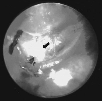
Careful microscopic examination revealed the existence of a Shah equalisation tube in the patient's right middle ear, lying behind an intact tympanic membrane (Figure 1). No middle-ear effusion was noted. The rest of the ENT examination was unremarkable.
Tympanostomy tube (arrow) observed in the middle ear, behind an intact tympanic membrane.

The patient underwent tympanography, which showed a type C pattern, and pure tone audiography, which revealed mild to moderate sensorineural hearing loss in the right ear.
The differential diagnosis, based on the clinical findings and investigation results, included middle-ear effusion and perilymphatic fistula.
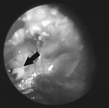
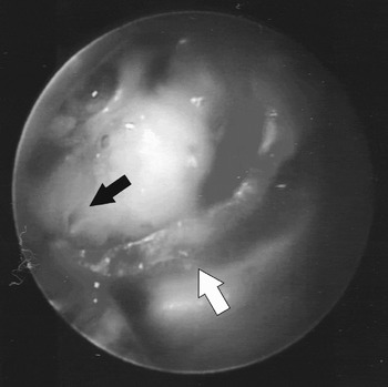
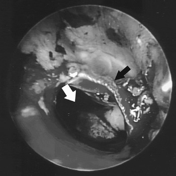
The patient underwent exploratory tympanotomy. A tympanomeatal flap was elevated and the grommet was recovered with ease, with no resistance encountered on removal (Figure 2). When the grommet was removed, a defect was noted in the bony medial wall against which the grommet had been lying (Figure 3). On further inspection, clear fluid was observed to flow from the defect into the middle-ear space. It was therefore assumed that the most likely diagnosis was traumatic perilymphatic fistula, presumably caused by gradual erosion of the bony wall. It was hypothesised that this erosion might be related to some type of radiotherapy-induced inflammatory process. The leak was repaired using tragal fat, temporalis fascia and Tisseel fibrin glue (Tisseel® Kit VH Fibrin Sealant; Baxter, Health Care Corporation, Irvine, CA) and the tympanomeatal flap was replaced (Figure 4).
Elevation of tympanic membrane, revealing the tympanostomy tube (black arrow) in the middle ear; the suction tip (white arrow) is also visible.

Removal of tympanostomy tube, exposing a perilymphatic fistula (black arrow); the tympanomeatal flap (white arrow) is also visible.

The fistula repaired (white arrow); the tympanomeatal flap (black arrow) is also visible.

Six weeks post-operatively, the patient was asymptomatic but his hearing remained unchanged from the pre-operative state.
Discussion
There are few clinical reports describing the incidence of perilymphatic fistula. House et al. Reference House, Morris, Kramer, Shasky, Coggan and Putter5 estimated the incidence of perilymphatic fistula to be less than 1 per 1000 otolaryngology out-patient visits. In all age groups, 50 per cent of perilymphatic fistulas are caused by otological surgery and trauma.Reference Seltzer and McCabe6
Clinical history and examination are the most important factors in the diagnosis of traumatic perilymphatic fistula.Reference Hughes, Sismanis and House7 The most common symptoms are hearing loss, vertigo, dizziness and tinnitus.Reference Goto, Ogawa, Kunihiro, Kurashima, Kobayashi and Kanzaki2
It has been suggested that exploratory tympanotomy should be performed if symptoms persist for more than 48 hours after the initial injury despite bed rest and head elevation.Reference Goodhill and Traumatic8 The authors of one series concluded that all patients with vertigo and/or a progressive, sudden onset or fluctuating hearing loss should undergo surgery.Reference Roman, Bourliere-Najean and Triglia9
If a leak is identified, the defect can be sealed by adipose tissue, temporalis fascia or a tragal perichondrium graft. Even if no leak is seen, some authors recommend placing a tissue graft over the two windows in case there is a small, unrecognised perilymphatic fistula. Symptoms will normally improve over a period of four to six weeks. The prognosis for the vestibular symptoms is usually good following surgery, but recovery of hearing is difficult to predict.Reference Roman, Bourliere-Najean and Triglia9
Migration of tympanostomy tubes into the middle ear is a rarely reported complication of tubes insertion. The proposed mechanisms are technical errors in insertion, eustachian tube dysfunction and biofilm formation on the medial surface of tubes.Reference Groblewski and Harley3, Reference Eken10
The optimal management of this rare but real complication of tympanostomy tube placement has not been well delineated. Some authors undervalue the severity of this complication and state that, in general, a medially migrated tympanostomy tube does not need to be surgically removed unless the patient is symptomatic.Reference Green, de Carpentier and Curley11, Reference Kumar, Khan and Davis12 However, other authors argue that a migrated tympanostomy tube should always be removed, based on the risk that a foreign body could cause an inflammatory reaction, leading to recurrent otitis media, cholesteatoma formation or ossicular chain destruction.Reference Groblewski and Harley3
• Medial migration of tympanostomy tubes into the middle-ear space is a rare complication of tympanostomy tubes insertion
• Proposed mechanisms are technical errors in insertion, eustachian tube dysfunction and biofilm formation on the medial tube surface
• Optimal management for this rare but real complication of tympanostomy tube placement has not been well delineated
• This is the first reported case of perilymphatic fistula caused by migration of a tympanostomy tube into the middle ear, probably assisted by the effects of radiation
• Given the potential sequelae of this complication, as well as the low morbidity associated with surgical removal, the authors recommend removal of a medially migrated tympanostomy tube soon after diagnosis, unless contraindicated
To our knowledge, the present case represents the first report of perilymphatic fistula, a serious complication, being caused by migration of a tympanostomy tube into the middle ear. However, we should not underestimate the effect of radiation treatment, which may have been a major contributing factor in this case. It is essential to follow up patients with a history of any operation involving the ear and tympanic membrane. Given the potential sequelae of our patient's complication, as well as the low morbidity associated with surgical removal, we would argue that removal of medially migrated tympanostomy tubes soon after diagnosis should be considered, unless contraindicated.