Introduction
Ejaculated spermatozoa must go through a process of capacitation before they are able to fertilize an oocyte. Capacitation takes place during the sequential sperm transport through the female genital tract, mainly in the oviduct, and primarily as an interaction between the spermatozoon and the tubal epithelium (see review by Suarez, Reference Suarez2002). Not only the epithelium itself but also the intraluminal fluid fulfils important functions including the facilitation of sperm capacitation, final zona pellucida (ZP) maturation, fertilization and nourishment of the ovulated oocyte and the early embryo (Parrish et al., Reference Parrish, Susko-Parrish, Handrow, Sims and First1989a; McNutt & Killian, Reference McNutt and Killian1991; Kawakami et al., Reference Kawakami, Hori and Tsutsui1998; Rodriguez-Martinez, Reference Rodriguez-Martinez2001; Rodriguez-Martinez et al., Reference Rodriguez-Martinez, Tienthai, Suzuki, Funahashi, Ekwall and Johannisson2001, Reference Rodriguez-Martinez, Saravia, Wallgren, Tienthai, Johannisson, Vazquez, Martinez, Roca, Sanz and Calvete2005; Hunter & Rodriguez-Martinez, Reference Hunter and Rodriguez-Martinez2004). The effect of oviductal fluid (ODF) on spermatozoa seems to depend on its origin, that is, from which anatomical region and at what stage of the estrus cycle, it is produced (McNutt & Killian, Reference McNutt and Killian1991; Grippo et al., Reference Grippo, Way and Killian1995; Tienthai et al., Reference Tienthai, Johannisson and Rodriguez-Martinez2004). A variety of substances within ODF are considered to be sperm-capacitating agents, including bicarbonate (Harrison et al., Reference Harrison, Ashworth and Miller1996; Tienthai et al., Reference Tienthai, Johannisson and Rodriguez-Martinez2004, Harrison & Gadella, Reference Harrison and Gadella2005), glycosaminoglycans (GAGs) (Lee et al., Reference Lee, Clayton, Bushmeyer, First and Ax1986; Kawakami et al., Reference Kawakami, Arai, Oishi, Hori and Tsutsui2000, Tienthai et al., Reference Tienthai, Johannisson and Rodriguez-Martinez2004), calcium (DasGupta et al., Reference DasGupta, Mills and Fraser1993; Visconti et al., Reference Visconti, Westbrook, Chertihin, Demarco, Sleight and Diekman2002), norepinephrine (Way & Killian, Reference Way and Killian2002) and progesterone (Libersky & Boatman, Reference Libersky and Boatman1995). Heparin, one of the sulphated GAGs (S-GAGs), is used routinely to induce capacitation of bull spermatozoa intended for in vitro fertilization. Generally, sperm capacitation is a stepwise process involving changes in the sperm plasma membrane such as a redistribution of lipids across and along the lipid bilayer, an increment in calcium influx, phosphorylation of specific proteins and other changes in the intracellular metabolism of the spermatozoon (Flesch & Gadella, Reference Flesch and Gadella2000; Chamberland et al., Reference Chamberland, Fournier, Tardif, Sirard, Sullivan and Bailey2001; Harrison and Gadella, Reference Harrison and Gadella2005).
Various methods have been applied to measure the membrane changes that occur during sperm capacitation. The most commonly used method for measuring capacitation is the CTC (chlortetracycline) assay (Fraser et al., Reference Grippo, Way and Killian1995). This fluorescent antibiotic binds to membrane-associated cations, especially Ca2+, and then exhibits enhanced fluorescence over the segments of the membrane where these cations accumulate. CTC has been shown to interact with mammalian spermatozoa of various species, depicting different regional binding patterns on the sperm head which are believed to reflect different stages of the capacitation process (mouse: Ward & Storey, Reference Ward and Storey1984; human: DasGupta et al., Reference DasGupta, Mills and Fraser1993; bull: Fraser et al., Reference Fraser, Abeydeera and Niwa1995; boar: Maxwell & Johnson, Reference Maxwell and Johnson1997). However, the definitive mode of action of CTC is still unclear and the changes that occur are considered to be a rather late step in the capacitation process (Harrison & Gadella, Reference Harrison and Gadella2005).
Different fluorescent probes have been used to detect capacitation by flow cytometry. Early plasma membrane lipid changes may be detected by the hydrophobic dye merocyanine-540. The higher the degree of disorder among phospholipids in the sperm membrane, the higher the amount of merocyanine-540 that binds, thus increasing the amount of fluorescence it releases when exposed to a laser beam (Harrison et al., Reference Harrison, Ashworth and Miller1996). Studies have indicated that the phospholipid scrambling detected by the shift in merocyanine-540 fluorescence (Mero-low to Mero-high) marks the earliest stages of sperm capacitation, before changes in Ca2+ distribution that can be detected by CTC (Rathi et al., Reference Rathi, Colenbrander, Bevers and Gadella2001; Rodriguez-Martinez et al., Reference Rodriguez-Martinez, Tienthai, Suzuki, Funahashi, Ekwall and Johannisson2001; Harrison & Gadella, Reference Harrison and Gadella2005).
Sperm exposure to total ODF from both the luteal and non-luteal cycle stages increases capacitation rate, compared with control media (McNutt & Killian, Reference McNutt and Killian1991). A later study indicated that ODF from the isthmic segment enhances the acrosome reaction (AR) more than ODF from the ampullar segment (Grippo et al., Reference Grippo, Way and Killian1995). It remains to be determined in greater detail how ODF collected in vivo from specific segments and stages of the estrous cycle affects capacitation of bull spermatozoa.
Several scientific reports suggest S-GAGs promote bull sperm capacitation in vitro (Miller & Hunter, Reference Miller and Hunter1986; Parrish et al., Reference Parrish, Susko-Parrish, Winer and First1988, Reference Parrish, Susko-Parrish, Handrow, Ax and First1989b). The component in ODF that causes sperm capacitation in vivo may be an S-GAG, such as heparan sulphate, which is closely related to heparin. However, S-GAGs other than heparin or heparan sulphate may also have capacitating ability, albeit at a lower rate, as indicated by Parrish et al. (Reference Parrish, Susko-Parrish, Handrow, Sims and First1989a). It may thus be of interest to investigate whether different GAGs already detected or presumably present in ODF have different roles during sperm capacitation. Some GAGs may also prevent capacitation. For instance, the non-sulphated GAG hyaluronan (HA; hyaluronic acid) may increase sperm survival of spermatozoa residing in the sperm reservoir of pigs (Tienthai et al., Reference Tienthai, Johannisson and Rodriguez-Martinez2004). Moreover, HA may be involved in boar sperm capacitation (Rodriguez-Martinez et al., Reference Rodríguez-Martínez, Han, Song, Funahashi and Niwa1997; Suzuki et al. Reference Suzuki, Asano, Eriksson, Niwa, Nagai and Rodriguez-Martinez2002; Tienthai et al., Reference Tienthai, Johannisson and Rodriguez-Martinez2004), or induce sperm AR in different species (Meizel & Turner, Reference Meizel and Turner1986; Kawakami et al., Reference Kawakami, Arai, Oishi, Hori and Tsutsui2000).
Bicarbonate triggers sperm capacitation in several domestic species such as the ram (Ashworth et al., Reference Ashworth, Harrison, Miller, Plummer and Watson1995), boar (Harrison et al., Reference Harrison, Ashworth and Miller1996; Harrison & Gadella, Reference Harrison and Gadella2005), stallion (Rathi et al., Reference Rathi, Colenbrander, Bevers and Gadella2001) and – presumably – bull (Sostaric et al., Reference Sostaric, van de Lest, Colenbrander and Gadella2005).
The objective of the present study was to investigate the effects of exposing ejaculated bull spermatozoa to: (1) ODF from different segments of the oviduct collected during well-defined stages of the estrus cycle, (2) particular GAGs including some confirmed to be present in ODF and (3) bicarbonate-enriched media. The methods used were the CTC assay and merocyanine-540 staining assessed by flow cytometry (FACS). Since only capacitated spermatozoa undergo AR when exposed to ZP proteins (Florman & First, Reference Florman and First1988), a challenge with solubilized homologous ZPs was issued to confirm the sperm capacitation status of the tested spermatozoa.
Materials and methods
Spermatozoa
Fresh spermatozoa used in this study were collected via an artificial vagina, from five progeny-tested Swedish dairy AI-bulls (of Swedish Red (SRB) breed and Swedish Holstein breed) allocated at Svensk Avel ek. för, Örnsro, Sweden. Semen was obtained at seven different occasions. The semen was extended 1:2 v/v immediately in a Tris-citrate-fructose buffer (Tris 250 mM, citrate 88 mM, fructose 14 mM; 325–350 mOsm; pH 7.0) after collection and transported to the laboratory in a warm (30 °C) container. The transportation time from the bull station to the laboratory was approximately 4 h. All chemicals were purchased from Sigma (St Louis, MO).
The frozen-thawed semen in experiment 5 came from four dairy bulls of the same breeds as those that provided the fresh semen. The collection and freezing of that semen were performed at Svensk Avel using standard procedures. Semen was diluted with a commercial extender (Triladyl, Minitüb, Germany) and packed into 0.25 ml plastic straws, containing 15 × 106 spermatozoa per straw.
Density gradient (Percoll) centrifugation
Centrifugation through two layers of Percoll (35–70%; Amersham Biosciences, Uppsala, Sweden) was used to cleanse the spermatozoa from seminal plasma and avoid eventual interference with the analyses (especially FACS). For the gradient centrifugation, extended semen was pooled (1–3 ejaculates or bulls), and 2 ml was layered on an upper cushion of Percoll (2 ml of 35% Percoll), which was layered over 2 ml of 70% Percoll in a 10 ml plastic tube. The tubes were centrifuged for 10 min at 700 g at room temperature. The sperm pellet was recovered and re-extended 1:2 v/v in a Tris-citrate-fructose buffer prior to determinations of sperm concentration, which was assessed in a counting chamber (Bürker haemocytometer). Sperm motility was estimated subjectively under a phase-contrast microscope equipped with a warm stage. Spermatozoa were further extended to approximately 20 × 106 sperm/ml in Fert-TALP without bicarbonate in 5 ml Falcon tubes, and subjected to the different treatments and incubation tines described in experiments 1–4 below.
Media, glycosaminoglycans, oviductal fluid and incubations
In this study, modified Fert-TALP was used as incubation media because, as seen in preliminary studies, sperm viability is better maintained in Fert-TALP compared with Tris-citrate-fructose-buffer, phosphate-buffered saline (PBS) or MBO medium. The Fert-TALP medium used in this study contained 114 mM NaCl, 3.2 mM KCl, 40 mM NaHCO3, 0.4 mM NaH2PO4, 2 mM CaCl2, 0.5 mM MgCl2, 10 mM sodium lactate and 1 mM phenol red (pH 7.8, 280–300 mOsm). All chemicals were purchased from Sigma. For Fert-TALP without bicarbonate, NAHCO3 was replaced with NaCl to reach the same osmolarity. The pH was then adjusted to the same level (7.8) as in Fert-TALP with bicarbonate, using NaOH (1 M). A modified Fert-TALP medium with extra bicarbonate (80 mM) was also used, mixed at equal amounts with a sperm sample containing Fert-TALP without bicarbonate to reach a 40 mM bicarbonate concentration (experiment 4 below). In experiment 5 (ZP protein incubation) Fert-TALP with 25 mM NaHCO3 was used.
ODF used in this study was collected chronically through indwelling catheters placed surgically in the lumen of the ampulla or isthmus of 10 SRB dairy heifers, according to Bergqvist et al. (Reference Bergqvist, Yokoo, Heldin, Frendin, Sato and Rodríguez-Martínez2005). The ODF was stored at −80 °C until used. After thawing, the ODF was filtered through Millipore filters with 0.2μm pores (Pall Corporation, Ann Arbor, MI). ODF samples from the same segments were pooled into standing estrus (i.e. pre-ovulatory) or ovulation day (i.e. immediately post-ovulation).
The GAGs used in this study were heparin (hep; Sigma), heparan sulphate (HS; Seikagaku, Tokyo, Japan), chondroitin sulphate C (CSC; Seikagaku), dermatan sulphate (DS; Sigma) and hyaluronan (HA; Sigma). All GAGs were diluted in SuperQ water (SVA, Uppsala, Sweden) to a concentration of 1 mg/ml, aliquoted in small vials and kept frozen at −20 °C until used.
Incubations were conducted in the dark at 39 °C and 5% CO2 except for the negative control samples, which were incubated without CO2. All media were incubated under these conditions for at least 1 h to reach equilibrium, before spermatozoa were added. Care was taken to maintain the above temperature level when samples were outside the incubator.
CTC (chlortetracycline) assay
The CTC-staining solution consisted of 0.7 mM CTC, 5.0 mM DL-cysteine, 130 mM NaCl and 20 mM Tris (Sigma; pH to 7.8). The ethidium homodimer-1 (EthD-1; Molecular Probes, Eugene, OR) was diluted 1:100 in PBS. Fresh staining solutions were prepared for each assay and protected from light by aluminium foil.
For the CTC assay, 49μl was taken from each sperm sample and mixed with 1μl EthD-1 (1:100). The samples were incubated for 3 min in the dark at room temperature, 25μl of CTC staining solution was added, and samples were then thoroughly mixed. Finally, 1μl of 4% paraformaldehyde was added for fixation. Equal amounts of anti-fade and sperm suspension were drop-mixed on a glass slide and mounted with a coverslip, pressed gently between sheets of tissue to remove excess fluid, and sealed with transparent nail vanish. All samples were made in duplicate. The glass slides were stored at 4 °C in the dark until read. Evaluation was accomplished within 12 h. The slides were observed under a Diaplan Leitz microscope (Wetzlar, Germany) equipped with epifluorescence optics. Cells were observed with a blue filter block H3 (excitation 420–490 nm) in duplicate. Two hundred morphologically normal EthD-1 negative spermatozoa per sample were classified according to Fraser et al. (Reference Fraser, Abeydeera and Niwa1995). Thus, F-pattern with green fluorescence over the entire sperm head indicated uncapacitated, acrosome-intact spermatozoa; B-pattern with a fluorescence-free band on the post-acrosomal region denoted capacitated, acrosome-intact spermatozoa; and AR-pattern was indicated by a fluorescence-free sperm head except for a thin line of fluorescence on the equatorial region. Spermatozoa that did not fit into any of these three categories were left uncounted.
Flow cytometric assessment of sperm plasma membrane stability
The following fluorochrome working solutions were used: merocyanine-540 (M 24571, Molecular Probes, Eugene, OR) 1 mM in DMSO (Sigma), Yo-PRO-1 (Y 3603; Molecular Probes) 25μM in DMSO, and Hoechst 33342 (Molecular Probes) 5 mg/ml in distilled water.
For staining, sperm samples were placed in 5 ml Falcon tubes containing 0.5μl Yo-PRO-1, 0.5μl merocyanine-540 and 1μl of Hoechst 33342 working solutions. Stained samples were incubated at 39 °C in 5% CO2, (except for the negative control) in the dark for 10 min before analysis.
Measurements were carried out on an LSR flow cytometer (Becton Dickinson, San Jose, CA), equipped with a HeCD ultraviolet laser (325 nm, 8 mW), an argon ion laser (488 nm, 20 mW) and a HeNe laser (633 nm, 17 mW) as excitation sources. Forward- and side-scatter values were recorded on a linear scale, while fluorescence values were recorded on a logarithmic scale. Acquisitions were made using CellQuest 3.3 software (Becton Dickinson, San Jose, CA). The flow cytometer was used at low flow rate (6–24 ml/min). Merocyanine-540 and Yo-PRO-1 were excited by an argon ion laser, while Hoechst 33342 was excited by a HeCD ultraviolet laser. Detector FL 1 (530/28 nm) was used to identify Yo-PRO-1 fluorescence. Merocyanine-540 fluorescence was identified with detector FL 2 (670 LP). Hoechst 33342 fluorescence was identified with detectors FL 4 (510/20 DF) and FL 5 (380 LP). Data acquisition and compensations were set as described above. All non-sperm events or non-DNA-containing events were gated out based on the Hoechst 33342 fluorescence. Acquisitions were stopped after recording 10 000 Hoechst-positive events. In the Yo-PRO-1/merocyanine-540 dot plots, the regions were set to differentiate viable and non-capacitated (i.e. Yo-PRO-1 negative and merocyanine-540 low fluorescence) spermatozoa from viable and capacitated (i.e. Yo-PRO-1 negative and merocyanine-540 high fluorescence) spermatozoa, and dead (i.e. Yo-PRO-1 positive) spermatozoa.
Experiment 1: Ejaculated, extended bull spermatozoa exposed to ODF
Extended spermatozoa were gradient-separated by Percoll, re-extended, then further extended in Fert-TALP without bicarbonate with 50% ODF (1:1 v/v) to a fixed concentration as described above. The following treatments were used in experiment 1: control, Fert-TALP without bicarbonate; Ista, ODF from the isthmic region at standing estrus; Asta, ODF from the ampullar region at standing estrus; Iov, ODF from the isthmic region at the day of ovulation; and Aov, ODF from the ampullar region at the day of ovulation. The samples were incubated for 30, 60 or 120 min (experiment 1a, b) or for 4 h (experiment 1c, d). Spermatozoa were then evaluated for occurrence of capacitation changes by:
1a: Examination with CTC assay after exposure for 30, 60 and 120 min.
1b: Examination with merocyanine-540 after exposure for 30, 60 and 120 min.
1c: Examination with CTC assay after exposure for 4 h.
1d: Examination with merocyanine-540 after exposure for 4 h.
Experiment 2: Ejaculated, extended bull spermatozoa exposed to GAGs
Extended spermatozoa were gradient-separated by Percoll and re-extended, then further extended in Fert-TALP without bicarbonate to a fixed concentration as described above. The treatments were a control consisting of Fert-TALP without bicarbonate or Fert-TALP without bicarbonate with the addition of HA (10μg/ml), hep (20μg/ml), CSC (20μg/ml), HS (20μg/ml) or DS (20μg/ml). Spermatozoa were incubated for 4 h (experiment 2a, b) and evaluated for occurrence of capacitation changes by:
2a: Examination with CTC assay.
2b: Examination with merocyanine-540.
Experiment 3: Ejaculated, extended bull spermatozoa exposed to bicarbonate-enriched media
Extended spermatozoa were gradient-separated by Percoll and handled as described above to be finally suspended in either Fert-TALP without bicarbonate (control) or Fert-TALP with 40 mM bicarbonate. Spermatozoa were incubated for 30, 60 or 120 min (experiment 3a, b) and evaluated for occurrence of capacitation changes by:
3a: Examination with CTC assay.
3b: Examination with merocyanine-540.
Experiment 4: Ejaculated, extended bull spermatozoa challenged with bicarbonate following exposure to ODF or GAGs
Extended spermatozoa were gradient-separated by Percoll as described above and resuspended in Fert-TALP without bicarbonate, prior to exposure to the same treatments as in experiments 1c, 1d, 2a and 2b, that is: Fert-TALP without bicarbonate (control), or Fert-TALP without bicarbonate with Asta-, Ista-, Iov- or Aov-ODF (diluted 50:50 with Fert-TALP without bicarbonate [1:1 v/v]), HA (10μg/ml), hep (20μg/ml), CSC (20μg/ml), HS (20μg/ml) or DS (20μg/ml) for 4 h and then challenged with bicarbonate (40 mM) for an additional 30 min to check whether bicarbonate was able to capacitate spermatozoa, even if some of the previous treatments would have prevented the process. Spermatozoa were then evaluated for the occurrence of capacitation changes by:
4a: Examination with CTC assay.
4b: Examination with merocyanine-540.
Experiment 5: Control experiment for sperm capacitation using the acrosome reaction as end point
To evaluate whether spermatozoa showing B-pattern (‘capacitated’) in the CTC assay were capacitated and able to acrosome react, we incubated these spermatozoa with solubilized ZP proteins. Offal bovine ovaries were collected at a local slaughterhouse and kept at 30 °C in a 0.9% NaCl solution. Visible antral follicles were aspirated using a vacuum pump. The COCs were transferred to Petri dishes with PBS supplemented with bovine serum albumin (BSA) and transferred, by pipetting, under a stereomicroscopy into 10 ml centrifuge tubes. ZP protein extraction was conducted essentially as described by Gil et al. (Reference Gil, Januskauskas, Håård, Håård, Johanisson, Söderquist and Rodriguez-Martinez2000). Aspirated oocytes were washed twice by centrifugation for 10 min at 800 g at room temperature in 2 ml of 5 mM ammonium bicarbonate solution (pH 8.2), and the supernatant was removed. The final pellet was resuspended in 150μl of the same solubilization media, heated to 70 °C for 30 min and finally centrifuged at 13 000 g for 5 min to remove debris. After ZP-solubilization was confirmed by microscopy (no visible ZP or ZP fragments were found in the pellet), aliquots of 100 ZPs per tube were stored at −80 °C, and thawed before use.
Frozen-thawed spermatozoa from four different bulls with documented good fertility were mixed and submitted to Percoll gradient separation, as previously described. After evaluation of sperm concentration, the sample was re-extended to 10 × 106 spermatozoa per millilitre in Fert-TALP with 25 mM bicarbonate. Two aliquots of the resuspended spermatozoa were incubated, either with the ZP protein equivalent to a rate of 1 oocyte per to 1000 spermatozoa (treatment) or with Fert-TALP with 25 mM bicarbonate (control), for 120 min at 39 °C and 5% CO2.
Statistical analysis
The statistical analyses were performed using SAS software, version 8.2 (SAS Institute, Cary, NC). Data were analysed by analysis of variance using the GLM procedure. The variables analysed for the CTC assay were percentages of alive, unreacted (F), capacitated (B) and AR spermatozoa. The variables analysed for the FACS assay were percentages of spermatozoa depicting low merocyanine-540 fluorescence (alive, unreacted), high merocyanine-540 fluorescence (alive, capacitated) and Yo-PRO-1 positive staining (dead).
Various statistical models and limitations of data were used. The statistical models included the effects of treatment (six treatments), incubation time (three durations: 30, 60, 120 min), run and interaction between treatment and incubation time (experiments 1a, b and 3a, b); the effects of treatment and run (experiments 1c, d and 2a, b); the effects of treatment (10 treatments), before and after 30 min bicarbonate addition, run and the interaction between treatment and before and after supplementation (experiments 4a, b). For experiment 5, a Student t-test was used (Microsoft Excel, 2003) to compare the means for control and ZP protein-treated samples. A p value < 0.05 was considered statistically significant.
Results
Progressive sperm motility after Percoll centrifugation was 80–90% for all experiments.
Experiment 1: Exposure to ODF elicits membrane changes in ejaculated, extended bull spermatozoa in vitro
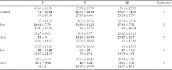
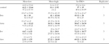
The data collected from the different assessments are summarized in Table 1a (CTC assay after 30–120 min exposure), Table 1b (merocyanine-540, after 30–120 min exposure), Table 1c (CTC assay after 4 h exposure) and Table 1d (merocyanine-540 after 4 h exposure). Values are the mean ± SD.
Experiments 1a–1d: means ± SD for three to six replicates. Percentages of ejaculated bull spermatozoa depicting the proportions (%) of CTC patterns F (uncapacitated), B (capacitated) and AR (acrosome-reacted) (a, c); of merocyanine-540 fluorescence intensity, Mero-low (uncapacitated) and Mero-high (capacitated); and of Yo-PRO-1 (dead) staining (b, d) when suspended in Fert-TALP without bicarbonate (control) or after exposure to Ista (oviductal fluid from the isthmic region at standing estrus), Asta (oviductal fluid from the ampullar region at standing estrus), Iov (oviductal fluid from the isthmic region at the day of ovulation) or Aov (oviductal fluid from the ampullar region at the day of ovulation) for (a, b) 30 min (italics), 60 min (bold) or 120 min (normal font) or 4 h (c, d) at 39 °C in 5% CO2 (control without CO2)


Experiment 1a
The number of alive, uncapacitated spermatozoa (F-pattern) varied significantly (p = 0.0017) between treatments. The different runs showed a weak significant difference (p = 0.030) in the number of F-pattern spermatozoa. Exposure to ampullar ODF at ovulation (Aov, p = 0.0041), to ampullar ODF at standing estrus (Asta, p = 0.0007), ovulation day isthmic ODF (Iov, p = 0.0003) and ODF from the isthmic region at standing estrus (Ista, p = 0.0010) significantly decreased the proportion of F-pattern spermatozoa compared with controls. There was also a significant difference (p = 0.022) in the percentages of spermatozoa with B-pattern spermatozoa, although smaller than that for the F-pattern spermatozoa, when comparing treated samples with each other. On further investigation we found that exposure to Aov-ODF did not differ significantly from the negative control, whereas the rest of the ODF treatments increased the proportion of capacitated spermatozoa (B-pattern), ampullar ODF at standing estrus (p = 0.0075), ovulation day isthmic ODF (p = 0.0030) and isthmic ODF at standing estrus (p = 0.012). The number of B-pattern spermatozoa varied significantly in the different runs (p = 0.014).
The number of alive, AR-spermatozoa was not dependent on treatments compared with each other or the individual ODF treatments compared with the controls, only on run (p < 0.0001). Incubation time (30, 60 or 120 min) was not significant for either pattern. There was no significant correlation between treatment and time.
Experiment 1b
The percentages of spermatozoa depicting low fluorescence (Mero-low, i.e. alive spermatozoa, with no lipid membrane shift) varied highly and significantly among treatments (p < 0.0001). The significance level was the same when comparing the ODF treatments one by one with the negative control. There were no significant differences between runs (p > 0.05). The percentages of spermatozoa showing high fluorescence (Mero-high, i.e. alive, capacitated) varied highly significantly with the different treatments (p < 0.0001), compared with both each other and controls. Spermatozoa exposed to ODF from standing estrus (either Asta or Ista) yielded the highest levels (p < 0.0001). Also, both Aov (p = 0.0002) and Iov (p = 0.0005) significantly increased these percentages compared with controls. The percentages of dead spermatozoa (Yo-PRO-1 positive) varied highly significantly with the different treatments (p < 0.0001), and in the different runs (p < 0.0001). Compared with the negative control, exposure to Aov, Asta and Iov significantly increased (p < 0.0001) the percentages of dead spermatozoa in the samples. Incubation time did not affect the outcomes of the ODF exposure, nor was there any (significant) correlation between treatment and time.
Experiment 1c
The percentage of alive, uncapacitated spermatozoa (F-pattern) did not vary significantly between the different treatments after 4 h of incubation. When comparing the ODF-challenged samples with controls, Iov did not significantly affect the number of F-pattern spermatozoa, while the other types of ODF did (Aov, p = 0.0087; Asta, p = 0.0023; Ista, p = 0.0137). Consequently, the proportion of B-pattern spermatozoa varied (p = 0.012) among treatments, increasing after exposure to Asta (p = 0.0032), Ista (p = 0.0076) and Aov (p = 0.0040), compared with controls. No significant difference between the different runs was noted for the proportion of F- and B-pattern spermatozoa. The proportion of alive, AR-spermatozoa did not vary significantly with the different ODF treatments compared with each other or with the negative control after 4 h of incubation. The number of AR-spermatozoa was slightly dependent on run (p = 0.034).
Experiment 1d
The proportions of Mero-low spermatozoa varied significantly among treatments following exposure for 4 h, compared with either each other or the controls (p < 0.0001). The Mero-low sperm population was also significantly dependent on the run (p = 0.0003). However, such lengthy exposure to ODF did not affect the percentage of spermatozoa with a shift in the phospholipid layer of the cell membrane (capacitated, Mero-high) when comparing treatments with each other. Only those samples treated with Ista had increased proportions of Mero-high spermatozoa (p = 0.018) when compared with controls. The shift to Mero-high was dependent on run (p = 0.048). The proportions of dead spermatozoa (Yo-Pro-1 positive) increased significantly after 4 h of exposure among treatments (p = 0.0029). Aov (p = 0.0043), Asta (p = 0.0012), Iov (p = 0.0090) and Ista (p = 0.0010) all increased these proportions compared with controls. The different runs showed significant variations (p = 0.0043).
Experiment 2: Exposure to GAGs elicits small membrane changes in bull spermatozoa
The data collected from the assessments are summarized in Table 2a (CTC assay after 4 h exposure) and Table 2b (merocyanine-540, after 4 h exposure). Values are the mean ± SD.
Experiments 2a and 2b: means ± SD for two to six replicates. Percentages of ejaculated bull spermatozoa depicting the proportions (%) of CTC patterns F (uncapacitated), B (capacitated) and AR (acrosome-reacted) (a); of merocyanine-540 fluorescence intensity, Mero-low (uncapacitated) and Mero-high (capacitated); and of Yo-PRO-1 (dead) staining (b) when suspended in Fert-TALP without bicarbonate (control) or hep (heparin), HS (heparan sulphate), HA (hyaluronan), CSC (chondroitin sulphate C) or DS (dermatan sulphate) after exposure for 4 h at 39 °C in 5% CO2 (control without CO2)

Experiment 2a
When comparing among treatments, no significant differences were observed in the proportions of F-pattern spermatozoa. When comparing the different treatments with controls, HA (p = 0.043) was the only GAG that significantly lowered the proportion of alive, uncapacitated spermatozoa (F-pattern). A significant difference between the different runs (p = 0.015) was noted for F-pattern proportions. There was no significant change in B-pattern when comparing among treatments. A significant increase was detected for B-pattern (capacitation) spermatozoa after HA exposure (p = 0.049) compared with controls. None of the treatments induced significant differences in the proportions of alive, AR-spermatozoa compared with controls. No dependence on run was seen for AR-spermatozoa.
Experiment 2b
When comparing among different treatments with each other, an incubation of 4 h did not significantly alter the proportion of Mero-low spermatozoa. There was a reduction, smaller than that for ODF-treated samples, incubated for the same period, in the proportion of intact spermatozoa (Mero-low) when spermatozoa were exposed to hep (p = 0.0087), compared with the negative control. The number of Mero-low cells was significantly dependent on run (p = 0.0019). Furthermore, no significant difference in the number of capacitated spermatozoa (Mero-high) was seen for any of the different treatments compared with each other. When compared with the negative control, DS caused a significant increase in the Mero-high sperm population (p = 0.035). The proportion of Mero-high spermatozoa was not significantly dependent on run. The different treatments did not change the proportions of dead spermatozoa, when compared with each other. When comparing the proportion of dead spermatozoa in treated samples with the negative control, only hep led to significant increases (p = 0.008). The proportions of dead spermatozoa were significantly (p = 0.0015) affected by run.
Experiment 3: Bicarbonate induces early membrane changes in bull spermatozoa
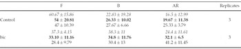
The data collected from the different assessments are summarized in Table 3a (CTC assay after 30–120 min exposure) and Table 3b (merocyanine-540 after 30–120 min exposure). Values are the mean ± SD.
Experiments 3a and 3b: means ± SD for three to seven replicates. Percentages of ejaculated bull spermatozoa depicting the proportions (%) of CTC patterns F (uncapacitated), B (capacitated) and AR (acrosome-reacted) (a); of merocyanine-540 fluorescence intensity, Mero-low (uncapacitated) and Mero-high (capacitated); and of Yo-PRO-1 (dead) staining (b) when suspended in Fert-TALP without bicarbonate (control) or Fert-TALP with 40 mM bicarbonate (bic) after exposure for 30 min (italics), 60 min (bold) or 120 min (normal font) at 39 °C in 5% CO2 (control without CO2)

Experiment 3a
Exposure to 40 mM bicarbonate clearly decreased the proportions of intact spermatozoa (F-pattern, p = 0.0005), but failed to induce a significant increase in the proportion of capacitated (B-pattern) spermatozoa compared with controls. However, proportions of alive, AR-spermatozoa increased significantly on exposure to bicarbonate (p = 0.0007). While the incubation time was not significant for F- or B-pattern spermatozoa, the longer the incubation time the more AR-spermatozoa were detected (p = 0.0072). Run was not a significant parameter for F-, B- or AR-pattern spermatozoa. There was no (significant) correlation between treatment and time for F-, B- or AR-pattern spermatozoa.
Experiment 3b
Bicarbonate significantly decreased the proportion of Mero-low spermatozoa (p < 0.0001) concomitantly with a significant increase in Mero-high (alive, capacitated) spermatozoa (p < 0.0001), compared with controls at all three incubation times (30, 60 or 120 min). There was a significant difference between the different runs for Mero-high and Mero-low (p < 0.0001). Incubation time was not significant for Mero-low or -high, which is in accordance with the results from experiment 3a. Bicarbonate supplementation significantly increased the proportion of dead spermatozoa compared with controls (p = 0.013), as did incubation time (p = 0.018). Run was a significant variable for all three parameters: Mero-low (p = 0.00017), Mero-high (p < 0.0001) and Yo-PRO-1 (p < 0.0001). There was no (significant) correlation between treatment and time, which is in accordance with the results from experiment 3a.
Experiment 4: Bicarbonate induces membrane changes in bull spermatozoa independent of the treatment before bicarbonate supplementation
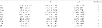
The data collected from the assessments are summarized in Table 4a (CTC assay after 4 h of exposure + challenge) and Table 4b (merocyanine-540 after 4 h of exposure + challenge). Values are the mean of difference ± SD.
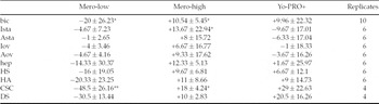
Experiments 4a and 4b: means of difference ± SD for four to 12 replicates. Difference in percentage before and after 30 min incubation with 40 mM bicarbonate of ejaculated bull spermatozoa depicting the proportions (%) of CTC patterns F (uncapacitated), B (capacitated) and AR (acrosome-reacted) (a), merocyanine-540 fluorescence intensity; Mero-low (uncapacitated), Mero-high (capacitated) and of Yo-PRO-1 (dead) staining (b) when suspended in Fert-TALP without bicarbonate (control) or after exposure to Ista (oviductal fluid from the isthmic region at standing estrus), Asta (oviductal fluid from the ampullar region at standing estrus), Iov (oviductal fluid from the isthmic region at the day of ovulation), Aov (oviductal fluid from the ampullar region at the day of ovulation), hep (heparin,), HS (heparan sulphate), HA (hyaluronan), CSC (chondroitin sulphate C) or DS (dermatan sulphate) at 39 °C in 5% CO2 (control without CO2)

Experiment 4a
After incubation for 4 h in experiments 1c, 1d, 2a and 2b, the sperm suspensions were challenged to 40 mM bicarbonate for an additional 30 min. Bicarbonate challenge significantly reduced (p < 0.0001) the proportions of F-pattern spermatozoa independently of the treatment before bicarbonate exposure. Comparison of individual treatments before and after bicarbonate supplementation showed that only Iov (p = 0.0028) and bicarbonate supplementation alone (p = 0.0035) decreased the proportion of F-pattern spermatozoa significantly (Table 4a). A significant difference between the different runs (p = 0.027) was noted for the F-pattern spermatozoa. Bicarbonate additions did not change the proportions of B-pattern spermatozoa, but there was a significance difference between the different runs (p = 0.0085).
AR-pattern spermatozoa increased significantly after bicarbonate treatment (p < 0.0001). When comparing the individual treatment with or without bicarbonate, the following samples had a significantly increased AR population: Aov (p = 0.0029), CSC (p = 0.012), Iov (p = 0.022) and bicarbonate treatment alone (p = 0.038), as shown in Table 4a. The number of AR-spermatozoa varied significantly with the different runs (p < 0.0001).
Experiment 4b
Bicarbonate challenge significantly reduced (p <0.0001) the proportion of Mero-low spermatozoa in the previously exposed samples. Comparison of the different treatments before and after bicarbonate addition showed that only CSC (p = 0.0021) and bicarbonate alone (p = 0.020) were significantly different (Table 4b). The different runs varied significantly (p < 0.0001) for Mero-low spermatozoa. Bicarbonate significantly increased the proportion of Mero-high spermatozoa (p < 0.0001), compared with before bicarbonate addition, and independent of the treatment used before the addition. Comparison of the different treatments before and after bicarbonate supplementation indicated that CSC (p = 0.028), Ista (p = 0.040) and bicarbonate alone (p = 0.046) significantly increased the Mero-high sperm population (Table 4b). There was no significant variation between the different runs for the number of Mero-high spermatozoa. Bicarbonate exposure did not increase sperm death (Yo-PRO-1 positive cells) significantly. The Yo-PRO-1 positive cells were significantly (p < 0.0001) affected by run.
Experiment 5
Challenge with solubilized ZP proteins significantly increased (p = 0.016) the proportions of alive, AR-spermatozoa, compared with controls. There was also a significant, though smaller, difference in the proportions of capacitated spermatozoa, which were larger in controls (p = 0.024).
Discussion
In this study we investigated the effects of the following on bull sperm capacitation in vitro: (1) ODF from well-defined stages of the estrous cycle and different regions of the oviduct, (2) different individual GAGs and (3) bicarbonate.
Capacitation includes reorganization of membrane components and stepwise changes in the intercellular metabolism of the spermatozoon. These processes, together with the consequent AR induced by the ZP glycoproteins, are part of irreversible preparatory events. These events are essential if a spermatozoon is to bind to and penetrate the ZP, fuse thereafter with the oolemma and be engulfed into the oocyte during fertilization (Yanagimachi, Reference Yanagimachi1989). Capacitation can be achieved in vitro in defined medium that attempts to mimic the oviductal milieu. In most cases capacitation media contain pyruvate, lactate, serum albumin, bicarbonate, calcium, potassium (Visconti et al., Reference Visconti, Westbrook, Chertihin, Demarco, Sleight and Diekman2002), sodium and, in the case of cattle, heparin. The mechanism of action by which these compounds promote capacitation is poorly understood at the molecular level.
Questions still arise concerning exactly how, where and when capacitation is elicited in the female genital tract. Some investigators have suggested that sperm capacitation occurs in the sperm reservoir of the oviduct (Grippo et al., Reference Grippo, Way and Killian1995; Lefebvre et al., Reference Lefebvre, Chenoweth, Drost, LeClear, MacCubbin, Dutton and Suarez1995; Lefebvre & Suarez, Reference Lefebvre and Suarez1996). Others suggest that the sperm reservoir (the utero-tubal junction and adjacent part of isthmus) prevents capacitation, and that capacitation occurs during transport of the spermatozoa towards the ampullar isthmic junction (AIJ) when they are exposed to increased amounts of bicarbonate (Rodriguez-Martinez, Reference Rodriguez-Martinez2001; Rodriguez-Martinez et al., Reference Rodriguez-Martinez, Tienthai, Suzuki, Funahashi, Ekwall and Johannisson2001, Reference Rodriguez-Martinez, Saravia, Wallgren, Tienthai, Johannisson, Vazquez, Martinez, Roca, Sanz and Calvete2005; Tienthai et al., Reference Tienthai, Johannisson and Rodriguez-Martinez2004). Bicarbonate is present at low levels in the sperm reservoir, at least in sows (Rodriguez-Martinez et al., 1998; unpublished results). The present investigation showed no overall difference between ODF from the ampullar or isthmic regions in eliciting bull sperm capacitation. The reason for this absence of difference may be that the oviductal isthmic catheters were placed closer to the ampullar isthmic junction (AIJ) than to the sperm reservoir (Bergqvist et al., Reference Bergqvist, Yokoo, Heldin, Frendin, Sato and Rodríguez-Martínez2005). We could not detect any major differences when comparing the two different time points investigated (standing estrus or ovulation day, i.e. pre- and post-ovulation). Some inconclusive results suggested that ODF from ovulation day seemed less potent in inducing capacitation (B-pattern at 30–120 min of incubation for ampullar ODF and at 4 h incubation time for isthmic ODF, when compared with controls).
These results were not in accordance with the FACS results, which yielded a significant increase in merocyanine-540 high fluorescence (alive, capacitated) spermatozoa exposed to ODF from ovulation day or ODF from standing estrus from both the isthmus and ampulla, at 30–120 min. The reason for these contrasting results may be that merocyanine-540 detects early changes in the sperm membranes, not yet detectable by CTC (Rathi et al., Reference Rathi, Colenbrander, Bevers and Gadella2001; Rodriguez-Martinez et al., Reference Rodriguez-Martinez, Tienthai, Suzuki, Funahashi, Ekwall and Johannisson2001; Harrison & Gadella, Reference Harrison and Gadella2005). Another reason may be that, considering that bicarbonate-induced capacitation could be detected without calcium increases (Harrison & Gadella, Reference Harrison and Gadella2005), this may mean that merocyanine-540 measures the ‘bicarbonate route’ of capacitation while CTC measures solely redistribution of calcium. Another explanation for these conflicting results between CTC and FACS could be that in the CTC assay spermatozoa are fixed before evaluation, while spermatozoa are still alive for the FACS analyses and therefore destabilize more promptly (including death) during the analytical process. AR-like events could also occur when spermatozoa are dying, and do not have to be preceded by capacitation. To exclude dead AR-spermatozoa from the CTC investigation in the present study, we used the live-dead dye ethidium monodimer.
Some previous studies have recorded that capacitation-like changes could be detected in frozen-thawed (bull) spermatozoa using CTC (Cormier et al., Reference Cormier, Sirard and Bailey1997; Thundathil et al., Reference Thundathil, Gil, Januskauskas, Larsson, Soderquist, Mapletoft and Rodriguez-Martinez1999; Bailey et al., Reference Bailey, Bilodeau and Cormier2000; Collin et al., Reference Collin, Sirard, Dufour and Bailey2000). We used fresh spermatozoa to avoid this problem. Recent studies in boar spermatozoa have shown a large difference between the number of fresh boar spermatozoa which attain merocyanine-540 measurable membrane changes (Mero-high) when triggered by bicarbonate, compared with frozen-thawed boar spermatozoa (Guthrie & Welch, Reference Guthrie and Welch2005). We recorded similar behaviour with bull (fresh vs frozen-thawed) spermatozoa supplemented with bicarbonate in preliminary studies (Bergqvist, unpublished results).
Previous studies have revealed that ODF can initiate bull sperm capacitation (Parrish et al., Reference Parrish, Susko-Parrish, Handrow, Sims and First1989a; McNutt & Killian, Reference McNutt and Killian1991). This is in accordance with our results. The present study used ODF from the ampullar or isthmic regions collected at standing estrus or the day of ovulation. All four types of ODFs increased the number of capacitated spermatozoa measured as a rise in B-pattern spermatozoa (although not significant for ODF from the ampullar region at ovulation), or as a shift in merocyanine-540 fluorescence, when incubating spermatozoa between 30 and 120 min. After 4 h of incubation an increase in the number of capacitated (B-pattern) spermatozoa was still seen for the four types of ODF (although not significant for ODF spermatozoa in the samples treated with ovulation day isthmic ODF). This may indicate that the ability of ODF to capacitate bull spermatozoa increases with the distance into the ampulla at which spermatozoa would be relocated. Unfortunately, this is not in accordance with the results of the present study obtained when exposing spermatozoa to isthmic ODF collected at standing estrus, which also significantly increased capacitation compared with controls. Only samples treated with ODF from the isthmic region during standing estrus had a significantly higher proportion of capacitated spermatozoa (Mero-high) compared with controls after 4 h of incubation. The diverging results in B-pattern spermatozoa and Mero-high fluorescence at 4 h of incubation may reflect the fact that CTC detects a later step in the capacitation process compared with merocyanine-540, as mentioned earlier. None of the ODF-treated sperm samples exhibited increased proportions of AR-spermatozoa. Already after 4 h of incubation, a significantly higher rate of dead spermatozoa was detected for all four types of ODF compared with controls. This may imply that the spermatozoa have already passed the stage of phospholipid scrambling in the membrane and are dead, or at least dying. This is confirmed by the significant increase in Yo-PRO-1 positive or dead spermatozoa in the ODF-treated samples compared with controls.
Several scientific studies report that S-GAGs promote capacitation in bull sperm in vitro (Parrish et al., Reference Parrish, Susko-Parrish, Winer and First1988, Reference Parrish, Susko-Parrish, Handrow, Ax and First1989b; Chamberland et al., Reference Chamberland, Fournier, Tardif, Sirard, Sullivan and Bailey2001; Therien et al., Reference Therien, Bergeron, Bousquet and Manjunath2005). In another study by our group we found an average concentration of 64μg/ml for all S-GAGs taken together in the tubal fluid of normally cycling dairy heifers during the functional period of the oviduct, that is, pro-estrus to meta-estrus (Bergqvist & Rodriguez-Martinez, Reference Bergqvist and Rodriguez-Martinez2006). We used a concentration of 20μg/ml for all S-GAGs in the present study, a figure close to that used in most IVF laboratories (10–20μg/ml). Although our intention was to try to use concentrations of the GAGs in the different experiments that were as close to physiological as possible, only one previous study (Lee & Ax, 1984) gives relative but not exact concentrations of the different GAGs in bovine ODF. This continues to be a problem, since no commercially available assay reliably separates and calculates the concentrations of the different S-GAGs in ODF. For these reasons we used the same concentration for all individual S-GAGs, which of course is not a true reflection of the situation in vivo.
Heparin is used routinely to capacitate bull spermatozoa in IVF procedures. Some studies suggest that heparin uses the same pathway as ODF to elicit capacitation, viewed as changes on the sperm surface (Mahmoud and Parrish, Reference Mahmoud and Parrish1996). However, other studies of the cAMP pathway have suggested the mechanism of action differs between heparin- and ODF-induced capacitation (Parrish et al., Reference Parrish, Susko-Parrish, Uguz and First1994). We did not find any significant increase in the proportion of capacitated spermatozoa when treated with heparin (20μg/ml) for 4 h. This is a discrepancy compared with many other studies (Januskaukas et al., Reference Januskaukas, Gil, Söderquist and Rodriguez-Martinez2000; Collin et al., 2002; Chamberland et al., Reference Chamberland, Fournier, Tardif, Sirard, Sullivan and Bailey2001). The reason for this discrepancy may be that the heparin-treated spermatozoa were already dead by 4 h, and thereby stained with ethidium monodimer, and were thus excluded from the counted spermatozoa. This is in agreement with the FACS results, which showed a significant increase in the Yo-PRO-1 positive (i.e. dead) sperm population for the heparin-treated samples. None of the other S-GAG-treatments significantly increased the dead sperm population, nor did any of the S-GAGs significantly increase the rate of AR-spermatozoa. Since heparin only exists in the granules of mast cells and not in ODF (Kjellen & Lindahl, Reference Kjellen and Lindahl1991) several studies have designated heparan sulphate, which is closely related to heparin, as the in vivo inducer of bull sperm capacitation (Parrish et al., Reference Parrish, Susko-Parrish, Handrow, Sims and First1989a; Kawakami et al., Reference Kawakami, Arai, Oishi, Hori and Tsutsui2000). However, in the present study we could not find evidence that heparan sulphate elicited capacitation in vitro. The reason for this may be that there is a large diversity between different types of heparan sulphate, making comparisons difficult, if not impossible, across studies.
Nevertheless, S-GAGs other than heparin and heparan sulphate may be involved in inducing capacitation (Parrish et al., Reference Parrish, Susko-Parrish, Handrow, Sims and First1989a). A recent study by Therien et al. (Reference Therien, Bergeron, Bousquet and Manjunath2005) concluded that chondroitin sulphate B (CBS) or DS, which consists of chondroitin sulphate B chains (Sugahara et al., Reference Sugahara, Mikami, Uyama, Mizuguchi, Nomura and Kitagawa2003), induced capacitation in bull spermatozoa. The results of Therien et al. (Reference Therien, Bergeron, Bousquet and Manjunath2005) are in accordance with the present study, where dermatan sulphate significantly increased sperm capacitation rates. Dermatan sulphate or CSB may be the GAG present in ODF that induces sperm capacitation in vivo, alone or together with other substances in the tubal fluid. Dermatan sulphate is one of the most heavily sulphated GAGs, and some investigators have suggested that the ability of an S-GAG to induce capacitation depends on its degree of sulphation (Parrish et al., Reference Parrish, Susko-Parrish, Handrow, Ax and First1989b). Our results support this hypothesis.
In a study by Miller & Hunter (Reference Miller and Hunter1986) chondroitin sulphate A failed to induce capacitation in bull spermatozoa. In the present study chondroitin sulphate C also failed to induce capacitation by itself, but was the GAG that achieved the highest increase in capacitated bull spermatozoa when comparing before and after bicarbonate addition, using merocyanine-540. However, we do not know which of the different chondroitin sulphate isomers (A, B, C, D and E) are present in tubal fluid and whether they react antagonistically or synergistically to bicarbonate exposure.
The hyaluronan concentration in the present study (10 μg/ml) was chosen to reflect the true concentration of hyaluronan in ODF according to our own results (Bergqvist et al., Reference Bergqvist, Yokoo, Heldin, Frendin, Sato and Rodríguez-Martínez2005). An older study by Lee & Ax (1984) found that hyaluronan comprised approximately 40% of the total GAG content in ODF from dairy cows. This is not in accordance with our findings in ODF from dairy heifers, where hyaluronan A comprised only 5–8% of the total GAG content (Bergqvist et al., Reference Bergqvist, Yokoo, Heldin, Frendin, Sato and Rodríguez-Martínez2005; Bergqvist & Rodriguez-Martinez, Reference Bergqvist and Rodriguez-Martinez2005). In the present study we found that HA significantly augmented the proportion of capacitated spermatozoa when measured by the CTC assay, without causing an increase in the percentages of either AR or dead spermatozoa. This is in agreement with a study on boar spermatozoa, in which hyaluronan was able to induce the B-pattern without leading to AR (Rodriguez-Martinez et al., Reference Rodríguez-Martínez, Han, Song, Funahashi and Niwa1997). Other authors have concluded that hyaluronan did not increase capacitation or AR in frozen-thawed bull spermatozoa (Januskaukas et al., Reference Januskaukas, Gil, Söderquist and Rodriguez-Martinez2000). In a study by Handrow et al. (1984) hyaluronan proved to be the least potent of all investigated GAGs at inducing AR. Hyaluronan in ODF may act as a naturally occurring sperm selector. Accordingly, studies from IVF laboratories show that hyaluronan works well as an in vitro sperm selector (Huszar et al., Reference Huszar, Willetts and Corrales1990; Shamsuddin et al., Reference Shamsuddin, Rodriguez-Martinez and Larsson1993; Cayli et al., Reference Cayli, Jakab, Ovari, Delpiano, Celik-Ozenci, Sakkas, Ward and Huszar2003).
In the present study individual GAGs had less ability to elicit sperm capacitation than the whole ODF. One explanation for this may be that S-GAGs in the ODF are present in the form of proteoglycans and thus may exert their functions differently. Another, more likely explanation is that other substances are present in ODF that cause or contribute to the capacitating process.
The bicarbonate concentration radically increases when spermatozoa are ejaculated, first in the seminal fluid and then in the female tract (Setchell et al., Reference Setchell1994; Yanagimachi, Reference Yanagimachi1994), including the oviduct (pig: Rodriguez-Martinez et al. unpublished results 1998). This dramatic change in extracellular ion concentration activates modulations in intracellular ion concentrations and leads to an altered membrane potential of the sperm plasma membrane. Several studies have demonstrated that capacitation is a bicarbonate-dependent event (Ashworth et al., Reference Ashworth, Harrison, Miller, Plummer and Watson1995; Harrison et al., Reference Harrison, Ashworth and Miller1996; Flesh et al., Reference Flesch, Brouwers, Nievelstein, Verkleij, van Golde, Colenbrander and Gadella2001; Tienthai et al., Reference Tienthai, Johannisson and Rodriguez-Martinez2004), at least in species other than cattle.
We found bicarbonate to be a potent inducer of bull sperm capacitation or phospholipid membrane changes measured as merocyanine-540 fluorescence intensity. This is in accordance with the findings of Tienthai et al., (Reference Tienthai, Johannisson and Rodriguez-Martinez2004) and Harrison & Gadella (Reference Harrison and Gadella2005) in pigs. Sostaric et al. (Reference Sostaric, van de Lest, Colenbrander and Gadella2005) found, using CTC, bicarbonate to be a more potent bull sperm capacitator than heparin. Also, in the present study we found bicarbonate to be more potent than any GAGs potentially present in ODF in inducing bull sperm capacitation in vitro. Moreover, and contrary to the results of Sostaric et al. (Reference Sostaric, van de Lest, Colenbrander and Gadella2005), bicarbonate did not increase the proportion of B-pattern spermatozoa but of AR-spermatozoa. The reason for this dissimilarity might be the different bicarbonate concentrations used (25 mM vs 40 mM), which may have accelerated the process. The finding that bicarbonate induces AR in spermatozoa agrees with other studies in the stallion (Rathi et al., Reference Rathi, Colenbrander, Bevers and Gadella2001), boar (Abeydeera et al., Reference Abeydeera, Funahashi, Kim and Day1997) and mice (Lee & Storey, Reference Lee and Storey1986). When comparing GAGs, ODF and a negative control, before and after 30 min of incubation in bicarbonate-enriched medium, exposure to ODF collected on the ovulation day (i.e. post-ovulatory) increased the AR-rate significantly. This may indicate that post-ovulatory ODF is more prone to induce AR in sperm than pre-ovulatory ODF when activated.
When comparing different IVF media, Tajik et al. (Reference Tajik, Wang, Okuda and Niwa1994) found that more bull spermatozoa penetrated more oocytes at bicarbonate concentrations of 46–49 mM compared with 34–43 mM. The present study used a concentration of 40 mM bicarbonate, which is more than the original formula for Fert-TALP. We decided to use a bicarbonate concentration similar to MBO+ medium (37 mM), which is used to capacitate boar spermatozoa (Tienthai et al., Reference Tienthai, Johannisson and Rodriguez-Martinez2004).
The reason for the relatively low viability in the semen samples in the present study could be the effect of a fluctuating temperature during transportation, handling and analyses. This may explain the low survival rate seen particularly in ODF- and bicarbonateexposed samples, since capacitated spermatozoa are more sensitive to temperature fluctuations and both ODF and bicarbonate facilitate sperm capacitation, a process of destabilization that ends – in vitro – with cell death if no fertilization occurs. Capacitated spermatozoa have, by definition, a shorter lifespan. This may also suggest that bull spermatozoa also need the oviductal epithelial cells for survival (Pollard et al., Reference Pollard, Plante, King, Hansen, Betteridge and Suarez1991).
Some authors claim that serum-derived albumin-like substances in ODF act as cholesterol acceptors during capacitation (Visconti et al., Reference Visconti, Westbrook, Chertihin, Demarco, Sleight and Diekman2002). Others conclude that protein supplementation may facilitate capacitation of bovine spermatozoa and/or penetration of oocytes but that it is not needed (Tajik et al., Reference Tajik, Wang, Okuda and Niwa1994; Keskintepe & Brackett, Reference Keskintepe and Brackett1996). The explanation for the low capacitation rate after GAG exposure in the present study may be that GAGs need the albumin-cholesterol route to achieve sperm capacitation, whereas bicarbonate is able to elicit capacitation without albumin. We did not use albumin (BSA) in the incubation medium in the present study because protein components have been shown to interfere with FACS readings (Peña et al., Reference Peña, Johannisson and Linde-Forsberg1999, Nagy et al., Reference Nagy, Jansen, Topper and Gadella2003). Another reason was that we did not want to add too many different components to the incubation medium, given our interest in the effects of the separate components. We wanted as few factors as possible to influence the sperm membrane and capacitation. For this reason, pyruvate, hypotaurine, penicillamine and epinephrine were also were excluded from the medium. This may also have influenced sperm viability.
The present study confirms previous findings that ODF stimulates sperm capacitation. We conclude that bicarbonate, a component of ODF, appears to be more potent in triggering bull sperm capacitation than other components of ODF, such as GAGs.
Acknowledgements
We would like to express our gratitude to Margareta Håård at Svensk Avel, Skara for access to bull semen samples and to Professor Lena Kjellén for the kind gift of the different S-GAGs. This study was supported by FORMAS, Stockholm. The flow cytometer used in this study was financed by the Swedish Research Council (Vetenskapsrådet), Stockholm.