Introduction
Research suggests that low birth weight and low fetal growth are associated with increased risk of stroke later in life.Reference Koupil, Leon and Lithell 1 – Reference Eriksson, Forsen and Tuomilehto 3 Fetal growth is influenced by in utero environmental factors, such as maternal malnutrition, and if such factors occur during critical periods of fetal development this may lead to an increased risk of morbidity in adulthood – a premise termed the fetal origins hypothesis.Reference Barker 4
Moreover, studies have shown that mothers with a flat pelvis produce offspring with lower birth weight,Reference Martyn, Barker and Osmond 5 and that these offspring have higher risk of stroke in adulthood.Reference Osmond, Kajantie and Forsen 2 , Reference Eriksson, Forsen and Tuomilehto 3 , Reference Martyn, Barker and Osmond 5 During pregnancy a flat pelvis may affect placental growth and fetal development,Reference Martyn, Barker and Osmond 5 as well as increase the risk of obstructed labour. A flat pelvis is thought to be the result of malnutrition in early childhood suggesting that intergenerational effects could play a role in the development of stroke.
In earlier analyses of Swedish men and women born between 1915 and 1929 and followed-up until the end of 2002,Reference Koupil and Leon 6 pelvic size measures were positively associated with birth weight and weight-for-gestational-age, and inversely associated with adult offspring’s risk of death from circulatory disease and stroke. The association between maternal pelvic size and death from circulatory disease was stronger in women. However, this study only included fatal cases and lacked power to investigate specific types of stroke separately.
Using data from the same Swedish cohortReference Koupil and Leon 6 followed-up for another 6 years until age 79–93 years, our aim was to investigate the associations of maternal pelvic size with risk of incident haemorrhagic stroke (HS), thrombotic stroke (TS) and other stroke (OS), focussing on potential gender differences and a mediating role of birth-weight-for-gestational-age.
Methods
Sample
Study participants belonged to the first generation of the Uppsala Birth Cohort Multigenerational Study (UBCoS Multigen; see http://www.chess.su.se/ubcosmg/) comprising 14,192 live births born at Uppsala University Hospital, Sweden, between 1915 and 1929.Reference Koupil 7 , Reference Koupil and Goodman 8 Deaths and emigrations were traced from parish records (~98% completeness to follow-up) until register data became available in the 1960s. Our sample was restricted to singleton births and those still alive and residing in Sweden in 1964 (n=11,580; Supplementary Fig. S1). The final analytical sample included 3296 men and 3066 women with complete data. The study was approved by the Regional Ethics Committee in Stockholm.
Variables
Our main exposure variables were pelvic measurements of the mother, birth characteristics of the offspring and family social class, obtained from obstetric records. Pelvic measurements were intercristal diameter (the maximal distance between the iliac crests), interspinous diameter (the distance between the anterior superior iliac spines) and external conjugate diameter (the difference between the spine of the fifth lumbar vertebra and the front of the pubic bone). Based on previous studiesReference Eriksson, Forsen and Tuomilehto 3 , Reference Martyn, Barker and Osmond 5 , Reference Barker, Osmond and Forsen 9 we defined flat pelvis as having an external conjugate diameter of ⩽18 cm and a difference between intercristal and interspinous diameters of <2.5 cm.
Birth characteristics analysed were gestational age (<36 weeks, 36–37 weeks, 38–39 weeks, 40–41 weeks and ⩾42 weeks) and birth-weight-standardized-for-gestational-age (z-score). Social class at birth was based on father’s occupation or mother’s occupation if she was single, and analysed as three categories as follows: high (higher and intermediate non-manual, entrepreneurs and farmers), intermediate (lower non-manual, skilled manual) and low (unskilled manual, house-daughters). Maternal age at birth and parity were also derived from obstetric records.
Highest level of educational attainment ever recorded for the offspring was obtained from censuses 1960 and 1970, and the Education Register from 1985 to 2008. Education was categorized into three groups as follows: low (less than upper-secondary education), intermediate (upper-secondary schooling) and high (any post-secondary education).
Incident TS, HS and OS were defined as the first recorded hospitalization or death with the respective diagnosis as the main or contributing diagnoses. Data were obtained from the Hospital Discharge Register and the Causes of Death Register. The following International Classification of Diseases (ICD) codes were used to define TS: ICD-7 [332–333 (no cases of 333 present)], ICD-8 (432–435, 437), ICD-9 (433–435) and ICD-10 [G45, I63–I66 (no cases of I64 present)]; HS: ICD-7 (330–331), ICD-8 (430–431), ICD-9 (430–432) and ICD-10 (I60–I62); and OS (unspecified): ICD-7 (334), ICD-8 (436, 438), ICD-9 (436–438) and ICD-10 [I64, I67–I69 (no cases of I68 present)].
Missing data
Comparisons of the final study population (n=6362) and the sample of excluded individuals (Supplementary Fig. S1) who lacked pelvic size measurements (n=4489) showed a statistically significant difference between the groups for mother’s age, parity and birth cohort (P<0.001). Compared with the excluded individuals, the analytical sample had mothers who were slightly younger [28.2 (6.4) years v. 28.6 (6.4) years], were more likely to be first born and born between 1925 and 1929.
Statistical methods
Descriptive statistics and Cox proportional hazards models were performed using STATA version 11. Follow-up commenced on 1 January 1964 and continued until diagnosis with stroke, emigration, death, or until 31 December 2008. All analyses were stratified by gender. In order to control for possible cohort effects and changes in diagnostic and recording practice over time, birth years were divided into three groups (1915–1919, 1920–1924 and 1925–1929). Birth cohort, age, parity and gender (where applicable), and social class at birth were adjusted for in hierarchical models. To explore potential interactions with gender of the offspring, we performed stratified analyses and combined the results when statistical tests did not indicate a significant effect modification by gender.
Results
Supplementary Table S1 displays the characteristics of the sample. Among the exposure variables, only own educational attainment was statistically different between men and women (P<0.001); men had higher education. Incident TS occurred at a rate of 436 cases per 100,000 person-years in males (95% CI 426–504) and 387 cases per 100,000 person-years in females (95% CI 354–425); incident HS at a rate of 131 cases per 100,000 person-years in males (95% CI 112–153) and 91 cases per 100,000 person-years in females (95% CI 76–110); and incident OS at a rate of 309 cases per 100,000 person-years in males (95% CI 279–343) and 243 cases per 100,000 person-years in females (95% CI 217–273) during the follow-up period from 1 January 1964 to 31 December 2008. Supplementary Table S2 presents the rates by main exposure variables.
Maternal pelvic measurements
Mean pelvic measurements were 28.3 (s.d. 1.7) cm for intercristal diameter, 25.3 (s.d. 1.8) cm for interspinous distance, 2.9 (s.d. 1.4) cm for the distance between intercristal and interspinous diameters, and 20.1 (s.d. 1.4) cm for external conjugate diameter. Only 182 (2.9%) mothers had a flat pelvis and they were more likely to produce offspring with lower birth weight and birth-weight-for-gestational-age compared with their counterparts: mean birth weight for offspring whose mothers had a flat pelvis was 3225 (s.d. 480) g and for those whose mothers did not have a flat pelvis it was 3470 (s.d. 503) g (P<0.001). Moreover, 8.2% of mothers with a flat pelvis produced offspring with low birth weight (<2500 g) compared with 3.0% of mothers without a flat pelvis (P<0.001).
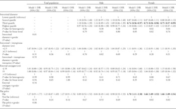
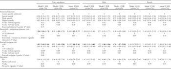
A statistical interaction between intercristal diameter and gender in their effects on TS (P<0.05) was observed (Table 1); intercristal diameter was associated with TS in females only. Flat pelvis was not associated with HS in our cohort (Table 2), but female offspring whose mothers had a flat pelvis had increased risk of TS. Test for statistical interaction of gender and flat pelvis in their effects on TS was of borderline statistical significance (P=0.06).
Hazard ratios (HR) for thrombotic stroke by maternal pelvic measurements for total population (n=6362) and stratified by gender (men: n=3296; women: n=3066)

Model 1 adjusted for birth cohort, age, gender and parity. Model 2 adjusted for birth cohort, age, gender, parity, gestational age and birth weight for gestational age. Model 3 adjusted for birth cohort, age, gender, parity and social class at birth. Gender not adjusted for in the stratified models.
Bolded font: significance at the 5% level (P<0.05).
Hazard ratios (HR) for haemorrhagic stroke by maternal pelvic measurements for total population (n=6362) and stratified by gender (men: n=3296; women: n=3066)

Model 1 adjusted for birth cohort, age, gender and parity. Model 2 adjusted for birth cohort, age, gender, parity, gestational age and birth weight for gestational age. Model 3 adjusted for birth cohort, age, gender, parity and social class at birth. Gender not adjusted for in the stratified models.
Bolded font: significance at the 5% level (P<0.05).
The difference between intercristal and interspinous diameters was associated with HS in the total population, but not in the gender-stratified analyses (Table 2); individuals whose mother’s difference was <2.5 cm had increased risk of HS. External conjugate diameter was associated with HS and this effect was stronger in men; men whose mothers had an external conjugate diameter of ⩽18 cm had a 1.83-fold increased risk of HS compared with individuals whose mothers had a diameter >19 cm. This effect was partly explained by birth-weight-for-gestational-age and social class at birth.
Interspinous diameter was not associated with any of the stroke types analysed (results not shown). No maternal pelvic measurements were associated with OS in the offspring (Supplementary Table S3).
Birth and social characteristics
Negative linear associations were seen between birth-weight-for-gestational-age and TS (P<0.01) and OS (P<0.05) in the total population (Supplementary Tables S4 and S5). When analyses were stratified by gender, the effect was apparent in men but not in women. In contrast, no association was observed between birth-weight-for-gestational-age and HS (Supplementary Table S6).
There was a statistical interaction between gestational age and gender in their effects on OS (P<0.001). Gestational age was associated with OS in females (P<0.01) but not in males (Supplemental Table S5). Females who were born before 36 weeks of gestation had double the risk of OS when compared with those who were born between 40 and 41 weeks gestation.
Social class at birth was associated with all stroke types, but to varying degrees (Supplemental Table S7). In both men and women, those from lower social groups had consistently increased risk of HS and OS compared with individuals from more advantaged social backgrounds. In contrast, the association between social class at birth and TS was observed only among women and the effect appeared to be partly mediated by own educational level.
Discussion
Summary of findings
In our cohort of adult men and women, only 3% of mothers had a flat pelvis. These mothers had offspring with a lower mean birth weight and birth-weight-for-gestational-age compared with offspring of mothers with a normal pelvis. Moreover, female offspring of mothers with flat pelvis had increased risk of TS.
Even a smaller variation in maternal pelvic measurements was associated with incident TS and HS in adult offspring, often in a gender-specific pattern, whereas no maternal pelvic measurements were associated with OS. A smaller difference between intercristal and interspinous diameters and a smaller external conjugate diameter (a combination of which define flat pelvis) were independently associated with HS; however, an inverse association of external conjugate diameter with HS was solely observed in male offspring.
Birth characteristics were also associated with stroke. Negative linear associations of birth-weight-for-gestational-age with TS and OS were noted in the total population, whereas no association was observed with HS. Low social class at birth was consistently associated with HS and OS in the total population, and with TS in women only.
Methodological considerations
The main strength of our study was using a large, well-established, historical longitudinal cohort with excellent completeness of follow-up; UBCoS Multigen Study enabled us to follow men and women to a very old age. It is unlikely we had misclassification of pelvic size measurements because all women were assessed in the same hospital where there would be standardized recording methods.
We were unable to adjust for possible confounding factors, such as maternal height and smoking, because these variables were not available in the obstetric records from Uppsala University Hospital. We were also unable to assess whether mothers with a flat pelvis had increased risk of obstructed labour because data on birth delivery methods was unavailable. Although our data included head circumference, which has been used in relation to low birth weight in similar studies investigating the association between maternal pelvic size and stroke, we did not include this variable as head circumference measured immediately after birth may not reflect the true head circumference, especially if there had been an assisted delivery or birth injury.Reference Simic Klaric, Tomic Rajic and Tesari Crnkovic 10
Our study population included only individuals with no missing data; from our possible restricted sample (Supplementary Fig. S1), only 58% of the sample had pelvic measurements recorded in the obstetric records. Consequently, through the process of selecting participants with no missing data, there appears to be a selection towards being from a primiparous pregnancy and be born between 1925 and 1929. However, we do not believe this has influenced our main findings.
Comparison with other studies
Overall, our findings support the limited existing research that suggests a link between maternal pelvic size and future stroke in the offspring. A British studyReference Martyn, Barker and Osmond 5 that investigated the association between impaired fetal growth and risk of stroke later in life among men born between 1907 and 1930 found the standardized mortality rate for stroke was greater in men whose mothers had a flat pelvis. Our findings showed that a flat pelvis increases the risk of TS among female offspring, with no effect for males.
Moreover, a Finnish studyReference Eriksson, Forsen and Tuomilehto 3 noted men with low birth weight adjusted for head circumference were more often born to mothers with flat pelvis, and were at increased risk of both HS and TS in adulthood. In another Finnish study,Reference Osmond, Kajantie and Forsen 2 men and women whose mothers had an external conjugate diameter of ⩽18 cm had increased risk of stroke compared with individuals whose mothers had a larger diameter. We found this association only for HS among men.
These earlier studiesReference Osmond, Kajantie and Forsen 2 , Reference Eriksson, Forsen and Tuomilehto 3 , Reference Martyn, Barker and Osmond 5 suggested that the aetiology for stroke may be intergenerational; poor nutrition in the mother’s early childhood may have affected the development of her pelvis, thereby affecting placental growth and thus fetal growth in late pregnancy. Although we did not examine the relationship between placental size and stroke, a recent studyReference Heshmati and Koupil 11 based on the Uppsala Birth Cohort born between 1915 and 1929 showed both placental weight and birth weight were negatively associated with ischaemic heart disease, which would support this hypothesis.
Another possible mechanism linking maternal pelvic size and stroke in the offspring could be due to the higher risk of obstructed labour in women with flat pelvis.Reference Konje and Ladipo 12 , Reference Dolea and AbouZahr 13 If labour is obstructed, then the delivery will require either a caesarean section or instrumentation such as forceps or vacuum extraction.Reference Dolea and AbouZahr 13 Studies have shown individuals who have experienced traumatic brain injury in adulthood have an increased risk of stroke.Reference Chen, Kang and Lin 14 , Reference Burke, Stulc and Skolarus 15 A traumatic brain injury may alter the blood supply to the brain as a clot may form at the injury site, leading to TS; there may also be leakage of blood owing to bleeding from an artery leading to HS.Reference Chen, Kang and Lin 14 We speculate that similar pathological changes may be affecting the development of brain structures and blood supply at early age.
Our finding of a socio-economic gradient on incident stroke, particularly for HS, is consistent with a systematic review that found childhood socio-economic conditions were strongly associated with mortality from HS.Reference Galobardes, Lynch and Davey Smith 16 However, recent studies have found similar social patterning for both TS and HS.Reference Galobardes, Lynch and Smith 17
In summary, our results support the hypothesis that a smaller pelvis in women, most often caused by malnutrition early in their life, may detrimentally impact the health of their offspring later in life. Even though the prevalence of flat pelvis is very low in the current Swedish population, these results are still relevant for other contexts in which early malnutrition among women is more prevalent.
Acknowledgements
The authors wish to thank Rawya Mohsen, Reidar Österman and Anna Goodman for assistance with data management and to Ulla Sovio for statistical advice.
Financial Support
The Uppsala Birth Cohort Multigenerational Study is supported by grants from the Swedish Research Council (Project No. 2013-5104, 2013-5474) and the Swedish Research Council for Health, Working Life and Welfare (Project No. 2013-1084). I.K. and P.C. funded by the Swedish Research Council for Health, Working Life and Welfare (Project No. 2006-1518).
Conflicts of Interest
None.
Ethical Standards
The authors confirm that the study does not include experiments, complies with the national laws and regulations on research ethics, and has been approved by the Regional Ethics Committee in Stockholm, Sweden.
Supplementary Material
To view supplementary material/s for this article, please visit http://dx.doi.org/10.1017/S2040174415007254.