Introduction
Interpretation of head and neck cancer after radiotherapy is difficult because radiotherapy and chemotherapy can result in oedema and fibrosis that can mimic tumour recurrence.Reference Chang, Wu, Yuan, Wu and Wu1–Reference Saito, Nadgir, Nakahira, Takahashi, Uchino and Kimura4 Differentiation between residual or recurrent tumour and post-radiation fibrosis is difficult with routine computed tomography (CT) and magnetic resonance imaging (MRI).Reference Shaikh, Handorf, Murphy, Mehra, Ridge and Galloway2–Reference Chikui, Kawano, Kawazu, Hatakenaka, Koga and Ohga6 Dynamic contrast MRI, dynamic susceptibility contrast perfusion-weighted MRI and proton magnetic resonance spectroscopy are used in the differentiation of recurrent head and neck cancer from post-radiation changes, but their parameters overlap.Reference Chikui, Kawano, Kawazu, Hatakenaka, Koga and Ohga6–Reference Abdel Razek and Poptani11 Contrast CT and perfusion CT are associated with the administration of contrast medium and radiation exposure.Reference Clavel, Charron, Bélair, Delouya, Fortin and Després12, Reference Razek, Tawfik, Elsorogy and Soliman13 Positron emission tomography (PET)/CT and PET/MRI are not routinely available and are expensive.Reference Kostakoglu, Fardanesh, Posner, Som, Rao and Park14, Reference Lee, Ramalho and Kessler15 Biopsy is the ‘gold standard’ for assessing a post-radiation neck, but the findings may be inconclusive or the sample inadequate.Reference Chang, Wu, Yuan, Wu and Wu1–Reference Saito, Nadgir, Nakahira, Takahashi, Uchino and Kimura4
Arterial spin labelling perfusion-weighted MRI allows for quantitative mapping of tissue perfusion, without the use of contrast agents. The magnetisation of arterial blood water is labelled by magnetic inversion or saturation, and the delivery of labelled blood water to tissues is observed.Reference Razek, El-Serougy, Abdelsalam, Gaballa and Talaat16, Reference Fujima, Kudo, Tsukahara, Yoshida, Sakashita and Homma17 A few recent studies have discussed the role of arterial spin labelling in head and neck cancer.Reference Fujima, Kudo, Tsukahara, Yoshida, Sakashita and Homma17–Reference Razek20 The pseudo-continuous arterial spin labelling is a variant of continuous arterial spin labelling, which utilises a series of discrete radiofrequency pulses to mimic the continuous arterial spin labelling method for spin labelling. The pseudo-continuous arterial spin labelling parameter of tumour blood flow reflects physiological information about tissue perfusion.Reference Fujima, Kudo, Tsukahara, Yoshida, Sakashita and Homma17–Reference Fujima, Kudo, Yoshida, Homma, Sakashita and Tsukahara19
Diffusion-weighted MRI is based on Brownian motion of water protons in the tissue, which are affected by the microstructure of tissue. Some previous studies have assessed diffusion-weighted MRI for differentiating and characterising primary head and neck tumours, and investigated its usefulness in discriminating between recurrent or residual tumour and post-radiation changes.Reference Razek20–Reference Fujima, Yoshida, Sakashita, Homma, Kudo and Shirato26
This study aimed to assess arterial spin labelling and diffusion-weighted MRI in the differentiation of recurrent head and neck cancer from post-radiation changes.
Materials and methods
Patients
Institutional Review Board approval was obtained; the requirement for patients’ informed consent was waived for this retrospective study.
The study included 49 consecutive patients with head and neck squamous cell carcinoma. All the patients included had completed a course of radiotherapy but had suspected recurrence clinically, and all had undergone imaging three months after treatment. Two patients were excluded from the study because of motion artefacts on MRI.
The images of 47 patients (33 males and 14 females), aged 52–74 years (mean age of 65 years), were evaluated. The sites of suspected recurrence were located in: the nasal cavity and paranasal sinuses (n = 27), the oropharynx (n = 11), and the oral cavity (n = 9). The final diagnosis was determined using surgical biopsy (n = 25), fine needle aspiration biopsy (n = 13) and core biopsy (n = 9).
Routine magnetic resonance imaging
All MRI scans were acquired on a 1.5 Tesla scanner (Ingenia; Philips Medical Systems, Best, the Netherlands) with a 16-channel neurovascular coil. T1-weighted images (repetition time/echo time = 800/15 ms) and T2-weighted fast spin-echo images (repetition time/echo time = 6000/80 ms) were acquired for all patients. The scanning parameters were: section thickness = 5 mm, interslice gap = 1.5 mm, field of view = 25–30 cm2, and acquisition matrix = 256 × 224. Coronal T2-weighted imaging was performed to a defined location of carotid artery bifurcation for the positioning of the labelling slab in pseudo-continuous arterial spin labelling. Post-contrast T1-weighted images were obtained after arterial spin labelling and diffusion-weighted MRI, using the same parameters as for pre-contrast T1-weighted images.
Diffusion-weighted magnetic resonance imaging
Diffusion-weighted MRI scans were obtained using a multi-slice, single-shot, spin-echo, echo-planar image sequence. Chemical shift selective fat-suppression was used to reduce diffusion-weighted MRI scan artefacts. The motion-probing gradient was applied before and after the 180-degree pulse with echo-planar imaging readout. The imaging parameters were: repetition time/echo time = 10 000/108 ms, number of excitations = 16, bandwidth = 300 kHz, field of view = 25–30 cm2, section thickness = 5 mm, interslice gap = 1 mm, and acquisition matrix = 256 × 128. The diffusion gradients were applied in three orthogonal directions (Y, X and Z). The diffusion-weighted MRI scans were acquired with b-values of 0, 500 and 1000 s/mm2, and the apparent diffusion co-efficient maps were reconstructed.
Arterial spin labelling
Pseudo-continuous arterial spin labelling was performed using single-phase arterial spin labelling, with a fast field echo-planar imaging sequence, to obtain control images and labelled images. The patients were instructed not to swallow, move their tongue or open their mouth during the scanning. The scanning parameters were: labelling duration = 1650 ms, post-label delay = 1280 ms, repetition time/echo time = 400/20 ms, flip angle = 35 degrees, slice thickness = 6 mm, interslice gap = 0.6 mm, field of view = 25 cm × 20 cm, sensitivity encoding (‘SENSE’) factor = 2.5, and scanning time = 4 minutes and 20 seconds. The labelling plane was located at the level of the common carotid artery, just below the bifurcation. A T1-weighted map was obtained to measure the longitudinal relaxation in the tumour tissue.
Image analysis
Image analysis was performed by a radiologist with 20 years of MRI experience (author AAK Abdel Razek), who was blinded to the clinical data and pathological results. The labelled images were subtracted from the control images to obtain a set of new subtracted images. A region of interest was placed on the subtracted image around the suspicious lesion, avoiding cystic and necrotic regions, using an electronic cursor (Figure 1). The tumour blood flow was calculated using a previously described equation.Reference Fujima, Yoshida, Sakashita, Homma, Tsukahara and Tha18 A copy of the region of interest was placed on the apparent diffusion co-efficient map.
Recurrent head and neck cancer after radiotherapy. (a) Axial T2-weighted magnetic resonance imaging (MRI) scan shows hypointense lesion (arrow) along the postero-lateral wall of the left maxillary sinus. (b) Axial contrast T1-weighted MRI scan shows mild inhomogeneous enhancement of the lesion (arrow). (c) Axial arterial spin labelling map shows localisation of the region of interest within the lesion. (d) Axial apparent diffusion co-efficient map shows restricted diffusion with a low apparent diffusion co-efficient value (0.91 × 10⁻³ mm2/second) for the recurrent cancer.

Statistical analysis
The statistical analysis of data was conducted using SPSS software version 16.0 (SPSS, Chicago, Illinois, USA). The mean and standard deviation values for tumour blood flow and apparent diffusion co-efficient, for recurrent head and neck cancer and post-radiation changes, were calculated. The data were analysed to determine statistically significant differences. The student's t-test was used to compare the tumour blood flow and apparent diffusion co-efficient values for recurrent cancer and post-radiation changes. A p-value of less than 0.05 was considered statistically significant. The receiver operating characteristic curve results for tumour blood flow, apparent diffusion co-efficient, and combined tumour blood flow and apparent diffusion co-efficient were used to differentiate recurrence from post-radiation changes, calculating the area under the curve, accuracy, sensitivity, specificity, and positive and negative predictive values.
Results
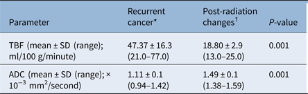
Table 1 shows the means, standard deviations and ranges of tumour blood flow (ml/100 g/minute) and apparent diffusion co-efficient (×10−3 mm2/second) values for recurrent head and neck cancer and post-radiation changes.
Tumour blood flow and apparent diffusion co-efficient values for recurrent head and neck cancer and post-radiation changes

*n = 31; †n = 16. TBF = tumour blood flow; SD = standard deviation; ADC = apparent diffusion co-efficient
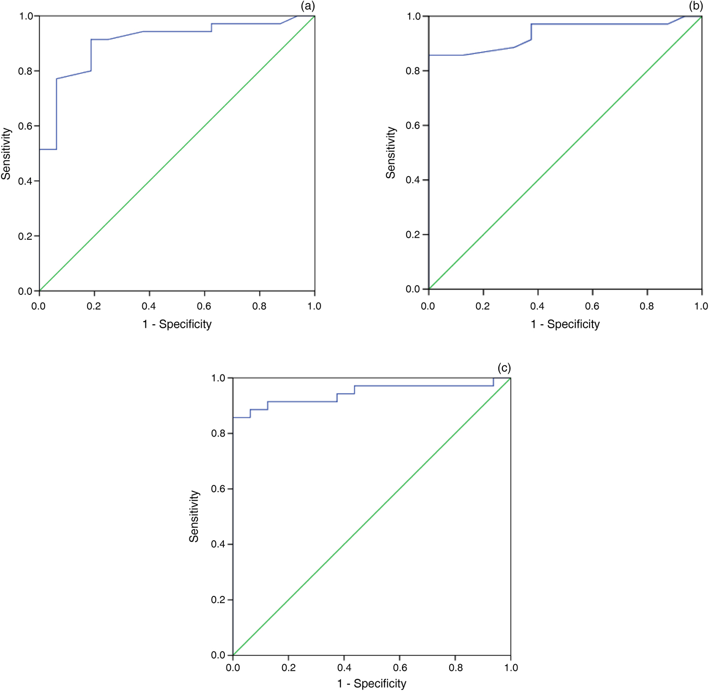
The tumour blood flow for patients with recurrent head and neck cancer (n = 31) ranged from 21.0 to 77.0 ml/100 g/minute, with a mean value of 47.37 ± 16.3 ml/100 g/minute. The tumour blood flow for patients with post-radiation changes (n = 16) ranged from 13.0 to 25.0 ml/100 g/minute, with a mean value of 18.80 ± 2.9 ml/100 g/minute. The difference in tumour blood flow between recurrent tumour and post-radiation changes was statistically significant (p = 0.001). The best results were obtained when a tumour blood flow value of more than 24.0 ml/100 g/minute was used for differentiating tumour recurrence from post-radiation changes, with an area under the curve of 0.94, accuracy of 88.2 per cent, sensitivity of 85.7 per cent, specificity of 93.8 per cent, a positive predictive value of 96.8 per cent and a negative predictive value of 75.0 per cent (Table 2 and Figure 2).
Receiver operating characteristic curve results for (a) apparent diffusion co-efficient, (b) tumour blood flow, and (c) combined apparent diffusion co-efficient and tumour blood flow values, used to differentiate recurrent head and neck cancer from post-radiation changes (the diagonal segments are produced by ties). (The thresholds used are detailed in the main text.)

Receiver operating characteristic curve results of tumour blood flow and apparent diffusion co-efficient for recurrent head and neck cancer versus post-radiation changes

AUC = area under the curve; PPV = positive predictive value; NPV = negative predictive value; TBF = tumour blood flow; ADC = apparent diffusion co-efficient
The apparent diffusion co-efficient for patients with recurrent tumours ranged from 0.94 to 1.42 × 10⁻³ mm2/second, with a mean value of 1.11 ± 0.1 × 10⁻³ mm2/second. The apparent diffusion co-efficient for patients with post-radiation changes ranged from 1.38 to 1.59 × 10⁻³ mm2/second, with a mean value of 1.49 ± 0.1 × 10⁻³ mm2/second. The difference between the apparent diffusion co-efficient values for recurrent tumour and post-radiation changes was statistically significant (p = 0.001). When an apparent diffusion co-efficient value of 1.21 × 10⁻³ mm2/second or less was used as the cut-off value for differentiating recurrence from post-radiation changes, the area under the curve was 0.90, accuracy was 88.2 per cent, sensitivity was 91.4 per cent, specificity was 81.2 per cent, the positive predictive value was 91.4 per cent and the negative predictive value was 81.2 per cent (Table 2 and Figure 2).
The combination of a tumour blood flow value of 24.0 ml/100 g/minute and an apparent diffusion co-efficient value of 1.21 × 10⁻³ mm2/second was used as a cut-off value for the differentiation of recurrence from post-radiation changes, with an area under the curve of 0.96, accuracy of 90.2 per cent, sensitivity of 88.6 per cent, specificity of 93.8 per cent, a positive predictive value of 96.9 per cent and a negative predictive value of 78.9 per cent (Table 2 and Figure 2).
Discussion
In this study, the main finding was that arterial spin labelling can be used for assessing head and neck cancer after treatment. Recurrent head and neck cancer was associated with higher tumour blood flow and restricted diffusion, with a reduced apparent diffusion co-efficient. In comparison, post-radiation changes were associated with lower tumour blood flow and no significant restriction in diffusion.
In this study, the tumour blood flow for recurrent head and neck cancer was significantly different to that for post-radiation changes. This is attributed to increased vascularity with increased capillary perfusion in recurrent or residual tumours compared to post-radiation changes. The tumour blood vessels are typically dilated and tortuous, with an abnormal branching pattern, dead ends, and no organisation into arterioles, capillaries and venules.Reference Razek, El-Serougy, Abdelsalam, Gaballa and Talaat16, Reference Fujima, Kudo, Tsukahara, Yoshida, Sakashita and Homma17 One study reported that tumour blood flow can be useful for determining local control, and the combined use of the percentage change of tumour blood flow and tumour volume had particularly high diagnostic accuracy.Reference Fujima, Yoshida, Sakashita, Homma, Tsukahara and Tha18 Another study showed that pre- and post-radiation tumour blood flow differed significantly.Reference Fujima, Kudo, Yoshida, Homma, Sakashita and Tsukahara19 Post-radiation tumour blood flow was significantly higher in patients with residual tumours than in those without. The tumour blood flow reduction rate was significantly lower in patients with residual tumours than in those without.Reference Fujima, Kudo, Yoshida, Homma, Sakashita and Tsukahara19
In this study, the apparent diffusion co-efficient for recurrent head and neck cancer was lower than that for post-radiation changes. Recurrent head and neck cancer showed restricted diffusion, with a low apparent diffusion co-efficient compared with radio-necrosis, presumably due to increased free water in necrosis and increased cellularity in recurrent tumours.Reference Abdel Razek21, Reference Varoquaux, Rager, Dulguerov, Burkhardt, Ailianou and Becker22 As diffusion within recurrent or residual tumours is impeded by the presence of cellular membranes and macromolecular structures, treatment with radiation and/or chemotherapy triggers cell death that can result in the loss of cell membrane integrity and reduced cell density, which can be detected as an increased mean diffusion value for the tumour.Reference Hwang, Choi, Kim, Kim, Lee and Yun23, Reference Abdel Razek, Kandeel, Soliman, El-shenshawy, Kamel and Nada24 One study reported that the apparent diffusion co-efficient value for residual or recurrent tumours was significantly lower than that of post-radiation changes.Reference Abdel Razek, Kandeel, Soliman, El-shenshawy, Kamel and Nada24 Another study found that bi-exponential fitting of the apparent diffusion co-efficient values on diffusion-weighted MRI were significantly lower in patients with residual tumours compared to those without tumour.Reference Hauser, Essig, Jensen, Laun, Münter and Maier-Hein25 Lastly, advanced diffusion fitting models such as diffusion kurtosis imaging and the stretched exponential model are the diagnostic tools for detecting residual tumours in post-treatment granulation.Reference Fujima, Yoshida, Sakashita, Homma, Kudo and Shirato26
In this study, combined tumour blood flow and apparent diffusion co-efficient values increased the diagnostic accuracy of MRI in the differentiation of recurrent head and neck cancer from post-radiation changes. A few studies have suggested that multi-parametric MRI, with the calculation of different biomarkers of diffusion and perfusion parameters, may help in differentiating recurrence from post-radiation changes.Reference Min, Lee, Lin, Holloway, Wijesekera and Gooneratne27–Reference King and Thoeny29
• There was a significant difference (p = 0.001) in tumour blood flow between recurrent head and neck cancer and post-radiation changes
• There was a significant difference (p = 0.001) in apparent diffusion co-efficient between recurrent head and neck cancer and post-radiation changes
• Arterial spin labelling is a non-invasive imaging method for assessing head and neck cancer after radiotherapy
• Combined tumour blood flow and apparent diffusion co-efficient values can predict head and neck cancer recurrence after radiotherapy
There are a few limitations to this study. First, there was no follow up of patients. Further studies, which monitor patients after radiotherapy, are recommended. Second, we utilised the calculation of tumour blood flow. Applications of advanced post-processing techniques for tumour blood flow, with the creation of parametric blood flow maps, and use of diffusion tensor imaging or kurtosis imaging, will improve the results.Reference Fujima, Yoshida, Sakashita, Homma, Kudo and Shirato26, Reference Abdel Razek, Elkhamary, Al-Mesfer and Alkatan30–Reference Razek36
Conclusion
The combination of tumour blood flow and apparent diffusion co-efficient values can play a role in differentiating recurrent head and neck cancer from post-radiation changes.
Competing interests
None declared