Introduction
Malignant mesodermal tumours of the larynx are rare malignancies compared with epithelial tumours: they account for less than 1 per cent of all malignant laryngeal tumours.Reference Paczona, Jóri, Tiszlavicz and Czigner1 Leiomyosarcomas usually originate in the uterus, stomach, extremities or retroperitoneum. Only 3 per cent of cases occur in the head and neck area.Reference Goldberg, Hanft and Ossakow2 The majority of leiomyosarcomas of the head and neck arise in the oral cavity, scalp, paranasal sinuses and jaws.Reference Marioni, Bertino, Mariuzzi, Bergamin-Bracale, Lombardo and Beltrami3 To the best of our knowledge, there have been approximately 50 laryngeal leiomyosarcomas reported in the English language literature.
In this report, we present two cases of laryngeal leiomyosarcoma occurring synchronously with squamous cell carcinoma.
Case reports
Case one
A 50-year-old man was referred to our clinic for evaluation of hoarseness which had been present for approximately three months. He had no history of alcohol consumption but had smoked one pack of cigarettes (20 cigarettes) daily for 25 years.
Indirect laryngoscopy revealed a lesion on the right vocal fold extending to the anterior commissure. Both vocal folds were mobile. Palpation of the neck was negative for lymphadenopathy.
Microlaryngoscopy was performed and showed a polypoid lesion arising from the right vocal fold and extending to the anterior commissure and 5 mm to the subglottic area on the right side. Pathological examination revealed the presence of both leiomyosarcoma and squamous cell carcinoma.
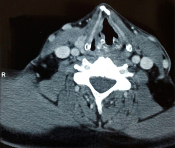
Cervical computed tomography examination demonstrated a soft tissue mass in the right vocal fold extending to the anterior commissure, with no evidence of lymphadenopathy (Figure 1). Investigation for distant metastases was negative. It was concluded that the tumour-node-metastasis (TNM) staging of the tumour was T1 N0 M0, according to the American Joint Committee on Cancer 2002 Staging Manual (6th edition).Reference Greene, Page, Fleming, Fritz, Balch and Haller4
Axial cervical computed tomography image of the first case, showing a soft tissue mass arising from the right vocal fold and extending to the anterior commissure.

Endolaryngeal CO2 laser resection was performed and the tumour completely removed. Frozen section analysis indicated that the surgical margins were clear.
Histological analysis revealed spindle cell proliferation with 12 mitotic figures per 10 high-power fields. The squamous component of the tumour was well differentiated. Immunohistochemically, the spindle cells were positive for vimentin and α-smooth muscle actin but negative for pancytokeratin.
The patient made an uneventful recovery and was discharged on the ninth post-operative day. Radiotherapy was not administered.
At three-year follow up, the patient did not show any evidence of recurrence or distant metastases, after physical and radiological examinations.
Case two
A 55-year-old man was admitted to our clinic with dyspnoea and hoarseness over the previous 9 months. He had smoked one pack of cigarettes daily for 35 years.
Fibre-optic laryngoscopy revealed a lesion on the right vocal fold extending to the ventricle, ventricular fold, anterior commissure and anterior one-third of the left vocal fold. The right vocal fold was paralysed. Palpation of the neck was negative for lymphadenopathy.
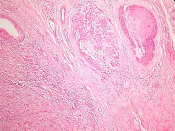
Microlaryngoscopy was performed, and showed that the lesion extended for approximately 10 mm to the subglottic area on the right side. Multiple punch biopsies were taken. Pathological examination revealed the presence of both leiomyosarcoma and squamous cell carcinoma (Figure 2).
Photomicrograph of biopsy specimen for case two, showing the presence of leiomyosarcoma (left side of image) and squamous cell carcinoma (upper right side of image). (H&E; ×200)

Investigation for distant metastasis was negative. The TNM staging of the tumour was T3 N0 M0.
Total laryngectomy, right lateral neck dissection and right total thyroid lobectomy were performed.
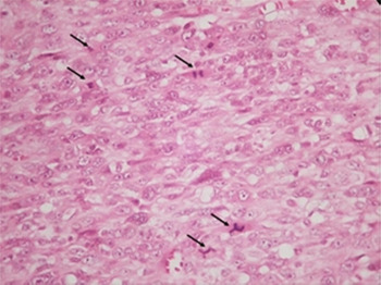
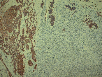
Histopathological examination revealed leiomyosarcoma and squamous cell carcinoma on both vocal folds. No lymphatic metastases were detected in the neck dissection specimens. No invasion was seen in the cartilage; however, angiolymphatic and neural invasion was established. Histological analysis showed spindle cell proliferation with 20 mitotic figures per 10 high-power fields (Figure 3). The squamous component of the tumour was well differentiated. Immunohistochemically, the spindle cells were positive for vimentin and α-smooth muscle actin but negative for pancytokeratin (Figure 4).
Photomicrograph of the surgical specimen for case two, showing increased mitotic activity (arrows) in the leiomyosarcoma component of the sample. (H&E; ×400)

Photomicrograph of the surgical specimen for case two, with immunohistochemical staining. Staining for Anti-pan Cytokeratin antibody is positive in the squamous cell carcinoma component but negative in the leiomyosarcoma component. (×200)

The patient made an uneventful recovery and was discharged on the 15th post-operative day.
At 15-month follow up, the patient was free of disease.
Discussion
Synchronous tumours of the larynx are extremely rare. To the best of our knowledge, only three cases have been previously described. The first was a chondrosarcoma with squamous cell carcinoma in situ,Reference Croce, Neri, Lattanzio and Angelucci5 while the second was a squamous cell carcinoma with adenocarcinoma.Reference Obermyer and Ramadan6 The third case was a synchronous tumour of leiomyosarcoma and squamous cell carcinoma, as in our case.Reference Tomidokoro, Hayashi, Yamasaki, Ishii, Kamijo and Ebihara7
The majority of malignant laryngeal tumours are squamous cell carcinoma. In the early stages, these can be successfully treated with partial laryngectomy or radiotherapy. Radiotherapy is preferred by most early-stage patients because of the resultant high speech quality. However, in patients with anterior commissure infiltration, large tumour volume, deep ulceration and subglottic expansion, radiotherapy may not be effective. Surgery should be the first treatment choice for these patients.
In addition to tumour stage, the degree of squamous cell carcinoma differentiation is also important and should not be ignored when deciding on patient management. Poorly differentiated tumours have more aggressive spreading patterns and must be treated with radical surgery.
In contrast to squamous cell carcinoma, laryngeal leiomyosarcoma is uncommon. The latter tumour type tends to present at sites such as the gastrointestinal tract, uterus and retroperitoneum.
If leiomyosarcoma occurs in the head and neck, it may originate from the erector pili muscles or the walls of blood vessels. However, the origin of head and neck leiomyosarcomas is still controversial. One theory speculates that they arise from aberrant undifferentiated mesenchymal tissue.Reference Kaya, Saydam and Ruacan8 Studies on the histopathogenesis of synchronous tumours of the respiratory tract have concluded that sarcomatoid tumours originate from complete or incomplete mesenchymal metaplasia of carcinomatous tumours.Reference Nappi and Wick9, Reference Nappi, Glasner, Swanson and Wick10 Another study found that synchronous tumours expressed the same antigens.Reference Atasoy, Bozdoğan and Sak11 It has been demonstrated that the same p53 mutations are present in both parts of these biphasic tumours.Reference Holst, Finkelstein and Colby12, Reference Bodner and Koss13 Völker et al. investigated the losses and gains on some chromosomes in a metachronous tumour of the larynx, and suggested that these changes might be related to transdifferentiation of the tumour from squamous cell carcinoma to leiomyosarcoma.Reference Völker, Zettl, Haralambieva, Blume, Hagen and Müller-Hermelink14
Based on all these data, we conclude that synchronous tumours originate from transdifferentiation of the same cell type. Another controversy centres on the reason for this differentiation. In our view, genetic and pathological research will help to clarify this subject.
Conventional light microscopy has some restrictions in the diagnosis of mesenchymal tumours. Diagnosis should be supported by immunohistochemical investigation, and seems to have become easier in the last two decades due to improvements in immunohistochemical staining techniques. On immunohistochemical staining, leiomyosarcoma is usually positive for smooth muscle actin and vimentin but negative for cytokeratins.
Because of the paucity of reported laryngeal leiomyosarcomas, there is no consensus on the treatment of this tumour. The most widely accepted treatment for extralaryngeal leiomyosarcoma is excision of the tumour with a wide surgical margin. Because of the unpredictable behaviour of this disease and the limited surgical experience, many surgeons treat laryngeal leiomyosarcoma with total laryngectomy.Reference Paczona, Jóri, Tiszlavicz and Czigner1, Reference Tewary and Pahor15–Reference Eggstone and Wolf17 However, endolaryngeal resection or partial laryngectomy techniques have been used in selected patients.Reference Paczona, Jóri, Tiszlavicz and Czigner1, Reference Marioni, Bertino, Mariuzzi, Bergamin-Bracale, Lombardo and Beltrami3, Reference Lippert, Schlüter, Claassen and Werner18–Reference Yoshizaki, Horikawa, Nonomura and Furukawa21
As described above, our exhaustive literature review disclosed only one previously reported case with both leiomyosarcoma and squamous cell carcinoma (Table I).Reference Tomidokoro, Hayashi, Yamasaki, Ishii, Kamijo and Ebihara7 Therefore, we had very limited experience available to guide patient management. In the previously reported case, leiomyosarcoma and squamous cell carcinoma were separated by non-neoplastic mucosa. The tumour of the left vocal fold consisted of moderately to poorly differentiated squamous cell carcinoma, while the tumour of the right vocal fold was a polypoid leiomyosarcoma. This case was treated with partial laryngectomy, and the patient had been free of disease for approximately nine months when the case was published. As regards the other published synchronous tumours, the T3 chondrosarcoma with squamous cell carcinoma in situ was treated with total laryngectomy, while the squamous cell carcinoma with laryngeal adenocarcinoma was treated with combined radiotherapy and surgical treatment.Reference Croce, Neri, Lattanzio and Angelucci5, Reference Obermyer and Ramadan6
Synchronous laryngeal leiomyosarcoma and squamous cell carcinoma: reported cases

Y = years; T = tumour; SCC = squamous cell carcinoma; M = male
With the increasing number of laryngeal leiomyosarcomas reported in recent years, there has been a trend toward using conservative treatment techniques. Partial laryngectomy or endolaryngeal CO2 laser resection seem to give good functional outcomes if the extent of the disease permits such approaches.Reference Paczona, Jóri, Tiszlavicz and Czigner1 There have been 23 reported cases treated with primary CO2 laser surgery.Reference Paczona, Jóri, Tiszlavicz and Czigner1, Reference Lippert, Schlüter, Claassen and Werner18, Reference Lan, Guo and Chu20, Reference Fusconi, Magliulo, Pizzuti, Ceccarini, Soldo and de Vincentiis22, Reference Gude, Tisch, Kraft, Danz and Maier23 Sixteen of these patients survived, with no report of disease at final follow up, three died of their disease, and three required total laryngectomy for survival. In the final case, no information on patient outcome was available.
There have been 16 reported cases of leiomyosarcoma treated with primary total laryngectomy. Of these, 12 patients survived and 4 died from their disease. According to these results, partial laryngectomy outcomes are no worse than total laryngectomy in selected patients. First and foremost, these patients need careful evaluation in order to select the optimum therapy.
In our cases, leiomyosarcoma and squamous cell carcinoma were identified together. In the first case, a small ‘island’ of squamous cell carcinoma was identified overlying the leiomyosarcoma, upon histopathological examination. In this case, we used endoscopic CO2 laser resection to remove the entire tumour. This was a glottic T1 tumour, so we did not perform elective neck dissection because of this tumour's reported low rate of occult metastasis. In the second case, leiomyosarcoma and squamous cell carcinoma occupied both vocal folds and extended for approximately 1 cm to the subglottic area, causing right vocal fold paralysis. Therefore, we performed a total laryngectomy together with right neck dissection, the latter due to the squamous component of the lesion. This was a T3 tumour of the glottis and also extended to the supraglottic and subglottic areas. Metastases from leiomyosarcomas are usually haematogenic but are rarely encountered.Reference Marioni, Bertino, Mariuzzi, Bergamin-Bracale, Lombardo and Beltrami3, Reference Lippert, Schlüter, Claassen and Werner18 Lymphatic metastasis to the head and neck area is very rare and is mostly seen in the late stages of disease. Therefore, in patients with such synchronous tumours of the larynx, treatment evaluation of the neck should be related to the stage of the squamous component of the tumour.
• Selecting adequate treatment for synchronous tumours is difficult due to different spreading rates and metastatic patterns
• Tumour location, stage and lymphatic metastases must be carefully evaluated to determine best treatment
• For combined squamous cell carcinoma and leiomyosarcoma, complete excision seems best
• Partial laryngectomy techniques may be preferred if disease extent permits
• Radiotherapy and chemotherapy are probably ineffective
Smoking is the main cause of squamous cell carcinoma. Both our patients had smoked at least 20 cigarettes daily for at least 25 years. This explains the existence of the squamous part of their tumours.
Radiotherapy is known to be an effective treatment for squamous cell carcinoma but seems ineffective for leiomyosarcoma. The literature reports four cases of leiomyosarcoma treated with primary radiotherapy. This treatment failed in three cases, requiring total laryngectomy. The one patient who survived primary radiotherapy was still alive three months after treatment, but there was no information on long-term follow up. Chemotherapy has been tried for uterine leiomyosarcoma and found to be ineffective.Reference Peckham, Pinedo, Veronesi, McGee, Isaacson and Wright24